한국표면공학회지 J. Kor. Inst. Surf. Eng.
Vol. 48, No. 5, 2015.
http://dx.doi.org/10.5695/JKISE.2015.48.5.211
<연구논문>
ISSN 1225-8024(Print) ISSN 2288-8403(Online)
Luminescence Properties of Ba 3 Si 6 O 12 N 2 :Eu 2+
Green Phosphor
Van Duong Luong
a, Dinh Phuong Doan
a, Hong-Ro Lee
b*a
Institute of Materials Science, Vietnam Academy of Science and Technology, 18 Hoang Quoc Viet Road, Cau Giay District, Hanoi, Vietnam
b
Department of Advanced Materials Engineering, Chungnam National University, Daejeon 305-764, Korea
(Received October 12, 2015 ; revised October 21, 2015 ; accepted October 28, 2015)
Abstract
To fabricate white LED having a high color rendering index value, red color phosphor mixed with the green color phosphor together in the blue chip, namely the blue chips with RG phosphors packaging is most favorable for high power white LEDs. In our previous papers, we reported on successful syntheses of Sr
2-Si
5N
8:Eu
2+and CaAlSiN
3phosphors for red phosphor. In this work, for high power green phosphor, green- emitting ternary nitride Ba
3Si
6O
12N
2:Eu
2+phosphor was synthesized in a high frequency induction furnace under N
2gas atmosphere at temperatures up to 1400
oC using EuF
3as a raw material for Eu
2+dopant. The effects of molar ratio of component and experimental conditions on luminescence property of prepared phos- phors have been investigated. The structure and luminescence properties of prepared Ba
3Si
6O
12N
2:Eu
2+phos- phors were investigated by XRD and photoluminescence spectroscopy. The excitation spectra of Ba
3Si
6O
12N
2:Eu
2+phosphors indicated broad excitation wavelength range of 250 - 500 nm, namely from UV to blue region with distinct enhanced emission spectrum peaking at ≈ 530 nm.
Keywords : Oxonitridosilicate phosphor, White LED, Ba
3Si
6O
12N
2:Eu
2+, Photoluminescence
1. Introduction
LED is not a new invention and most of us are used to LEDs being red or green signal markers on Hi-Fi or television set. But these are so called low power LEDs. During the last couple of years high power LEDs, i.e. LEDs operating at powers of around 2 ~ 100 W, have reached a level of cost and performance that make them attractive to the general lighting industry. However, white LED which is essential to lighting industry has a still low color rendering index (CRI) problem for high light source efficiency(>180 lumen/watt) because energy-efficient LEDs often sacrifice color quality for efficiency.
While many LEDs on the market today focus on maximum efficiency, objects illuminated by them can
appear unsaturated, washed out, or just outright strange. To increase color rendering index value, red color mixed with the green color of the adapted phosphors separatively in the blue chip, namely the blue chips with RG phosphors packaging is most favorable for high light source efficiency of white LEDs. In our previous papers, we reported successful syntheses results of Sr
2Si
5N
8:Eu
2+and CaAlSiN
3:Eu
2+as a promising red phosphors
1-7). In this work, for high light source efficiency of green phosphor, green- emitting ternary nitride Ba
3Si
6O
12N
2:Eu
2+phosphor was synthesized in a high frequency induction furnace under N
2gas atmosphere at temperatures up to 1400
oC by using solid state second-step heat treat- ment process. SiN
4-base covalent nitride materials, including nitridosilicates, nitridoaluminosilicates and CaAlSiN
3:Eu
2+etc. are good host lattices for high power red phosphors. Ba
3Si
6O
12N
2:Eu
2+is a layer-like oxonitridosilicate green phosphor and consists of vertex-sharing SiO
3N-tetrahedra forming as fundamental
* Corresponding Author : Hong-Ro Lee
Department of Advanced Materials Engineering, Chungnam National University
E-mail : [email protected]
building units. These nitride materials have strong absorption of the light emitted by the LED chips, i.e.
UV (350 - 410 nm) or blue (440 - 480 nm) light. In general, the stronger absorption, the higher emitting efficiency. Another strong point is higher quantum or conversion efficiency. The other good point is chemical stability which refers to the stability in chemical composition and crystal structure of phosphor. Low chemical stability of a phosphor will not only make the production process more complex and costly, but also reduce the luminous efficiency and seriously shorten the lifetime of LED products.
From these point of view, in this work, we focused on the synthesis of Eu
2+activated Ba
3Si
6O
12N
2phosphors by the solid-state reaction method using multi-step heat treatment. Additionally these properties were compared with recently reported other reports on properties of phosphor synthesized from the ammonia nitridation of an oxide precursor in aqueous-solution. The excitation spectra of Ba
3Si
6O
12N
2:Eu
2+phosphors indicated broad excitation wavelength range of 250 - 500 nm, namely from UV to blue region with distinct enhanced emission spectrum peaking at ≈530 nm. With an increase of Eu
2+ion concentration, the peak position of emission in spectra was red-shifted from 520 to 540 nm.
Through multi-step heat treatment process, prepared phosphor showed excellent luminescence properties, such as high emission intensity and low thermal quenching. EuF
3was used as a raw material for Eu
2+dopant with N
2gas flowing instead of using commercial EuN chemical for Ba
3Si
6O
12N
2:Eu
2+synthesis. In this work, we report on Eu
2+activated Ba
3Si
6O
12N
2oxonitridosilicate green phosphor for white LEDs synthesized by solid state reaction
method using the second-step heat treatment. We have discussed the effect of the synthesis method on the relative emission characteristics by using photoluminescence (PL) and investigated the crystal structure by using powder X-ray diffraction (XRD).
2. Experimental
2.1 Synthesizing equipment and prepared chemicals Raw material compositions for Ba
3Si
6O
12N
2:Eu
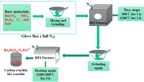
2+synthesis are indicated in the Table 1. Synthesis process for phosphors by high frequency induction heating was shown in Fig. 1. The high frequency induction furnace have temperature range from R.T.
to 2000
oC and temperature rate with 100
oC/min (increase) and 300
oC/min (decrease) respectively.
Vacuum was controlled as 10
−3Torr with 1000 ml/
min N
2gas flow rate.
2.2 Synthesis procedure and analysis 2.2.1 Synthesis of Ba
3Si
6O
12N
2:Eu
2+phosphors Ba
3Si
6O
12N
2:Eu
2+phosphors were synthesized by using a multi-step heating process from raw materials mixtures of barium carbonate (BaCO
3, 99.98%), silicon
Table 1. Prepared chemicals for Ba
3Si
6O
12N
2:Eu
2+synthesis
Chemical Molecular
Formula
Purity (%) Barium carbonate BaCO
399.98 Silicon dioxide (Nano Powder) SiO
299.50 Silicon nitride Si
3N
499.90 Europium (III) fluoride EuF
399.99
Activated carbon C 99.00
Fig. 1. Synthesis process for phosphors by high frequency induction heating.
dioxide (SiO
3, 99.50%), europium (III) fluoride (EuF
3, 99.99%), activated charcoal nano powder (C, 99.99%) and an excess of α-silicon nitride (α-Si
3N
4, 99.90%).
The concentration of Eu
2+varied in a range of 0 - 10 at% with respect to an excess of α-silicon nitride.
They were stoichiometrically weighed and mixed thoroughly in an agate mortar. Mixtures were charged into a graphite crucible inside a glovebox under Ar atmosphere and placed in a radio-frequency (RF) induction furnace at maximum temperatures of 2000
oC under N
2atmosphere with a flow rate of 1000 ml/
min during the heating process. For the first heating step, temperature was rapidly raised to 900
oC and maintained for 1 h to decompose BaCO
3completely.
After then, temperature was increased to 1200
oC and maintained for 3 h to form Ba
3Si
6O
12N
2:Eu
2+phosphor, which is called 1
stheat treated specimen. Finally, the 1
stheat treated specimens were transferred into a molybdenum crucible to obtain more homogeneous powder product of highly crytalline state, and sintered again at 1200
oC and 1400
oC respectively under N
2gas flow for 3 h to synthesize remaining small impurities of different Ba oxosilicate, BaSi
2O
2N
2:Eu
2+to re-synthesize as Ba
3Si
6O
12N
2:Eu
2+accor- ding to the reaction below
8);
BaSi
2O
2N
2:Eu
2++ 2BaO + 4SiO
2+ α-Si
3N
4→ Ba
3SiO
6O
12N
2:Eu
2++ α-Si
3N
4After firing, the obtained 2
ndheat treated phosphors were cooled down to 650
oC with a rate of about 0.33
oC/min to offer best conditions for good crystallinity in a furnace under a continuous flow of N
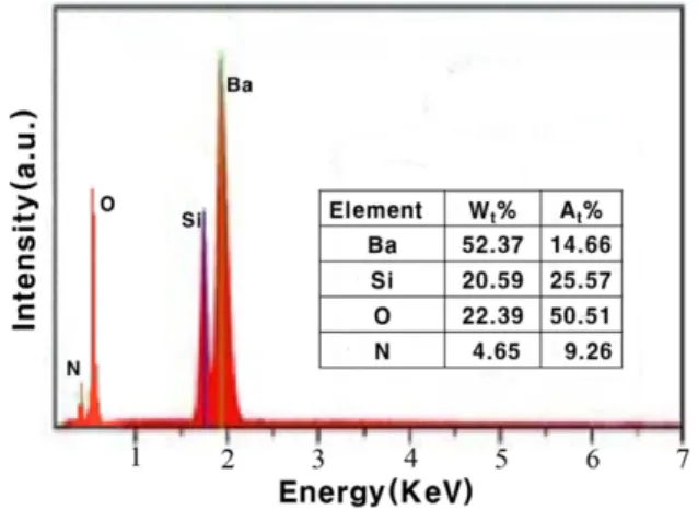
2gas. The atomic ratio Ba:Si:O:N = 3:6:12:2 of Ba
3Si
6O
12N
2was confirmed by EDX (Energy-Dispersive X-ray Spectroscopy) analysis (Fig. 2).
2.2.2 Structure and photoluminescence of Ba
3Si
6O
12N
2:Eu
2+The crystalline structure of Ba
3Si
6O
12N
2:Eu
2+powder was measured using X-ray powder diffrac- tion (SIEMENS X-ray diffractometer) with Cu K
αradiation (λ = 1.5406 Å . The data were collected in the 2θ range from 10 to 70° with a scanning rate of 3°/min. Diffuse reflection spectra were obtained using a BaSO
4powder calibrated UV–Vis spectro- photometer (UV-2200, SHIMADZU). Photolumine- scence (PL) measurement was carried out at room temperature using 405 nm as the excitation wave- length with a Perkin Elmer LS-45 luminescence spec- trometer. The temperature dependence of photolu- minescence was measured with a multichannel spectrophotometer (model MCPD7000; Otsuka Elec- tronics) equipped with temperature-controlled sample holders and a Xe lamp. Oxygen, nitrogen, and carbon residual impurity content of obtained phosphors were measured by using an oxygen/nitrogen analyzer (EMGA-930; HORIBA) and carbon/sulfur analyzer (SLE-CS-8520; SPECTRO).
3. Results and Discussion 3.1 Structure and residual concentration
Figure 3 gives the X-ray diffraction pattern of the 1
stand 2
ndheat treated specimen at 1200
oC and 1400
oC respectively for comparison. Most of the diffraction peaks of synthesized Ba
3Si
6O
12N
2:Eu
2+phosphors showed identical peaks to the standard specimen and no apparent impurity phases were found. Moreover, it was also indicated that the doped
Fig. 2. EDX spectrum of 2
ndheat treated Ba
3SiO
6O
12N
2:Eu
2+phosphor.
Fig. 3. XRD patterns of synthesized Ba
3Si
6O
12N
2:Eu
2+phosphors (a) 1200
oC,2
nd, (b) 1200
oC,1
st, (c)
1400
oC,2
ndand (d) 1400
oC, 1
stheat treatment.
Eu
2+ions did not caused any significant change in the host structure, because of close ionic radii of Ba
2+, Si
2+and Eu
2+. Activated carbon nano powder which was used as reductant to decompose oxide raw materials was also not detected. After the 1
stheat treatment finishing, specimens were heat treated again each at 1200
oC and 1400
oC respectively to eliminate residual impurities as low as possible (Table 2). The crystal structure of obtained Ba
3Si
6O
12N
2:Eu
2+phosphors showed an trigonal crystal structure with lattice space group of (No.147) and lattice parameters of a=7.5046 Å and c=6.4703 Å
8). 3.2 Photoluminescence properties of Ba
3Si
6O
12N
2:Eu
2+In this work under near UV to blue light excitation a saturated green emission band with a peak wavelength of ≈ 530 nm was observed. The broad excitration band enables efficient excitation at wavelengths below 450 nm. Fig. 4. shows emission band peak at ≈ 530 nm of the Ba
3Si
6O
12N
2:Eu
2+(2 at %) phosphor at the excitation of 405 nm. Photolumine- scence (excitation/emission) spectra of the Ba
3Si
6O
12N
2:Eu
2+(2 at %) phosphor exhibits an intense green body color due to absorption of Eu
2+in the blue
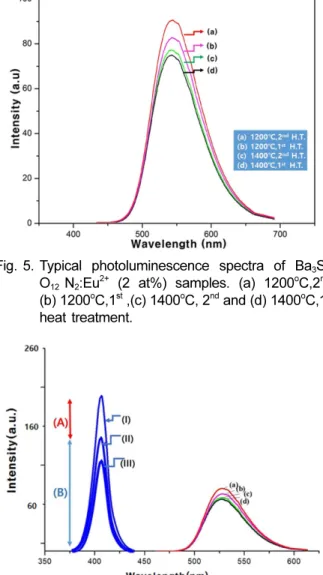
to green spectral range. Due to the high covalent environment around Eu
2+ion, emission of Ba
3Si
6O
12N
2:Eu
2+phophors synthesized at 1200
oC (Fig. 5(a), (b)) showed a fairly high intensity value but syn- thesized at 1400
oC showed a little decreased intensity, which may be owing to mainly a decomposition of Ba
3Si
6O
12N
2into BaSi
4O
6N
2or to sintering behavior of this material
8-9). Through multi-steps heat treatment process residual impurities could be lowered as low as possible (Table 2), which result in fairly enhanced luminescence intensity.
3.3 Reflection spectra and emission intensity The optical reflection spectra of Ba
3Si
6O
12N
2:Eu
2+phosphors with different Eu
2+ion concentration are shown in Fig. 6 (I, II, III). Reflection means total number of photons reflected and not absorbed by the P3
Table 2. Oxygen, nitrogen, and carbon residual con- tents (wt%)
Ba
3Si
6O
12N
2:Eu
2+O N C 1
stheat treated sample 36.56 3.65 0.08 2
ndheat treated sample 37.72 3.69 0.01
Ideal 38.09 3.70 0.00
Fig. 4. Typical photoluminescence spectra of excitation (a) and emission. (b) of 1200
oC, 2
ndheat treated Ba
3Si
6O
12N
2:Eu
2+(2 at%) samples.
Fig. 5. Typical photoluminescence spectra of Ba
3Si
6O
12N
2:Eu
2+(2 at%) samples. (a) 1200
oC,2
nd, (b) 1200
oC,1
st,(c) 1400
oC, 2
ndand (d) 1400
oC,1
stheat treatment.
Fig. 6. Reflection spectra of (Ba
3-xEu
x)Si
6O
12N
2pho-
sphor relation to emission intensity. (a) 2.0,
(b) 4.0, (c) 6.0 and (d) 1.0 at % of Eu
2+.
phosphor. According to reflection light curves (II, III) become decreased comparing to excited light curve (I), namely absorption part (A) increase relatively to reflection part (B), the efficiency of the phosphor performance in absorbing an external excitation light increase. This high absorbing efficiency as well as consistency are required for high quantum efficiency of phosphor. The absorption is related to the electronic transition of the Ba
3Si
6O
12N
2host and the 4f
7(
8S
7/2) → 4f
65d
1transition of Eu
2+ion
8). When applying excited light (curve (I)), as the reflection intensity changed from curve (II) to curve (III), absorption intensity (A) was increased and reflection intensity (B) was decreased. This may be caused by strong absorption according to increasing Eu
2+concentration up to 2 at% (i.e. x = 0.02), which means to become gradually distinct green-emitting state. At 2 at % Eu
2+concentration, Ba
3Si
6O
12N
2:Eu
2+phosphors showed lowest reflection intensity (III) but highest emission intensity (Fig. 4 curve (a)).
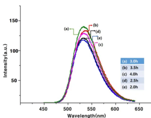
Dependence of emission intensity and peak emission wavelength on Eu
2+concentration of the (Ba
3-xEu
x)Si
6O
12N
2phosphor is shown in Fig. 7. With increasing Eu
2+content, emission intensity of (Ba
3-x