결장암에 대한 활성 자연살해세포의 항암효농
성 혜 란 * • 김지연 • 박민경 • 김일회 • 이 동 욱 • 한상배 • 이종길 • 송 석 길 ^ 층 복 대학교 약학대학, Cancer Center and Research Institute (Received May 10, 2010: Revised May 18, 2010: Accepted May 18, 2010)
Anticancer Effect of Activated Natural Killer Cells on Human Colorectal Tumor
Hyeran Sung*, Jee Youn Kim, M in Gyeong Park, Il-Hoi Kim, Dong Wook Lee, Sang-Bae Han, Chong-Kil Lee and Sukgil Song끓
College of Pharmacy, Chungbuk National University, Cheongju 361-763, Korea
^Department of Comprehensive Melanoma Research Center (CMRC),
H. Lee Moffitt Cancer Center & Research Institute, 12902 Magnolia Drive, Tampa, Florida 33612
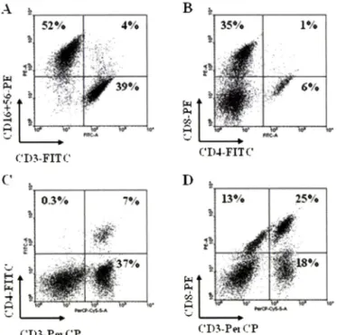
A b stra ct —— Colorectal cancer is one of the most common alimentary malignancies. In this study, the antitumor activity of activated human natural killer (NK) cells against human colorectal cancer was evaluated in vivo. Human NK cells are the key contributors of innate immune response and the effective functions of these cells are enhanced by cytokines. Human peripheral blood mononuclear cells (PBMC) were cultured with interleukin-2 (IL-2)-containing medium for 14 days and resulted in enriched NK cell population. The resulting populations of the cells comprised 7% CD3■느CD4■느 cells, 25%
CD3'"CD8'" cells, 13% CD3 CD8'' cells, 4% CD3''CD16/CD56'" cells, 39% CD:TCD16/CD56' cells, and 52% CD3 CD16/
CD56두 cells. Tumor necrosis factor alpha (TNF-a), interferon gamma (IFN-y), IL-2, IL-4, and IL-5 transcripts of the acti
vated NK cells were confirmed by RT-PCR. In addition, activated NK cells at doses of 2.5, 5 and 10 million cells per mouse inhibited 10%, 34% and 47% of SW620-induced tumor growth in nude mouse xenograft assays, respectively. This study sug
gests that NK cell-based immunotherapy may be used as an adoptive immunotherapy for colorectal cancer patients.
Keyw ords □ natural kille r cells, adoptive immunotherapy, SW620 colorectal cancer
결 장 암(Colon cancer)은■ 남녀 모두에게 흔히 발 병하는 악성중 양으로써 암 사 망 률3위에 해 당 된 다 .^ 결 장 암 의 발 생 은 개발도 상 국 과 선 진 국 모 두 과거에 비해 증 가 하 는 추 세 이 며, 소 화계에 새로이 진 단되는 암 중 40%를 차지한다고 보 고 되 었 다.2* 결장암 은 수 술 및 보 조 화 학 요 법 으 로 치 료 한 다.^' 이 러한 치료법이 확 설히 암 치료의 향상에 도 움 을 주 지 만, 2기 흑 은 3기의 결 장 암 환자의 경우 치 료 3년 내에 재 발하거나 전이가 발 생 한 다:® 사 망 자의 대부분이 간 과 같은 기관으로의 전 이가 발 생 하 였 으 며, 이 는 암 환 자 사 망 원인 중 50% 에 해 당 된 다 .® 결 장 암 환자의 생 존 율 을 높이기 위 해 서 는 효 과적인 대체 보 조 요법에 대 한 개발 이 절 실하 다. 면 역요법은 기존의 화학요법제인 항암제 가 가졌던 득성 문제률 해결하고 암세포에 특이적으로 작용하 며, 전이된 암
본 논문에 관한 문의는 저자에게로 (전화) 043-261-2817 (팩스) 043-268-2732 (E-mail) songs(a'chungbuk.ac.kr
세포까지의 치료가 가능하다는 장점을 갖고 있어 새로운 치료 요 법으로 부각되 고 있 다.
자 연 살 해 세 포(N K cell)는 세포 득성 림프구로서 자 국 이 나 면 역 요 법(예방주사)없이 러스 감염 세 포 나 종 양 세 포를 용해시 키 며,® 표적에 대항하여 직접으로 세 포 득 성을 나 타 내 거 나, 세포 매개형 세포 득성 (antibody-dependent cell-mediated cytotoxicity, ADCC), T h l-typ e cytokine의 분 비, 대 식 세 포(DCs)와의 상호 작 용 파 같은 다 양 한 매 커 니 즘 으 로 조 절 된 다. 일 단 자 연 살 해 세 포 (N K cell)가 활 성 화 되 면, 이들은 perforin 및 granzyme 같은 세 포득 성 과 럽 을 분 비 하 거 나, T R IA L 흑 은 FasL 그리 고 T h l-typ e cytokine, chemokine 같은 세 포사멸 유 도 인 자률 발현시켜 표적 세포에게 손상을 입 힌 다. 또 한, 자 연 살 해 세 포(N K cell)의 세포득 성 활성은 세포표면의 수용체의 활 성 과 억제 신 호 로 조 절 된 다. 자연살해세포의 활성을 억제하는 대표적인 수용체로는 inhibitory kille r immunoglobulin-like receptors(KIRs)가 있 다. 표적세포에 게서 self-MHC class I 분 자 인식이 이 루 어 지 면, 이 러한 수용체
NKM 의 Colon Cancer 항암효과 193
돌이 억제 신 호 률 전달하여 활 성 화 신호 를 중 지 시 키 고, 자연살 해세포의 용 해와 매개된 normal M H C class I 발 현 한 다. 활성화 수 용 체 들 로 는 natural cytotoxicity receptors(NCRs)와 NKG2D 가 포 함 되 며, 표적 세포 표면의 리 간 드 로 분산되어 세포득성에 대해 균형 을 이 룬 다 .^ 자 연 살 해 세 포 의 역할 은 부 분 적 으 로 낮 은 수준의 M H C class I 분자의 발 현 하 는 종양에 대 한 보 조 면 역치료법으로의 사용의 가능성을 보 였 다.1오^
이 연구를 통하여 인간 결장암에 대한 활성화된 자연살해세포 의 항 암 능 력 을 시 험 하 였 다. 우 러 는 IL -2률 이 용 하 여 human peripheral blood mononuclear cells 에서 ex vivo expansion을 통해 자연 살 해 세 포 룰 세 포 수 룰 증 가 시 키 고, phenotypei:결정 지었으며 nude mouse xenograft model옴 이용하여 N K M의 항 암능력을 평 가 하 였 다.
실험방법
세포배양
SW620(ATCC # CCL-227) 세 포 는 51세의 코 카 시 안 남성 으 로부터 분리되어 확립된 colorectal adenocarcinoma celL로, 10%
fetal bovine serum, 100 U/m/ penicillin, 100 mg/ml strepto- mycin(Invitrogen, CA, USA)를 함 유 한 RPMI-1640 medium에 서 배 양하였다. N K세포의 증폭 은 건강한 지원자의 말초혈액 단 핵세포(peripheral mononuclear cells, PBMC)로 만 돌어 졌 다. 지 원자에게는 사전의 층분 한 설명과 자유의사에 의한 동의 률 거?!
후, heparin과 함께 4()m/의 혈 액 을 채 취 하 였 다. 말초혈액 단핵 세 포 는 buffy coats를 포 함 하 고 있기 때 문 에 Ficoll-Hypaque density centrifugation 분리법과 PBS틀 이용하여 세척하였다. 세
^ 5% human serum(Biowhittaker-Cambrex, Walkersville, M D)가 함유된 Lymphomedia로 1 x 10\:ells/m/로 현탁하였으며 , 고정화시킨 anti-CD3 antibody(OKT-3 10 ng/ml; BD Pharmingen, NJ, USA) 와 recombinant human IL-2(Proleukin 500 U/m/, Chiron, Em eryville, CA, USA) 함께 5일 동 안 배 앙 하 였 다. 5인 배양후 0K T -3를함유한 배지를버 리고, rhIL-2(500 U /W)와 5%
human serum이 함유된 새로운 배지로 교 체 한 뒤, rhIL-2가 힘■유 된 배지로 매 배앙주기 때마다 보충하뇌 준 다. 배양주기 동 안 세 포수는 약 1x10^cells/mm를유지하도록 한 다. 이와 같은 배양 파 정을통해 언은 세포의 생존률은 14일 배양후 73%로확인되었다.
세포 표현형 분석
인간항체인 anti-CD3-FITC/CD16+CD56-PE, anti-CD4-FITC/
CD8-PE/CD3-PerCP(BD Biosciences, CA, USA)를 이 용 하 여 enriched N K세포의 표현형을 분 석 하 였 다, 약 1x10^ cells를 1%
bovine serum albumin(BSA)를 첨가한 PBS/BSA buffer로 한번 세척한 후, PBS/BSA buffer 100 ^ 9H1 현 탁 시 켰 다. 이들 세포
에 항체를 첨가한 후 4"C에서 20분 동 안 반응시킨 후, PBS로 2 번 세 척 하 고 400 10/의 PBS로 다시 현 탁 시 켜 FACSCanto flow cytom eter(BD Biosciences, CA, USA)로 죽 정 하 여 W inM D I statistical software(Scripps, La Jolla, CA, USA)률 이 용하여 분 석하 였 다. N K M외 생 존 률 은 FACSCanto flow cytom eter(BD Biosciences, CA, USA)을 통해 죽 정 했 다. Propidium iodide(PI, 틀 이 용하 여 세 포 를 10분 간 염 색 하 였 으 며, PI positive 세 포 는 죽 은 세포로 간 주 하 였 다.
RNA분리 및 RT-PCR에 의한 m RNA 증폭
RT-PCR(Reverse transcriptase-polymerase chain reaction) 은 이 연구에서 사 용 하 였 던 활 성 화 된 N K M의 cytokine 발 현 을 분 석 하 기 위해 진 행 하 였 다. 세 포 는 phytohemagglutinin(PHA;
3 |ig /m/)에 0, 0.3, 1, 5시간 노 출 시킨 뒤, TR IZO L™ reagent (Molecular Research Center, Cincinnaati, OH, USA)를 이 용 하 여 total RNA를 추 출 하 였 다. 얻 어 낸 total R N A는 AccuPower RT PreM ix PCR kit(Bioneer, Daegeon, Korea)률 이 용 하 여 semiquantitative reverse transcription-polymerase chain reaction(RT-PCR)를 진 행 하 였 다. RT-PCR반 응 은 얻 은 cD NA 1 |a/를 template로 하 여, 94°C에서 30초 년 성(denaturation), 5 4 X에서 (IL-2, IL-4, IL-5, and p-actin) 흑 은 5 7 X (T N F -a and IFN -y) 에서 30초 어닐링(annealing), 72°C 에서 30초 신 장 (extension)의 과정으로 이 루어지는 1사 이 클 을 30회 반복 수 행 하 였 다(Applied Biosystems, Foster City, CA, USA).
PCR반응에 사 용 된 prim er의 염 기 서 열 파 증 폭 산 물 의 크 기 는 hIL-2, sense 5'-AACAGTGCACCTACTTCAAG-3', antisense 5'-GTTGAGATGATGCTTTGACA-3', 3 9 8 bp; hIL-4, sense 5 - TCTCACCTCCCAACTGCTTCC-3', antisense 5'-CGTTTCA- GGAATCGGATCAGC-3\ 321 bp: hIL-5, sense 5'-TGCCTAC- GTGTATGCCATCCC-3', antisense 5'-CTTGGCCCTCATTCT- CACTGC-3', 4 3 8 bp; hTNF-a, sense 5-GAGTGACAAGCCT- GTAGCCCATGTTGTAGCA-3', antisense 5'-GGCAATGATC- CCAAAGTAGACCTGCCCAGACT-3', 445 bp; hlFN-y, sense 5 '-G C A T C G T T T T G G G T T C T C T T G G C T G T T A C T G C -3 ', antisense 5'-C TC C TTTTTC G C TTC CCTG TTTTAG C TG C T- GG-3', 427 bp; hb-actin, sense 5'-GGGTCAGAAGGA TTC C - TATG-3', antisense 5'-GGTCTCAAACATGATCTGGG-3', 238 bp이 다. Human (3-actin은 cD N A 합성의 대 조 군(control)으로써 PCR을 진 행하였다. 증폭된 PCR 산 물은 1% agarose gel에서 전 기 영동을 하여 분 석 하 였 다.
Nude mouse xenograft 분석
Human tum or xenograft 살험을 위해 SLC Japan, Inc에서 생 산 된 Specific pathogen-free female BALB/c-nu/nu mice(nude
Fig. 1 - The phenotypic characterization of the activated NK cells, which was used in nude mouse xenograft assay. Human PBMCs were cultured in the presence of IL-2 for 14 days and the resulting NK cell populations were stained with human antibodies, such as anti-CD3-FITC/CD16+ CD56- PE (A) and anti-CD4-FITC/CD8-PE/CD3-PerCP (B, C, and D), followed with FACS analysis.
mice)를 사 용 하 였 다. M ice는 ( 6 - 8 weeks old) 중 북대학교 실험 동물연구지원센터의 SPF시설에서 1주일 동 안 순화시킨 후 진행 하 였 다. 시험 당 일(0 day) SW620을 2 x lO S xlls/m ouse의 농 도 로 nude mice에 괴 하 이 식 하 였 다. N K M은 1주일에 한 번 씩(day 0, 7, 14) 2.5, 5, 1 0 x l0 \:e lls /m o u s e로 정맥 투 여 하 였 다. 양성 대 조 군 인 Adriamycin(ADR; Sigma-Aldrich, St. Louis, USA)도 매주 1회씩 총 3회 투 여하였다. Tumor vo lu m e fr 가 로(mm) x세 로(m m )x높이 (mm)/2로측정하였다. 최중일(16 day)에 종양을: 분 리 한 후 무 게 률 측 정 하 였 다. N K M의 득 성 을 확 인 하 기 위하여 nude mouse의 체중변화를 죽 정 하 였 다.효^^
자료분석 및 통계처러
In vivo 실 험 결 과 는 설험 당 9마리 mice를 분 석 하 였 으 며, in vitro 결 과 는 three samples의 mean values로 나 타 내 였 다. 표준 편 차(SD) 와 /M ^ u e s는 Student's ^-test 및 ANOVA(GraphI설d Prism, GraphPad Software, CA, USA)& 사용하뇌 산출하였다.오그^
실험결과
Activated human NK cell(NKM)의 표현형
인간말초혈액에서 C DJC D56'" N K cells은 약 5% 이 내를 차
52%
4
4%
r " "
CTO-FITC
0.3% 7%
,■평
35%
g
1%§ 6%
to* <0*
CD4-FITC
13%
i
25%
€ w
1활 18%
V w C m .P e i CP
지 하 는 것 으 로 알 려 져 있 다. 하 지 만, IL-2를 함 유 하 는 배지에서 14일 배앙 후, 5배 이상의 면역세포의 증 가 가 나 타 났 다. 배양한 세 포 률 fluorescence-activated cell sorting(FACS) 분 석 을 수행 하여 N K M의 표 현 형 을 확 인 하 였 다. N K M은 7% CD3'^CD4'"
cells, 25% CDS^CDS'" cells, 13% C D 3C D 8우 cells, 4%
CD3우CD16/CD56+ cells, 39% CD3'"CD16/CD56' cells, 52%
CD3CD16/CD56^ cells로 구 성 되 어 었 었 다(Fig. 1). Fresh PBMC의 CD3 CD56^ cells은 보통 15% 미 만 이 지만, IL-2와 함 께 14일 배양한후에는 73%로 증가했다. 대부분의 N K M & CD3' C D 16/C D 56\ CD3우CD16/CD56- cells으 로 거의 CD3누CD16/
CD56^ ce lls fr 아 니 었 다. 즉, N K M은 인체내에서 발견되는 N K cell의 표현형적 특 성 을 반영하고 있 었 다.
Activated human NK cell(NKM)의 cytokine transcript 발현
투 여 한 N K M 의 human TNF-a, INF-y, IL-2, IL-4, IL -5의 mRNA transcript level의 분 석 을 위해 PHA-stimulated 흑 은 unstimulated N K cells에서 주 출 한 total RNA를 이 용 하 여 semiquantitative RT-PCR를 시 행 하 였 다. Normal N K cells에서 의 IL-2, IL-4, IL-5 transcripts는 거의 발 견 할 수 없 었 지 만, PH A에 노 출 된 N K cells에 서 는 현 저 하 게 증 가 하 였 다. 하 지 만, normal N K cells 파 PHA-stimulated N K cells에 서 의 TNF-a, INF-y transcripts을 발 현 하 였 다(Fig. 2). INF-y, TNF-a 같 은
EL-2
0.3
IL
IL 5
TN F-a
IFN-7
P-iicrtii
Fig. 2 - Cytokine expression of the activated NK cells. The activated N K cells were stimulated with 0, 0.3, 1 /ml of phytohemagglutinin (PHA) for 5 h. Human TNF-ct, INF-y, IL-2, IL-4, and IL-5 gene expression levels were analyzed by semiquentitative RT-PCR.
B
M-sa>
D
i
%
nr
\
, AM9;;+9T<L>c
8 10 12 14 16 D a ys after tu m o rtra n s p la tls tio n
18
황 총 * A
m m • 츰 획
w w w w
m m % %
# 셜 # %
m m % m m
m % m m
m m 0 % #
# 4m
L 2 L
"o n tio l
N K c m
(2.5X10^ cells/mouse)
N K celLs
(5X10® cdls/m ouse)
N K cetl.<
(10X10® cells/mouse)
A D R
I)
Days after tum or transplantation
2 , 5 5 1 0 A D R
N K cel l s ( x l C f c e l l s / m o u s e )
Fig. 3 - Inhibition of SW620-induced tumor growth by activated NK cells in nude mouse xenograft models. Nude mice (n=9) were implanted subcutaneously with two million SW620 cancer cells. The activated NK cells at doses from 2.5 to 10 million cells per mouse were injected intravenously once a week. Adriamycin (ADR) was injected intravenously at 2 mg/kg. Tumor volumes were estimated by the formula: length (mm) x width (mm) x height (mm)/2. Statistical significance was determined using the Student's ^-test versus PBS- treated control group (*p<0.05, ***/)<0.001) (A). On day 16, the mice were sacrificed and the tumor weights were measured (B).
Representative photographs are shown (C). Statistical significance was determined using the Student's /-test versus PBS-treated control group (**/x0.01). The body weights of the tumor-bearing nude mice were measured to estimate toxicity (D).
NKM 의 Colon Cancer 항암효과 195
Th 1-type cytokines은 주 도 activated N K cells에서 생 산 된 다. activated N K c e lls ^ 또한 perforin, granzyme B 같은 cytolytic m oleculesi:발현함 을 확 인 하 였 다(data was not shown).
N K cell의 In vivo a n titu m o r 효과
Nude mouse xenograft 분석을 통하여 N K M의 항암효과를 검
증 하 였 다. 예 비 실 험 을 통하식 3 x 1 0 " cells(유 효 용량 의 약 30배) 의 N K M을 nude mice에 투 여 할 경우에 도 탈 모, 이 상 행 동, 체중 감소의 유 의 한 득 성 은 관찰되지 않 았 다.
SW620 세포를 nude mice 에게 2 x 10^ cells/mouse를 피 하 이 식하 여 16임_ 후 종 양 크 기 가 2 4 7 ± 4 7 m m크(w =9)로 성 장 하 였 다 (Fig. 3A). N K M은 2.5, 5, 1 0 x1 0산 cells/mouse의 능 도 로 정맥
려 여 ^면r나
0— ■써**증*"
00
— 0 — C o n
— HK.
- h r - f l K - - y t - N k - _ 0 ^ a o f
S36U23Apog
o
jj
<3
o
cly
8
o
o 0 0 0-0 0
0
o
@
C o
8.
O
o o B
— o — (
— t
- A - f
— f
— o ~ t
늑 35f m
5050
2
2
1
1
{아ELLO aEnloA JOEnl
50
주사하였으며, in vivo 종양성장의 각각 10%, 34%, 47%룰 억제 하였다. Adriamycin(ADR)는 양성 대조군으로 사용하였으며 , 이 는 SW620 중양성장에 강한 억제눙력을 지녔다.
최종일(16일)에 nude mouse에서 종양을 분리한 루 무게를 측 정하여 SW620 종양에 대한 NKM의 영향을 증명하였다(Fig. 3B and C). SW620 종양의 무게는 주입 16일 후 429 mg으로 무게 가 증가하였다. NKM을 5 X 10® cells/mouse 투여군에서는 25%
의 종양 성장 억제를, NKM을 10X10®cells/mouse 투여군에서 는 44%의 종양 성장 억제 효과률 나타내었다. 양성대조군으로 사용한 ADR 투여군에서는 52%의 종양 성장 억제 효과를 나타 내었다.
선반적으로, NKM 투여군에서 109-112%의 체중증가를 보였 으며, 이는 NKM이 체중에 미치는 영향이 없고 득성이 없옴을 시사한다(Fig. 3D).
결 론 및 고 찰
본 연구에서는 활성 자연살해세포의 인간 결장암(SW620)에 대한 항암효과를 nude mouse xenograft model을 이용하여 검 증하였다. 활성 자연살해세포의 득성을 확인하기 위하여 16일 동 안 nude mouse의 체중번화와 행동변화를 관찰하여 유효용량에 서의 득성이 없옴을 검증하였다. Nude mouse에 이식된 SW620 세포는 7일째부터 육안으로 관찰되기 시작하였으며 16일째까지 꾸준한 성장을 보였다. 최중일에 종양을 분리한 후 무게룰 측정 한 결과 NKM을 2.5, 5, 10x10® cells/mouse 투여에 의해 10%, 34%, 47%의 종양성장 억제률 확인하였다. 이러한 결2!■로부터 활 성 자연살해세포는 인간 결장암(SW620)에 대한 항암효과가 있 옴을 확인하였으며, 유효용량에서 득성이 없옴을 검증하쉬 세포 면역치료 법 로의 가능성을 제시하였다.
결장암(Colon cancer)은 사망에 이르는 대표적인 암으로 면역 성 종양으로 알려져있으며, 결장암•의 기존 치 료 법 ^ 는 수술과
화학요법이 있으나 이 치료법에 따른 생존률은 높지 않다. 5-
flurouracil-based regimens 나 irinotecan 같은 새로운 물질을 이 용하여 진행하는 Adjuvant systemic chemotherapy는 고 위험 질병의 환자둘의 생존율을 향상시켰다."®"I*"* 그럼에도 불구하고 결장암으로 진단받은 환자의 10년 후 생존율은 60%에 불과하다.
따라서, 수술과 화학요법을 대체할 보다 효과적인 치료법이 필 요하다.
지난 수십년간, 종양 면역학에 대한 연구가 활발히 이루어졌 다. 몇몇 mechanism은 결장암과 그에 대한 cellular 및 humoral immune responses에 대해 규명해주었다. 첫째, 많은 종앙 세포 들은 human leucocyte antigen(HLA) class I 발현을 억 제하거 나 감소시키기 때문에 cytotoxic T lymphocyte(CTL)이 필요로 하육T peptide룰 제시하지 못해 항원과 결합하지 못한다. 둘째,
결장암에서 peptide transporting molecules(TAP)의 번성이 관 찰되었으며, 이는 T cell epitope과 억제된 cellular immunity의 결과로 초래되었다고 보여진다.2® 셋째, 결장암에서 기존 종양과 전이 종앙 세포에서의 FasL 발현이 보다 더 번 번 했 다 T세포 표면에서 FasL이 Fas(Apo-VCD95)이 결합하는 것은 활성화 된 T세포의 apoptotic cell death을 야기한다. 넷째, 결장암 환자에 게서 peripheral blood lymphocyte 와 T cell infiltrates(TIL) 의 T 세포 수용체인 CD3 zeta chain의 발현 감소가 증명되었다.22>
면역세포에 기초한 암 치료법은 ex vivo로 확장되고 활성화 된 면역세포 이식을 통한 것이다. 대식 세포(dendritic cell), 자연살 해세포, 림포카인활성 살해세포(LAK cell), 싸이토카인 유도 살 해세포(CIK cell), 세포득성 T세포(cytotoxic T cell)와 같은 면 역세포는 암에 대한 active immunotherapy로 연구되었다.으고 하지만, DC therapy는 이식된 대식 세포가 activate effector T cells로의 번화가 어렵고 화학요법에 의해서 보통 억제된다.®*
CTL therapy는 MHC-restricted mechanism, tumor-associated antigen의 억제, tumor-specific CTL의 적은 수에 의해 저해된다.™
Solid tumors에 대한 NK와 LAK cell therapy의 효능은 cancer cell에 대해 제한된 세포득성 활성을 나타내었다.
NK cell activity가 낮다면 암으로 발전하기 쉬우며 ,® 암환자 의 NK cell activity는 건강한 사람에 비해 현저하게 낮 았 다.^
NK cell infiltration은 gastric carcinoma, squamous cell lung carcinoma, colorectal cancer 같은 몇몇의 암 종류에서 positive prognostic parameter로 이용된 다.그^체게다가, human malignant cells은 human NK cells 투여 후 약화되거나 전이가 억제되어진 다는■ 것■을: severe combined immunodeficiency(SCID) mouse model을 통 한 설험으로 규 명 하 였 다.^®'3®> 이 러 한 결 과 가 tumor ImmunosurveillanceS]- NK cell의 Immunotherapy strategiesS.
의 가능성을 제 시 해 준다.
전이성 신장세포암에 대한 autologous NK cells을 이용한 치 료 시도 역시 놀랄만한 중앙뢰화를 나타냈다. Brain tumor에서 도 역시 궁정 적인 치료 영향이 보 고 되 었 다 .샌 이 와 반대로, autologous NK cell의 adoptive transfer 효능은 제한적이라고도 증명되고 있다.
Solid tumors에 대한 NK cells 치료 효과는 시험에 사용하는 NK cell에 따라 다른 효눙을 나타냈다. 이는 고활성 NK cells틀 충분히 배양하기 어렵다는 것을 의미한다. 따라서, 안전한 임상 결과를 얻을 수 없었으며 실용적인 NK cell expansion 방법 연 구가 될요하다. 최근, GMP 조건하에 IL-2와 함께 a; ot'to에서의 NK cell expansion이 증명되었으며 이를 통해 제공되는 activated NK cells은 대체 요법으로 사용되어지고 었다/ 2
NK cell의 향암■효능은 다양한 종류의 동물 모형에서 증명되었 왔으며^46-48) 현재 임상단계에서 평가되고 있다;*®> vivo expanded human NK cells은 고 득성을 띄지 않을 뿐 아니라
NKM 의 Colon Cancer 힘^^효파 197
T h l-ty p e cytokines(e.g., IFN-y and T N F -a)을 다 량 생 산 하 며, L A K cells에 비 하 여 10배 이 상 에 달 하 는 cytotoxic activity와 absolute number를 나 타 냈 다 .페 따 라 서, 이견 실 험 방 법 과 비교 하 여, 높은 항암*효과룰 기대해 볼 만 하 다.
본 연구에서는 결 장암 유 발 nude mouse xenograft 모델에 활 성 화 된 N K cell의 항 암 효 과 룰 관 찰 하 였 으 며, N K cell im munotherapy 기능성을 증명하였다. Human PBMC를 IL-2가 함 유된 배지에서 14임 배양함으로써 CDJCD56"" 표 현형을 나타내 는 N K cell을 증 폭 시켰 다. mouse xenograft model을 이 용 한 N K M의 결장암에 대 한 항 암 효 과 는 향 후 결 장 암 치 료 를 위 한 cell immunotherapy로 가능성을 증 명 하 였 다.
감사의 말씀
본 연구는 2008년 충 북 대 학교 학술연구지원에 의하여 연구되
었 옴.
참고문헌
1) Davis, D. L., Hoel, D., Fox, J. and Lopez, A. : International trends in cancer mortality in France, West Germany, Italy, Japan, England and Wales, and the USA. Lancet 336, 474 (1990).
2) Jemal, A., Siegel, R., Ward, E., Hao, Y., Xu, J., Murray, T. and Thun, M. J . : Cancer statistics, 2008. CA Cancer. J. Clin. 58, 71 (2008).
3) Bar-Sela, G. and Haim, N . : Abnoba-viscum (mistletoe extract) in metastatic colorectal carcinoma resistant to 5-fluorouracil and leucovorin-based chemotherapy. Med. OncoL 21, 251 (2004).
4) de Gramont, A., Tournigand, C., Louvet, C., Maindrault- Goebel, E and Andr^, T. : First-line therapy for advanced colorectal cancer. Curr. OncoL Rep. 7, 167 (2005).
5) Joosten, J., Jager, G., Oyen, W, Wobbes, T. and Ruers, T. : Cryosurgery and radiofrequency ablation for unresectable colorectal liver metastases. Eur. J. Surg. OncoL 31, 1152 (2005).
6) Gravalos, C., Garcia-Sanchez, L., Hernandez, M., Holgado, E., Alvarez, N., Garda-Escobar, L, Martmez, J. and Robles, L. : Surgical resection of a solitary pancreatic metastasis from colorectal cancer: a new step to a cure? Clin. Colorectal.
Cancer. 7, 398 (2008),
7) Renouf, D., Kennecke, H. and Gill, S. ; Trends in chemotherapy utilization for colorectal cancer. Clin. Colorectal.
Cancer. 7, 386 (2008).
8) Kiessling, R., Klein, E. and Wigzell, H. : "Natural" killer cells in the mouse. I. Cytotoxic cells with specificity for mouse
Moloney leukemia cells. Specificity and distribution according to genotype. Eur. J. Immunol 5, 112 (1975).
9) Lanier, L. L. : N K cell recognition. Annu. Rev. Immunol 23, 225 (2005).
10) Bryceson, Y. T., March, M. E., Ljunggren, H. G. and Long, E. 0. : Activation, coactivation, and costimulation of resting human natural killer cells. Immunol Rev. 214, 73 (2006).
11) Ljunggren, H. G. and Malmberg, K. J. : Prospects for the use of NK cells in immunotherapy of human cancer. Nat Rev.
Immunol 7, 329 (2007).
12) Han, S. B,, Lee, C. W, Jeon, Y. J., Hong, N. D., Yoo, I. D., Yang, K. H. and Kim, H. M , : The inhibitory effect of polysaccharides isolated from Phellinus linteus on tumor growth and metastasis. Immunopharmacology 41, 157 (1999).
13) Han, S. B., Moratz, C., Huang, N. N., Kelsall, B., Cho, EL, Shi, C. S., Schwartz, O. and Kehrl, J. H. : Rgsl and Gnai2 regulate the entrance of B lymphocytes into lymph nodes and B cell m otility within lymph node follicles. Immunity 22, 343 (2005).
14) Greenlee, R. X, Murray, T , Bolden, S. and Wingo, R A. : Cancer statistics, 2000. CA Cancer. / Clin. 50, 7 (2000), 15) Moore, H. C. and Haller, D. G. : Adjuvant therapy of colon
cancer. Semin OncoL 26, 545 (1999).
16) Chung, K. Y. and Saltz, L. B. : Antibody-based therapies for colorectal cancer. Oncologist. 10, 701 (2005).
17) Kelly, H. and Goldberg, R. M . : Systemic therapy for metastatic colorectal cancer: current options, current evidence. J. Clin.
OncoL 23, 4553 (2005).
18) Pozzo, C., Barone, C., Szanto, J., Padi, E., Peschel, C., Bukki, J., Gorbunova, V, Valvere, V, Zaluski, J., Biakhov, M., Zuber, E., Jacques, C. and Bugat, R. : Irinotecan in combination with 5- fluorouracil and folinic acid or with dsplatin in patients with advanced gastric or esophageal-gastric junction adenocarcinoma:
results of a randomized phase II study. Ann. OncoL 15, 1773 (2004).
19) Todryk, S. M., Chong, H., Vile, R. G., Pandha, H. and Lemoine, N. R. : Can immunotherapy by gene transfer tip the balance against colorectal cancer? Gut 43, 445 (1998).
20) Yip, D., Strickland, A. H,, Karapetis, C. S., Hawkins, C. A. and Harper, R G. : Immunomodulation therapy in colorectal carcinoma. Cancer. Treat Rev. 26, 169 (2000).
21) Mann, B., Gratchev, A., Bohm, C., Hanski, M. L., Foss, H. D., Demel, G., Trojanek, B., Schmidt-Wolf, L, Stein, H., Riecken, E. 0., Buhr, H. J. and Hanski, C. : FasL is more frequently expressed in liver metastases of colorectal cancer than in matched primary carcinomas. Br. J. Cancer. 79, 1262 (1999).
22) Nakagomi, H., Petersson, M., Magnusson, I., Juhlin, C., Matsuda, M., Mellstedt, H., Taupin, J. L., Vivier, E., Anderson, E and Kiessling, R. : Decreased expression of the signal- transducing zeta chains in tumor-infiltrating T-cells and NK
cells of patients with colorectal carcinoma. Cancer. Res. 53, 5610 (1993).
23) Bremers, A. J., Kuppen, P J. and Parmiani, G. : Tumour immunotherapy: the adjuvant treatment of the 21st century?
Eur. J. Surg. Oncol. 26, 418 (2000).
24) Kalinski, R, Nakamura, Y., Watchmaker, R, Giermasz, A., Muthuswamy, R. and Mailliard, R. B. : Helper roles of NK and CD8+ T cells in the induction of tumor immunity. Polarized dendritic cells as cancer vaccines. Immunol Res. 36, 137 (2006).
25) Raja, Gabaglia, C., Diaz, de Durana, Y., Graham, F. L., Gauldie, J., Sercarz, E. E. and Braciak, T. A. : Attenuation of the glucocorticoid response during Ad5IL-12 adenovirus vector treatment enhances natural killer cell-mediated killing of MHC class I-negative LNCaP prostate tumors. Cancer Res. 67, 2290 (2007).
26) Takashima, K., Fujiwara, H., Inada, S., Atsuji, K., Araki, Y., Kubota, T. and Yamagishi, H. : Tracking of green fluorescent protein (GFP)-labeled LAK cells in mice carrying B16 melanoma metastases. Anticancer Res. 26, 3327 (2006).
27) Thome, S. H., Negrin, R. S. and Contag, C. H. : Synergistic antitumor effects of immune cell-viral biotherapy. Science 311, 1780 (2006).
28) Wang, W, Epler, J., Salazar, L. G. and Riddell, S. R. : Recognition of breast cancer cells by CD8+ cytotoxic T-cell clones specific for NY-BR-1. Cancer. Res. 66, 6826 (2006).
29) Kim, H. M., Kang, J. S., Lim, J., Park, S. K., Lee, K „ Yoon, Y. D., Lee, C. W, Lee, K. H., Han, G., Yang, K. H., Kim, Y. J., Kim, Y. and Han, S, B. : Inhibition of human ovarian tumor growth by cytokine-induced killer cells. Arch. Pharm. Res. 30, 1464 (2007).
30) Grabert, R. C., Cousens, L. P., Smith, J. A., Olson, S., Gall, J., Young, W. B., Davol, R A. and Lum, L. G. ; Human T cells armed with Her2/neu bispecific antibodies divide, are cytotoxic, and secrete cytokines with repeated stimulation.
Clin. Cancer. Res. 12, 569 (2006).
31) Bums, L. J., Weisdorf, D. J., DeFor, T. E „ Vesole, D. H., Repka, T. L., Blazar, B. R., Burger, S. R., Panoskaltsis-Mortari, A., Keever-Taylor, C, A., Zhang, M. J. and Miller, J. S .: IL-2-based immunotherapy after autologous transplantation for lymphoma and breast cancer induces immune activation and cytokine release: a phase I/II trial. Bone. Marrow. Transplant. 32, 177 (2003).
32) Imai, K., Matsuyama, S., Miyake, S., Suga, K. and Nakachi, K. : Natural cytotoxic activity of peripheral-blood lymphocytes and cancer incidence: an 11-year follow-up study of a general population. Lancet 356, 1795 (2000).
33) Bobek, V, Boubelik, M., Fiserova, A., L'uptovcova, M., Vannucci, L., Kacprzak, G., Kolodzej, J., Majewski, A. M. and
Hoffman, R. M. : Anticoagulant drugs increase natural killer cell activity in lung cancer. Lung. Cancer. 47, 215 (2005).
34) Whiteside, T. L. and Herberman, R. B. : Role of human natural killer cells in health and disease. Clin. Diagn. Lab. Immunol 1, 125 (1994).
35) Ishigami, S., Natsugoe, S., Tokuda, K., Nakajo, A., Che, X., Iwashige, H., Aridome, K., Hokita, S. and Aikou, T. : Prognostic value of intratumoral natural killer cells in gastric carcinoma. Cancer. 88, 577 (2000).
36) Takanami, L, Takeuchi, K. and Giga, M . : The prognostic value of natural killer cell infiltration in resected pulmonary adenocarcinoma. J. Thorac. Cardiovasc. Surg. 121, 1058 (2001).
37) Coca, S., Perez-Piqueras, J., Martinez, D., Colmenarejo, A., Saez, M. A., Vallejo, C., Martos, J. A. and Moreno, M. : The prognostic significance of intratumoral natural killer cells in patients with colorectal carcinoma. Cancer. 79, 2320 (1997).
38) Dewan, M. Z., Terunuma, H., Toi, M., Tanaka, Y., Katano, H., Deng, X., Abe, H., Nakasone, T , Mori, N., Sata, T. and Yamamoto, N. : Potential role of natural killer cells in controlling growth and infiltration of AIDS-associated primary effusion lymphoma cells. Cancer, Sci. 97, 1381 (2006).
39) Dewan, M. Z., Terunuma, H., Takada, M., Tanaka, Y., Abe, H., Sata, T , Toi, M, and Yamamoto, N. : Role of natural killer cells in hormone-independent rapid tumor formation and spontaneous metastasis of breast cancer cells in vivo. Breast Cancer. Res. Treat 104, 267 (2007).
40) Escudier, B., Farace, E, Angevin, E., Charpentier, E, Nitenberg, G., Triebel, E and Hercend, T. : Immunotherapy with interleukin-2 (IL2) and lymphokine-activated natural killer cells: improvement of clinical responses in metastatic renal cell carcinoma patients previously treated with IL2. Eur.
J. Cancer. 30A, 1078 (1994).
41) Ishikawa, E., Tsuboi, K., Saijo, K., Harada, H., Takano, S., Nose, T. and Ohno, T. : Autologous natural killer cell therapy for human recurrent malignant glioma. Anticancer. Res. 24, 1861 (2004).
42) Carlens, S., Gilljam, M., Chambers, B. J., Aschan, J., Guven, H., Ljunggren, H. G., Christensson, B. and Dilber, M. S. : A new method for in vitro expansion of cytotoxic human CD3- CD56+ natural killer cells. Hum. Immunol 62, 1092 (2001).
43) Klingemann, H. G .: Natural killer cell-based immunotherapeutic strategies. Cytotherapy 7, 16 (2005).
44) Alici, E., Sutlu, T, Bjorkstrand, B., Gilljam, M., Stellan, B., Nahi, H., Quezada, H. C., Gahrton, G., Ljunggren, H. G. and Dilber, M.S. Autologous antitumor activity by NK cells expanded from myeloma patients using GMP-compliant components. Blood. I l l , 3155 (2008).
45) Guven, H., Gilljam, M., Chambers, B. J., Ljunggren, H. G.,
NKM 의 Colon Cancer 후^^효과 199
Christensson, B., Kimby, E. and Dilber, M. S. : Expansion of natural killer (NK) and natural killer-like T (NKT)-cell populations derived from patients with B-chronic lymphocytic leukemia (B-CLL); a potential source for cellular im
munotherapy. Leukemia. 17, 1973 (2003).
46) Alici, E., Konstantinidis, K. V, Sutlu, T , Aints, A., Gahrton, G., Ljunggren, H. G. and Dilber, M. S. : Anti-myeloma activity of endogenous and adoptively transferred activated natural killer cells in experimental multiple myeloma model. Exp. Hematol 35, 1839 (2007).
47) Basse, P H., Whiteside, T. L. and Herberman, R. B. : Cancer immunotherapy with interleukin-2-activated natural killer cells. Mol BiotechnoL 21, 161 (2002).
48) Siegler, U., Kalberer, C. R, Nowbakht, R, Sendelov, S., Meyer-
Monard, S. and Wodnar-Filipowicz, A . ; Activated natural killer cells from patients with acute myeloid leukemia are cytotoxic against autologous leukemic blasts in NOD/SCID mice.
Leukemia. 19, 2215 (2005).
49) deMagalhaes-Silverman, M., Donnenberg, A., Lembersky, B., Elder, E., Lister, J., Rybka,W, Whiteside, T. and Ball, E. : Posttransplant adoptive immunotherapy w ith activated natural killer cells in patients with metastatic breast cancer. /.
Immunother. 23, 154 (2000).
50) Terunuma, H., Deng, X., Dewan, Z., Fujimoto, S. and Yamamoto, N. : Potential role of N K cells in the induction of immune responses: implications for N K cell-based immuno
therapy for cancers and viral infections. Int. Rev. Immunol 27, 93 (2008).