약학회 지 제 46 권 제 2 호 113~119 (2002) Yakhak Hoeji Vol. 46, No. 2
-
藥 學 含 확
홍삼 산성 다당체의 마크로파지 및 자연살해세포의 활성화에 의한 항암작용
김영숙 • 박경미 • 신한재 ■송 경식* • 남 기 열. 박종대# 한국인삼연초연구원,*경북대학교 농과대학 (Received January 8, 2001; Revised February 14,2002)
Anticancer Activities of Red Ginseng Acidic Polysaccharide by Activation of Macrophages and Natural Killer Cells
Young-Sook Kim, Kyeong-Mee Park, Han-Jae, Shin, Kyung-Sik Song*, Ki-Yeol Nam and Jong-Dae Park#
Korea Ginseng & Tobacco Research Institute, Taejon 305-345, Korea
^College of Agriculture, Kyungbook National University, Taegu 720-701,Korea
Abstract —— The composition of monosaccharides of acidic polysaccharide isolated from ethanol-insoluble and water-soluble fractions of red ginseng roots was analysed and its immunological activities were investigated. Red ginseng acidic polysac
charide (RGAP) was composed of glucose (26.1 mole %), arabinose (1.6 mole %), glucuroninc acid (51.8 mol %) and galac- turonic acid (5.1 mole %) as determined by gas liquid chromatography. Addition of RGAP increased production of nitric oxide (NO) and tumor necrosis factor (TNF)-a in the rodent macrophage cultures. Peritoneal macrophages from RGAP-treated mice exhibited potent tumoricidal activities toward P815 and WEHI 164 tumor cells. It was also observed that con
centrations of NO and TNF-a were high in the culture medium of macrophages from the mice administered with RGAP Moreover, treatment of RGAP in vivo stimulated tumoricidal activities of natural killer (NK) cells. Treatment with RGAP increased life span of sarcoma 180-bearing mice and decreased tumor weights of B 16-tumor-bearing mice. These results suggest that activation of macrophages and NK cells serve to enhance in vivo anticancer activities of RGAP
Keywords □ Panax ginseng, red ginseng acidic polysaccharide, macrophage, natural killer cell, anticancer activities.
인삼(Panax ginseng C. A. Meyer).0•로부터분리된다당성분 은항암작용을비롯한다양한면역조절작용을함이보고되고있 다.i-4) 일반적으로 산성다당체는 galacturonic acid, glucuronic acid 및 mannuronic acid가결합된분자량이 15,000 이상의 다 당체이며중성다당체에비해면역체계에미치는영향이크다.5〕백 삼으로부터분리된 저혈당작용을갖는 21종의 Panaxan A-U 중 산성다당체는8종이고나머지는중성다당체로알려져 있다.6ᅵ9) 인삼뿌리에서도세망내피계를활성화시키고항보체작용을갖는 수종의 다당체가분리되었으며 최근세망내피계를활성화하는 4 종의 ginsenan이분리되어 일부그구조가밝혀졌다.10) 인삼잎 으로부터 분리된세포벽 성분의 rhamnogalacturonan II을함유 하는다당체는 macrophage의 Fc receptor 발현을 촉진시켜 면
#본 논문에 관한 문의는 저자에게로 (전화) 042-866-5534 (팩스) 042-861-1949 (E-mail) [email protected]
역복합체(immune complex)의제거룰증강시킴이 보고되었다.u) 백삼의물주출물에서분리된 ginsan은 T hl 세포와 macrophage 유래의 사이토카인을유도하여 activated killer cell을 생성시켜 암세포를사멸시킬 수있음이 보고되었다.12) 여러연구를통하 여인삼에서 분리된다당체 성분은인삼의 약리 효능을 나타낼 수있는주요한 성분임이 입증되고있으나화학적 조성과구조 및작용기작이 아직 정확히 밝혀지지 않고있다.
면역계에서 macrophage는선천적 면역을담당히는주요한 세 포로서미생물등을식작용하고 NO, 반응성산소종및효소를분
비하여미생물과암세포* 죽이고감염의 확산을조절한다.13) 또
한 antigen-presenting cell(APC)로작용하여 T세포에의한특이 적면역을 개시하고조절한다. 활성화된 macrophage에서 생성 되는 tumor necrosis factor-(x(TNF-oc)는종양세포의 용혈성 괴 사를 일으키고 septic shock와국소염증조직에서 병태생리를 매기ᅵ하는사이토카인이다. TNF-a가표적세포의 표면에 수용체 와 결합하면 수용체의 death domain에 TRADD(TNF-R1
114 김영숙 • 박경미 • 신한재 . 송경식 • 남기열 • 빅종대
associated protein with death domain), FADD(Fas-associated death domain protein) 또는 RIP(Receptor inhibitory protein) 과 같은 death factor가결합하여 일련의신호전달을통해서표적세 포를 사멸시킨다.14’15) 한편 natural killer(NK) cell는 major histocompability(MHC) antigen의제한이나감작없이도암세포나 미생물과같은표적세포를죽일수있는임파구의한종류로서말 초혈액임파구의약 15%를차지하며선천적 면역을매개하는세 포이다.16’17) 또한 NK 세포는면역반응초기에사이토카인을 제 공하며 특이적 면역에 영향을미친다.18)
홍삼은수삼을증숙후건조하며 제조한것으로이수치과정 에서수삼또는백삼과는다른성분이 생성됨이 알려져있다. 그 예로서백심에존재하는 malonyl-ginsenoside Rbl, Rb2, Re, Rd 는 malonyl기가 떨어져서 ginsenoside Rbl, Rb2, Rc, Rd가 생 성되며 ginsenoside의 C-20위에서 glycosyl 잔기가이탈되거나, C- 20위에서 수산기가이성화되어 20(S)-ginsenoside Rg3, 20(R)- ginsenoside Rg2, 20(S)-ginsenoside Rh2, 20(R)-ginsenoside
R hl로 전환된다.19) 그러므로흥삼의 제조과정에서 화학성분의
변화가일어나백삼과는다른생리적활성성분의 함량차이를나 타내고있다. 다당체의 경우도백삼에서분리된 다당체와홍삼 에서분리되는다당체는화학적성상이다를것으로예상되나아 직홍삼으로부터 분리된 다당체의 구조및활성에 대한연구는 거의 없다.
저자둥은 홍삼으로부터 분리한 15 kDa 이상의 산성 다당체 (Red Ginseng Acidic Polysaccharide, RGAP) 를마우스에 투여 시 macrophage를증가시키며 nitric oxide synthase를유도시킴 을보고하였다. 또한 RGAP의면역조절작용이 macrophage에서 유래되는 NO에의해 매개되어짐을제시하였다.20)
본연구에서는 RGAP의이화학적특성을알아보기위해 당조 성을분석하였고항암작용을일아보기 위해 in vitro 또는 in vivo 에서 macrophage와 NK 세포의암세포살해능을조사하였다. 또 한 sarcoma 180 이식 쥐에서수명연장율및 B16 melanoma 이
식쥐에 RGAP를투여시 나타나는항암효과를관찰하였다.
실 험 재 료 및 방법
십험동물
BALB/c계 마우스 암 ■수컷과 C57BL/6계마우스 수컷 및 ICR계 d K•스수컷을 대한바이오링크(충북, 음성)에서 구입하 여 실험에 사용하였다. 4-5주령의 동물을 입수하여 무작위로 폴리카보네이트 cage에 5마리씩 분리 수용하여 최소한 1주이 상 순화시켰으며, 사료(삼육)와 수돗물을 자유로이 섭취시켰 다. 동물실의 환경조건은 실내온도 23±30C, 상대습도는 40- 60%를 유지하였고, 150-300 Lux의조도로 12시간씩 명암을 조절하였다.
시약
Earle's balanced salt solution(EBSS), RPMI 1640,Dulbecco's modified Eagle's medium(DMEM), Minimum essential medium (MEM), Ca2"'"/Mg2"'"-free PBS, penicillin, streptomycin, new born calf serum 및 fetal bovine serum(FBS) 은 Gibco사 (Grand Island, NY, 미국) 제품을 사용하였다. Cytosine p-D- arabino-furanoside(ara-C), actinomycin D, TNF-a, A^-mono- methyl-L-arginine(NMNA), lipopolysaccharide(LPS) 은 Sigma (St. Louis, USA)제품을사용하였다.
R G A P의분리
6년근홍삼(양삼 30지) 600 g을분말로 하여 5배량의 85%
EtOH을가하고 950C에서 5회추출하여 사포닌둥비수용성물 질을 제거하고남은잔사에 물을가하고 80oC에서 4회주출하 여수용성추출물을 얻었다. 이추출물을흐르는수돗물에서 7
일간투석하여, 15 kDa 이상의 고분자가함유된 투석 내액을
8000 rpm에서 원심분리하였다. 얻어진상등액에 EtOH를가해 생성된침전을동결건조하며산성다당체(RGAP)를제조하였다.
산성다당체 분획의 이화학적 특성조사
산성다당체분획의 중성당,산성당 및단백질의 조성비 - 총당
은 Phenol-H2S04법21>으로 glucose를표준품으로하여 정량하였 으며, 산성당은 Carbazole-H2S04법215으로 glucuronic acid를표 준품으로 하여 정량하였다. 단백질은 Lowry 법으로22) bovine serum albumin을표준품으로정량하고, Libermann-Burchard 반 응으로사포닌함유여부를조사하였다.
단당류의 몰조성비 조사23>
1) 완전산가수g•해및단당류의 환원
시료 10 mg을튜브에 넣고 2N trifluoroacetic acid를 10 ml
가하고밀봉하여 1 2 1 0C에서 1시간가수분해하였다. 얻어진가수
분해물을여과하고농축하였다. 잔사에 NaBH4 10 mg 을함유 한 1.0 M NH4OH 2 m/를가하고반응액을냉장고에서 12시간 반응시켰다. 반응이 끝난후 sodium borohydride를제거하여 잔 사를 얻었다.
2) Alditd과 Aldonic acid의분리
1)에서 얻어진 잔시를물 5 m/에녹이고 IRA- 6 8 음이온교환 수지 10 요을가해실온에서 30분간반응한후여과지로 여과하 고수지는물로2번세척하였다. 여액과세척액을증발건조시켜 alditol을분리하였다. alditol에무수초산과피리딘을가해실온에 서하루동안방치하여아세틸화한후이로부터 얻은아세틸화 물은아래의 GLC조건으로분석하여 중성당의 mole 조성을측 정했다. Aldonic acid 는수지를 1.0 M HC1 10 mZ로분산시켜 실온에서 1시간동안저으면서용리시키고수지는여과지로여과 하여 여액을얻었다.
J. Pharm . Soc. Korea
홍삼 신성 다당체의 항암3)용 115
3) Aldonic acid의유도체화
2)에서 얻어진여액을농축하여 KOH pellet의존재하에진공 농축기에서방치하고 남은염산을제거하였다. 건조시킨시료는 10 mM sodium borate 에 용해하고 sodium borohydride 20 m g i:첨가하였다. 반응물을냉장고에서 1 2시간반응시키고증발 농죽하였다. 농축후 잔사를 pyridine과 acetic anhydride로 acetylation 시켜 alditol acetate를얻었다.
4) GLC 분석조건
다음과 같은조건에서 단당류의유도체를분석하였다.
G .L .C 분석 조건
Varian 3400GC
fused silica capillary colum n (Supelco, SP-2330, 0.32 m m x 30 m )
F lam e Ionization D etector(F ID ) 260°C
250°C
after 2 m in of intial heating at 200°C, increased at a rate 4°C/m in to 250°C and m aintained 10 m in
N itrogen at a flow rate of 30 rr^/min
세포배양
B16-F10 melanoma 는 10% heat-inactivated FBS, 2 mM glutamine, 100 ^ig/ml streptomycin, 100 U/m/ penicillin 및 4.5 g/L glucose가함유된 DMEM에서배잉하였고 P815 mastocytoma, WEHI 164 fibrinosarcoma 및 YAC-1 lymphoma는 10% heat- inactivated fetal bovine serum, 2 mM glutamine, 100 [ig/ml streptomycin 및 100 U/m/ penicillin이함유된 RPMI complete 배지에서 배양하였다. WEHI 164 fibrinosarcoma는 10% heat- inactivated FBS, 2 mM glutamin, 100 [Lg/ml streptomycin 및 100 U/m/ penicillin이함유된 MEM으로배양하였다.
복강 macrophage의분리24>
5주령의 BALB/c 마우스에 1 m/의 thioglycollate medium (Difco)을 3일전에복깅주사하거나 RGAP를 1일 1회연속 7일간 복강주사하였다. BALB/c 마우스의 복강으로부터 10 U/m/
heparin을함유히는 RPMI 1640 complete 배지로복강삼출세포 를회수하고 hemacytometer로세포수를측정하여총복강세포 수(PEC)로하였다. Teflon으로코팅한 plastic dish에서 2시간배 양하여부작되지 않는세포를제거하고순수한 macrophage를얻 었다. 분리한 macrophage는 phenol red를함유하지 않는 RPMI 1640 complete 배지에 현탁시켜 다음실험에사용했다.
N O 정량 및 N O 매개성 암세포 살해능25〉
분리한 macrophage를 96-well 세포배양 dish에 well 당 2X 10⁵ 개의세포로분주하고 370C의 C02 배양기에서 2시간동안
배양한다음,P815 암세포를 well 당 1X10⁵ 개의 세포로분주 하여 100 마의 RPMI complete 배지로 18시간 배양했다. 배양 액중의 NO는 70 |i/의배양상등액을동량의 Griess 시약과섞
어 550 nm에서 흡광도를측정함으로써 정량했다. 이때 표준물
질로는 sodium nitrite를사용했다. NO 매개성 암세포의 살해 는 macrophage와암세포를 함께 배잉하고, 배양종료후 2 0 의 PMS/MTS 용액을 배양액에 첨가하고 37°C의 C 02 배양기 에서 4시간동안배양하여 490 nm에서흡광도를측정하여 정량 했다.
T N F-a 정량 및 TNF-oc 매개성 암세포살해능26’27)
배양상등액중의 TNF-oc는 TNF-a 민감성 W EHI 164 세포의 세포사를이용하는 bioassay방법을사용하여 정량하였다. W EHI 164 세포를 96-well 세포배양 dish에 well 당4X 10⁴ 개의 세포 로 분주하고 37°C의 C02 배양기에서 18시간 배양한 다음, macrophage 배양상등액 (thioglycollate medium을주사한 BALB/
c 미우스로부터 macrophage를분리하여 96-well 세포배양 dish 에 well 당2X1 0 5 개의세포로분주하고 RGAP를처리하여 24 시간배양하여 얻음) 50 ji/, 2 |ig/m/ actinomycin D 용액 50 \\1 와 MEM complete 배지 50 ja/를첨가하여 18시간배양하였다. 배양종료후 배지를 제거하고 0.05% crystal violet으로 10분간 염색한후 100% methanol로세척하고 595 nm에서흡광도를측 정하였다. 검링곡선은표준물질로써사람재조합 TNF-oc를 W EHI 164 세포에 첨가하여 18시간배잉하고 crystal violet으로 염색한 후 595 nm에서흡광도를측정하여 작성하였다. 처리군의 TNF- o핑은검량곡선에서 구하였고단위는 pg/1 0 6 세포로표시하였다. TNF-a 매개성 암세포살해능은 RGAP를투여한 BALB/c 마우 스로부터 분리한 macrophage를 W EHI 164 세포와 6시간배양 하여측정했다. 먼저 WEHI 164 암세포를 107 세포당 100 나Ci
의 Na2 5 1Cr04를첨가하여 1시간배양히여방사능을표지시킨다
음 96-well 세포배양 dish에 well 당 lx 105 개의세포로분주하 고 1 jig/m/ actinomycin D의존재하에서 3시간배양했다. 암세 포 : macrophage의비율이 1 :1, 1 :5,1:10되게 macrophage를 첨가하여 1 |ig/m/ actinomycin D의존재하에서 총2 0 0 ji/의용 적으로 6시간 배양시켰다. 배양종료후 상등액 1 0 0 |0/를 취해 scintillation vial 에담고丫-counter로유출방시능을측정하고활 성은 다음의 식을 이용하여 계산했다. % specific lysis = (experimental release cpm-spontaneous release cpm)/(maximum release cpm-spontaneous release cpm) x 1 0 0
N a t u r a l K ille r C e ll 활성
Effector cells로서 C57BL/6 마우스 비장세포를 사용하고 target cell로서는 100 |iCi의 Na2 5 1Cr04로 1시간표지한 YAC-1 세포( lx io4)를 사용하였다. Effector : target 세포의 비율이
G C
C olum n D etection D etector temp.
Injection temp.
C o lum n temp.
Carrier gas
116 김영숙 • 박경미 • 신한재 • 송경식 • 남기열 • 빅종대
0 4 40 400 400/NMMA ^
R G A P (ug /m l)
Fig. 1 - Effect of RGAP on killing of P815 cells \n vitro. Coculture of P815 cells (1 x 1 0 5 cells/well) with peritoneal macrophages
(2 X 10⁵ cells/well) was performed as described in the materials and methods. R G A P (4—400 |ig/m/) with or without N M M A (1 m M ) was added at the start of coculture. The proliferation was determ ined at 18 h absorbances at 490 n m and expressed as cytotoxicity. N itrite was also determ ined in
the culture supernatant of the respective groups. * and **
indicate the significant differences at p < 0 .0 5 and p < 0 .0 1
from zero. ##indicates the value significantly different at
p < 0 .0 1 from R G A P 400 (ig/m/.
galacturonic acid 9.2%이었다. RGAP 구성당의 mol%를종합하 면 Table I과같다.
NO 생성과암세포살해에 미치는영향
NO는미생물또는암세포를직접적으로사멸시킬수있기때 문에 RGAP에의한 macrophage의 NO의생성이 암세포를사 멸시키는가를 in yzYn?와 in 에서 조사하였다. 복강 macrophage에 RGAP를첨가하고 P815 비만세포종과함께 배 양했을때 NO는 RGAP를 0,4, 40,400 |xg/m/의농도로첨가 시각각 2.2,7.9, 10.6, 33.4 (iM로증가되었으며,P815 세포의 사멸은각각 0,10.5,23.9, 35.8%로 증가되었다. 또한, RGAP 400 과 NO 합성억제제인 NMMA 1 mM를동시 처리시 NO 생성은 2.3 |iM로감소되었으며 P815 세포의사멸도현저 하게 억제되어(Fig. 1),RGAP에의한 암세포의 사멸이 NO를 매개로일어남을확인하였다. RGAP를 BALB/c 미우스에복강 투여 후분리한복강 macrophage를 P815 암세포와 함께 배양 시 배지내의 NO 농도는 대조군에서 4.6 jiM이었으나,RGAP 100,300 mg/kg투여로각각 10.3 나M, 26.8 증가되었고 P815 세포의사멸도각농도에서 15.3%, 25.9%로유의성 있게 증가하였다(Fig. 2).
TNF-a 생성과암세포살해에미치는영향
TNF-oc의정량은복강 macrophage의배양액에 RGAP를 0, 4,
40,400 의농도로 처리하고 24시간배양한후배양상등
액을 WEHI 164 세포에가하여 WEHI 164 세포의사멸을정량 100 : 1,50 : 1,25 :1이되도록 96-well plate에분주하여 4시간
동안 370C 의 C02 incubator 에서 반응시켰다. Spontanous release의즉정은 target cell에배지만가한경우이고, maximum release는 10% NP-40 100 峰 가함경우의 cpm이다. 배잉종료
후1 0 0 0 rpm에서 1 0분간원심분리한후상등액을 1 0 0 취하
여 y-counter로방사능측정하였고,활성은다음과같은식으로
계산하였다. % specific lysis= (experimental release cpm- spontanous release cpm)/(maximum release cpm-spontanous release cpm)x 1 0 0
항암활성(수명연장및항종양효과)
Sarcoma 180세포는 ICR 미우스의 복강에서 계대배양하여유 지하였으며 1X10⁶개의 세포를복강에 이식하고 24시간후부터 RGAP를 1일 1회 7일간복강투여한후 30일간수명을관찰하 였다. B16 melanoma는 1、: 105개의 세포를 C57BIV6 마우스의 등에피하미식 24시간후에 RGAP를 1일 1회 7일간복강투여 하며 생성된종양의무게를 측정하였다.
통계분석
실험결과는평균±표준편차로나타내었으며 처리에 의한결과 의유의성 검정은 students /-testS. 분석하였다.
실 험 결 과
RGAP의이화학적특성
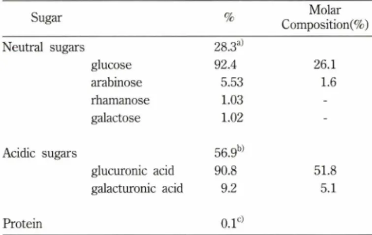
RGAP는중성당<>1 28.3%, 산성당이 56.9%, 단백질이 0.1%이 하로함유된다당분획으로 GLC로분석한결과,중성당은 glucose 92.4%, arabinose 5.53%, rhamnose 1.03%, galactose 1.02% 이 었다. 산성당의 GLC 분석결과는 glucuronic acid 90.8%,
Table I - Analysis of neutral and acidic sugars of R G A P
Sugar % M o lar
C om position(% )
N eutral sugars 28.3a)
glucose 92.4 26.1
arabinose 5.53 1.6
rham anose 1.03 -
galactose 1.02 -
Acidic sugars 56.9b)
glucuronic acid 90.8 51.8
galacturonic acid 9.2 5.1
P rotein 0 . l c)
a)d e te rm in e d b y phenol-sulfuric acid assay, b)d e te r m in e d by carbazol assay, 신d e te r m in e d by L o w ry m e th o d . A naly tical m e th o d an d c o n d itio n for G L C w e re described in M a te ria ls and M e th o d s .
40
s o l
20
3 B E izᄋooo4321
□(%)AloxcncoAo
J. Pharm . Soc. Korea
홍삼신성다당체의 힘암작용 117
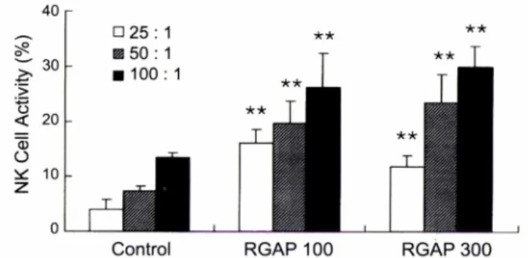
F ig . 4
- Effects of R G A P on natural kille r cell activity. C 57 B L /6 m ic e were adm inistered intraperitoneally w ith R G A P 100 or 300 m g/kg for consecutive 7 days. N atural kille r cell activities were determ ined at various ratio o f effector cells to target cells (25 : 1,5 0 : 1,1 0 0 :1 ). ** indicates the value significantly different at p < 0 .0 1 from each ratio o f control.
S u rv iv a l D a ys
- A n titu m o r avtivity o f R G A P in sarcom a 180 tum or-bearing IC R m ice. T he m ice w ere adm inistered intraperitoneally w ith R G A P 100 m g/kg, 300 m g/kg or Ara-C 5 m g /k g for consecutive 7 days after the intraperitoneal inoculation o f 1 x 10⁶ sacoma 180 cells. Life span w as observed for the n e x t 30 days.
□ 25 : 1 孩 50 : 1
■
100
:1
J a l l
C o n tro l R G A P 1 0 0 R G A P 3 0 0
N K 세 포 활 성 에 미 치 는 효 과
NK 세포활성에 미치는 영향을 알아보기 위해 RGAP를 100,300 mg/kg 용량으로 7일간 C57BL/6 마우스에 복강투 여 한후 비장세포를 effector 세포로 하고 YAC-1 lymphoma 세포를표적세포로 사용하여 NK 세포의 활성을 측정하였다. RGAP 투여는 effector 세포와 표적세포의 각 비율(25 : 1, 50 : 1,100 : 1)에서 NK 세포의 활성을 유의적으로 증가시켰 다(Fig. 3).
R G A P의 항 암 효 과
RGAP의항암작■용을알아보기 위해 sarcoma 180 세포를 ICR 미우스에 복강으로이식한후 30일간의 생존율을관찰한 결과,
대조군은 20일이후에는생존한마우스가없었으나 RGAP 100, 300 mg/kg 투여군에서는 생존율이 각각 57.1%,85.7% 였으며 ara-C 5 mg/kg 투여군에서는2 8.6%였다(Fig. 4). B16 melanoma
세포를 C57BL/6 마우스에피하주사하여 고형암의생성 정도를
조사해 본결과, RGAP 100 또는 300 mg/kg 투여군에서는 고
R G A P (m g /kg )
Fig. 2 - Effect of RGAP on killing of P815 cells in vivo. Coculture of
P815 cells (1 x 10⁵ cells/well) w ith the R G A P (100 or 300 mg/kg)-administered peritoneal macrophages (2 x 10⁵ cells/
well) was perform ed as described in m aterials and methods.
T he proliferation was determ ined at 18 h absorbances at 490 n m and expressed as cytotoxicity. N itrite was also determ ined in the culture supernatant of the respective groups. **indicates the value significantly different at pcO .O l from zero.
Table II - TNF-a levels in the medium of peritoneal macrophages
treated w ith R G A P
Treatm ent \xg/ml pg/106 cells
Control _ 42 + 28
R G A P 4 347 ± 82
40 1,072 + 94
400 32,626 ±47
LP S l 15,670 ±47
Peritoneal m acrophages from B A L B /c m ice w ere incubated for 24 h w ith various co ncentratio n o f R G A R T NF-a levels w ere de term in e d w ith bioassy u sing T N F a-sensitive W E H I 164 cells.
T able I I I - W E H I 164 tu m o r cell killing o f m acrophages treated w ith R G A P
T reatment Dose
(mg/kg)
T um or Killing (% )
Control - 9.16 ± 3.3
R G A P 100 19.18 ± 2.9
300 64.1 ± 4.3
Peritoneal macrophages were isolated from BALB/c mice administered w ith R G A P for consecutive 7 days. T N F a-sensitive W E H I 164 cells w ere labeled w ith 51C r an d coincubated for 6 h u nd e r the presence o f 1 [ig/ml actino m y cin D . T um or k illin g represents specific activities.
하는 bioassay 방법을사용했다. 양성대조물질로사용한 LPS는 1 의농도로처리시 TNF-oc 생성이 15,670±47 pg/106 cells 이었으며 RGAP는농도의존적으로 TNF-oc의생성을현저하게 증가시켰다(Table II). 또한 RGAP를 BALB/c 미우스에복강튜며 하고복강 macrophage를분리하여 WEHI 164세포의사멸을측 정한결과,WEHI 164세포의사멸이 RGAP의투여용량의존적 으로현저히증가되었다(Table I II) .
F i!
1 20
100
80 60 40 200 0/
) 0
aj e cr
f fl
>
!
co
o ooo
4
3
2
1
{%)Aiov
l o
XN
1
(i
)eiNᄋᄋooᄋ 4
3
2
1 oᄋooo
4
3
2
1
D {%
) AAocno!;
118 김영숙 • 박경미 • 신한재 • 송경식 • 남기열 • 박종대
<0.05
<0.05
<0.05 Control - 2.82 ± 0.50
RGAP 100 1.75 ± 0.61 300 1.32 ± 0.98 Ara-C 10 0.95 ± 1.28
C57BL/6 mouse was transplanted subcutaneously with B16 melanoma cells ( 1 x 1 05) and administered intraperitoneally with RGAP from the following day to seven days. The tumor weights were determined at the 19th day after tumor inoculation.
형암의생성이 39%, 53% 억제되었으며 ara-C 10 mg/kg 투여군 에서는 67% 억제되었다(Table IV).
고 찰
홍삼에서 분리된 산성 다당체(RGAP)는 glucose 92.4%, arabinose 5.53%, rhamnose 1.03%, galactose 1.02% 로 구성된 중성당과 glucuronic acid 90.8%, galacturonic acid 9.2%로구성 된산성당으로이루어져 있었으며 mole % 조성비는 glucuronic acid 51.8%, galacturonic acid 5.1%, glucose 26.1%, arabinose
1 . 6 1% 이었다. 또한백삼으로부터 면역증강작용을갖는 ginsan
은산성당이 43.1% 함유에비하여 RGAP는산성당이 56.9%를 함유하고 ginsan의중성당조성은 glucose와 galactose였으나,3’12) RGAP는 glucose, arabinose, rhamnose, galactose 를 함유하여
ginsan과는화학적 조성과구조가다른새로운산성 다당체임을
제시하였다. 본연구결과, RGAP를미우스에투여시 macrophage 를활성화시켜 NO 와 TNF-a의생성이증가되고 NK 세포의활 성화가일어나암세포를사멸시킴으로써 항암작용을나타낼 수 있음을나타내었다.
NO는반감기가매우짧은가스로여러종류의세포에서 nitric oxide synthase(NOS)에의해서 L-arginine으로부터 생성된다. Macrophage에서분비되는 NO는 inducible NOS에의해다량이 합성되며28) 박테리아와암세포를파괴시키고 mitogen의세포증 식과 T 세포의활성을조절함이알려져 있다.29) 저자등은 RGAP 가 in vitro와 in 에서 NO 생성을 촉진시킴을 밝혔으며,
RGAP를투여한마우스에서 concanavalin A 유도에 의한비장 세포의증식과면양적혈구에 대한항체생성반응이 NO를매개 로하여 조절되어짐을보고한바있다.20)
본 연구에서는 RGAP의 항암활성을 B16 melanoma와
sarcoma 180 세포를이식한마우스에서 조사한결과,현저하게
종양생성이 억제되고수명이 연장되어 RGAP가흥삼을 이용한 새로운 의약품 소재로서의 개발가능성을 시사하였다. 이러한 RGAP의항암효과는 macrophage 및 NK 세포의 활성화에의해 기인됨을나타내었다. RGAP는일차적으로선천적 면역계의주 Table IV - Antitumor activity of RGAP in B16 melanoma-bearing
C57BL/6 mice
G Dose Tumor Weight Median Significance
r°Up (mg/kg) (g) 6 mn (p)
요한 세포인 macrophage와 NK 세포에 작용하여 이들 세포로丁 터 사이토카인을 생성하거나 기능을 활성화시키는 기작을 통해 서 항암작용을 나타내고, NK 세포의 활성이 NO에 의해 촉진된 다는 보 고 들 은 RGAP에 의한 NO의 생성이 NK 세포의 활 성화를 촉진할 가능성을 시사한다.
인삼에서 분리된 다당 성분들의 면역계에 미치는 연구들은 인 삼의 면역기능에 대한 약리적 효능이 다당에 기인될 수 있으며 암치료에서 사용되는 화학요법제,방사선 치료, 수술요법둥의 부 작용을 감소시키고 치료효과를 높이기 위해 숙주의 면역을 증강 시킬 수 있는 치료방법에 기여할 수 있는 유용한 자원임을 제시 하고 있다. 버섯류에서 분리된 다당체 성분의 lentinan, polysaccharide K등은 암치료의 보조제로서 활용이 되고 있으며 홍삼으로부터 분리된 산성다당체도 선천적 면역계의 가장 주요 한 macrophage와 NK 세포를 활성화시켜서 면역 증강과 항암 효과를 갖는 의약품으로서의 개발가능성을 보이고 있다. 이와 같 은 개발을 위하여 RGAP의 지표물질로서 보다 정제된 분획을 분
리하여 구조를 연구중에 있으며 올러 면역 증강과 항암 작용
에 관련된 기작을 연구하고자 한다.
문 헌
1) Kim, Y. S., Kang, K. S. and Kim, S. I. : Study on antitumor and immunomodulating activities of polysaccharide fractions from Panax ginseng: comparison of effects of neutral and acidic polysaccharide fraction. Arch. Pharm. Res. 13,330 (1990).
2) Kim, Y. S., Kang, K. S. and Kim, S. I. : Effects of ginseng components on immunotoxicity of cyclophosphamide. Korean J. Ginseng Sci. 15,13 (1991).
3) Lee, Y. S., Chung, I. S., Lee, I. R., Kim, K. H.,Hong, W S. and Yun, Y. S .: Activation of multiple effector pathways of immune system by the antineoplastic immunostimulator acidic polysaccharide ginsan isolated from Panax ginseng. Anticancer Res. 17,323 (1997).
4) Park, K. M.,Jeong, T. C.,Kim, Y. S., Shin, H. J., Nam, K. Y. and Park, J. D. : Immunomodulatory effect of acidic polysaccharide fraction from Korean red ginseng (Panax ginseng). Natural product sciences 6,31 (2 0 0 0).
5) Srivastava, R. and Kulshreshtha, D. K. : Bioactive polysaccharides from plants. Phytochem. 28,2877 (1989).
6) Konno, C., Sugiyama, K.,Kano, M.,Takahashi, M. and Hikino, H .: Isolation and hypoglycemic activity of panaxans A, B, C, D and E, glycans of Panax ginseng roots. Planta Medica 50, 443 (1984).
7) Hikino, H.,Oshima, Y., Suzuki, Y. and Konno, C .: Isolation and hyploglycemic activity of panaxans E G. H. I. J. K. and L, glycans Panax ginseng roots. Shoyakigaku Zasshi 39,331 (1985).
1 8
9
2 0
7
8
2 3L.
0. 0.
J. Pharm . Soc. Korea
홍삼산성디당체의 항암작용 119
8) Konno, C. and Hikino, H. : Isolation and hypoglycemic activity of panaxans M, N, 0 ,and FJ glycans of Panax ginseng roots. Int.
J. Crude Drug. Res. 25,53 (1987).
9) Kanno, C.,Murakami, M.’ Oshima, Y. and Hikino, H .: Isolation and hypoglycemic activity of panaxans Q, R, S, T and U, glycans of Panax ginseng roots. J. Ethnopharmacol 14,69 (1985).
10) Sonoda, Y., Kasahara, T,Mukaida, N., Shimizu, N.’ Tomoda, M.
and Takeda, T. : Stimulation of interleukin- 8 production by acidic polysaccharides from the root of Panax ginseng.
Immunopharmacol. 38,287 (1998).
11) Shin, K. S., Kiyohara, H.,Matsumoto, T. and Yamada H. : Rhamnogalacturonan II from the leaves of Panax ginseng C. A.
Meyer as a macrophage Fc receptor expression-enhancing polysaccharide. Carbohydrate Res. 300,239 (1997).
12) Kim, K. H., Lee, Y. S., Jung, I. S., Park, S. Y., Chung, H. Y.,Lee, I. R. and Yun, Y. S .: Acidic polysaccharide from Panax ginseng, ginsan, induces Thl cell and macrophage cytokines and generates LAK Cells in synergy with rIL-2. Planta Medica 64, 110 (1998).
13) Abbas, A. K., Lichtman, A. H. and Pober, J. S. : Cellular and molecular immunology 3rd ed., W. B. Saunders Co., p.26 (1997).
14) Fraser, A. and Evan, G .: A License to kill. Cell 85,781 (1996).
15) Nagata, S. : Apoptosis by Death Factor. Cell 8 8,355 (1997).
16) Cerwenka, A. and Lanier, L. L. : Natural killer cells, viruses and cancer. Nature Rev. Immunol. 1, 41 (2001).
17) Cooper, M. A., Fehniger T. A. and Caligiuri M. A .: The biology of human natural killer-cell subsets. Trends in Immunology. 22, 633 (2001).
18) Cifone, M. G.,Ulisse, S. and Santoni, A. : Natural killer cells and nitric oxide. Int. Immunophamacol. 1,1513 (2001).
19) Park, J. D. : Recent studies on the chemical constituents of Korean ginseng (Panax ginseng C. A. Meyer). Korean J.
Ginseng Sci. 20,389 (1996).
20) Park, K. M.,Kim, Y. S., Jeong, T. C., Joe, C. O., Shin, H. J., Lee, Y. H.,Nam, K. Y. and Park, J. D. : Nitric oxide is involved in the immunomodulating activities of acidic polysaccharide from
Panax ginseng. Planta Medica. 67, 122 (2001).
21) Chaplin, M. J. and Kennedy, J. E : Carbohydrate Analysis.
Oxford: IRL Press, p.2 (1994).
22) Lowry, O. H.,Rosebrough, N. J. Farr, A. L. and Randall, R. J.
: Protein measurement with the Folin phenol reagent. J. Biol.
Chem. 193,265 (1951).
23) Albersheim, R, Nevins, D. J., English, R D. and Karr, A. : A method for the analysis of sugars in plant cell-wall polysaccharides by gas-liquid chromatography. Carbohydrate Res. 5’ 340 (1967).
24) Klimetzek, V and Remold, H. G. : The murine bone marrow macrophage, a sensitive indicator cell for murine migration inhibitory factor and a new method for their harvest. Cell.
Immunol. 53,257 (1980).
25) Green, L. C., Wanger, D. A., Glogowski, J., Skipper, R L., Wishnok, J. S., Tannenbaum, S. R. : Analysis of nitrate, nitrite, and [15N] nitrate in biological fluids. Anal. Biochem. 126,131 (1982).
26) Garrelds, I. M.,Zijlstra, E J., Tak, C. J., Bonta, I. L., Beckmann, I., Ben-Efraim, S. : A comparison between two methods for measuring tumor necrosis factor in biological fluids. Agents Actions, 38,C89 (1993).
27) Colotta, E, Peri, G., Villa, A., Mantovani, A. : Rapid killing of actinomycin D-treated tumor cells by human mononuclear cells. I. Effectors belong to the monocyte-macrophage lineage.
]• Immunol. 132,936 (1984).
28) Nathan, C. and Xie, Q. W : Nitric oxide synthases: roles, tolls, and controls. Cell 78,915 (1994).
29) Farias-Eisner, R.,Sherman, M. R, Aeberhard, E. and Chaudhuri G .: Nitric oxide is an important mediator for tumoricidal activity in vivo. Proc. Natl. Acad. Sci. 91,9407 (1994).
30) Park, K. G., Hayes, R D., Garlick, R J., Sewell, H. and Eremin, O. : Stimulation of lymphocyte natural cytotoxicity by L- arginine. Lancet 337, 645 (1991).
31) Cifon, M. G., Festuccia, C.,Cironi, L., Cavallo, G.,Chessa, M.
A. and Pensa, V : Induction of the nitric oxide-synthesizing pathway in fresh and interleukin 2-cultured rat natural killer cells. Cell. Immunol. 157,181 (1994).