Intermittent Exotropia
Nam-Kyun Koo, MD
1, Young-Chun Lee, MD
2, Se-Youp Lee, MD
1Department of Ophthalmology, School of Medicine, Dongsan Medical Center, Keimyung University
1, Daegu, Korea Department of Ophthalmology, School of Medicine, Uijongbu St. Mary Hospital,
The Catholic University of Koera
2, Uijongbu, Korea
Purpose: The surgical technique for intermittent exotropia 「X(T)」 is quite simple. However, in many cases, the condition recurs due to any one of a number of causes, including undercorrection. This study examined the factors associated with undercorrection on X(T) patients.
Methods: The study examined 199 X(T) patients who underwent bilateral recession of the lateral rectus muscle or unilateral recession of the lateral rectus muscle and resection of the medial rectus muscle, and who were followed-up for more than a year. Patients whose near and far distance angles of deviation were 9 prism diopters (PD) or more at one year after surgery were designated as group 1. Those whose PD was 8 or below or who had orthophoria were assigned to group 2. Various factors were compared and analyzed.
Results: One day after surgery, group 1 showed an average overcorrection of 1.9 and 4.1 PD at near and far, respectively, and group 2 showed an average overcorrection of 6.3 and 7.6 PD at near and far, respectively. A statistically significant difference was observed between the two groups (p<0.05). Factors such as the age of onset of strabismus, age at the time of surgery, the interval from the onset of strabismus to surgery, the preoperative angle of deviation, the dissociated vertical deviation, amblyopia, anisometropia and vertical strabismus had no influence on the undercorrection of X(T) patients (p>0.05).
Conclusions: Of the many factors that might influence the surgical results of X(T) patients, the angle of deviation during the initial postoperative period is the most important factor. Korean Journal of Ophthal- mology 20(3):182-187, 2006
Key Words: Intermittent exotropia, Overcorrection, Strabismus, Undercorrection
Received: January 18, 2006 Accepted: June 15, 2006
Reprint requests to Se-Youp Lee, MD. Department of Ophthal- mology, School of Medicine, Dongsan Medical Center, Keimyung University, #194 Dongsan-dong, Jung-gu, Daegu 700-712, Korea.
Tel: 82-53-250-7720, 7707, Fax: 82-53-250-7705, E-mail: lsy3379@
dsmc.or.kr
*This study was presented at the Korean Ophthalmological Society’s 94nd Annual Meeting, Korea, October, 2004.
The cause of intermittent exotropia 「X(T)」 is unclear but X(T) is the most frequently acquired exotropia which develops in Korean children. Surgery is mainly used to treat X(T). X(T) can be cured through surgery but there is a 20- 50% recurrence rate depending on the follow-up period.
1In Korea, Kim and Cho
2reported the incidence of postoperative undercorrection of X(T) to be 27%. In other countries, Richard and Parks
3reported an incidence of 38% and Clarke and Noel
4an incidence of 50%.
Among the factors associated with the outcome of surgery for X(T), the surgical methods, type of strabismus, preopera- tive deviation angle, and deviation angle in the initial postoperative period have been reported to have significant
influence. Many studies on the factors associated with the success of surgery have been performed but there are few reports on undercorrection.
Therefore, this study examined the factors associated with undercorrection in 199 patients who were diagnosed with X(T), who underwent surgery, and were available for an average long-term follow-up of more than 2 years. The rela- tionship of undercorrection with the following factors was examined: the age of onset, age at the time of surgery, near and far preoperative deviation angle, amblyopia, anisometro- pia, dissociated vertical deviation, inferior oblique overaction, the presence of concomitant vertical strabismus, and the deviation angle at the initial postoperative period.
Materials and Methods
The study was performed from January 1999 to February
2004 on 199 patients who were diagnosed with X(T) at our
hospital and were available for follow-up for more than 1
year. There were 83 males and 116 females. Of the types of
X(T) encountered, the basic type was most prevalent and
occurred in 151 cases. This was followed by 29 cases of the
pseudodivergence excess type, 5 cases of the convergence insufficiency type, and 14 cases of the divergence excess type. Among the patients who underwent unilateral recession of the lateral rectus muscle and resection of the medial rectus muscle (60 cases) or bilateral recession of the lateral rectus muscle (139 cases), at the last follow-up, the group with deviation angles more than 9 prism diopters (PD) at near and far distance was designated as group 1 (undercorrection group). The group with orthophoria or deviation angles less than 8 PD was designated as group 2 (success group). Eighty patients were included in group 1 and 119 patients were included group 2.
The age at onset was assessed during the examination and age at the time of surgery was also recorded. In all patients, a refraction was performed after applying 1% Cyclogyl
Ⓡ. The refractive error, was calculated in diopters as the spherical equivalent, or the addition of the spherical power and half the magnitude of the cylinder power. Anisometropia was defined as an absolute difference of the spherical equivalent of 2 or more diopters between the right and left eyes. In cases where the uncorrected visual acuity was less than 20/30, the refractive error was corrected and the visual acuity was assessed with an alternate prism cover test to measure the deviation angle. The function of the extraocular muscle, its overaction and underaction were assessed by monocular and binocular movements. The same surgeon performed the surgical procedure for all patients. The deviation angle at near and far distance was measured 1 day, 1 week, 1 year, and at the last follow-up after surgery. In the two groups, the relationship between the postoperative deviation angle and the age at onset, age at the time of surgery, near and far preoperative deviation angle, amblyopia, anisometropia, dissociated vertical deviation, inferior oblique overaction, and the presence of concomitant vertical strabismus were examined. For statistical analysis, the near and far deviation angle was analyzed using a 2-sample t-test, and the other factors were analyzed by a Chi-square test. A p value <0.05 was considered significant.
Results
The average postoperative follow-up period was 26 months (12-80 months). Table 1 shows the age of onset in the under- correction and success groups at the last follow-up. There was no statistically significant difference between the two groups.
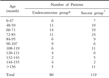
Regarding the age at the time of surgery, there were 6 patients younger and 74 patients older than 4 years in the undercorrection group, and 7 patients younger and 112 patients older than 4 years in the success group. There was no statistically significant difference between the two groups (p=0.385) (Table 2).
One day after surgery, an average overcorrection of 1.9 and 4.1 PD at near and far distance, respectively, were observed in the undercorrection group. On the other hand, the
Age (month)
Number of Patients
Undercorrection group* Success group
†0-12
13-24 25-36 37-48 49-60 61-72 73-84 85-96
>96
25 16 7 7 7 5 6 3 4
19 16 26 17 14 5 1 3 18
Total 80 119
P value=0.375, 2 sample t-test.
*: The group whose near and far angle of deviation 1 year after surgery was 9 or more.
†: The group whose near and far angle of deviation was 8 or below or who had orthophoria.
Table 1. Age at onset
Age (month)
Number of Patients
Undercorrection group* Success group
†0-47
48-59 60-71 72-83 84-95 96-107 108-119 120-131 132-143 144-155
>156
6 13 14 11 6 9 6 4 2 4 5
7 19 19 24 9 6 11 6 5 2 11
Total 80 119
P value=0.385, 2 sample t-test.
*: The group whose near and far angle of deviation 1 year after surgery was 9 or more.
†: The group whose near and far angle of deviation was 8 or below or who had orthophoria.
Table 2. Age at the time of surgery
success group had an average overcorrection of 6.3 and 7.6 PD at near and far distance, respectively, which was statis- tically significant (p=0.000). Table 3 shows the distribution of each group 1 day after surgery according to the near deviation angle. In the undercorrection and the success groups, 15 patients (19%) and 48 patients (40%) respectively showed overcorrection of more than 10 PD (Table 3).
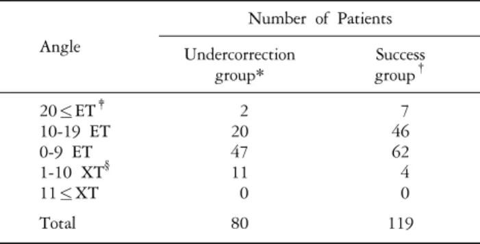
Regarding the distribution of the far angle of deviation at 1
day after surgery, in the undercorrection group, 2 patients had
an esodeviation at 20 with 20 PD or greater, 20 patients had
an esodeviation at 10 with 19 PD, 47 patients had ortho-
phoria or the esodeviation at 1 with 9 PD, and 11 patients
had exodeviation at 1 with 10 PD. However, in the success
group, the distribution of the far deviation angle among the
above categories was 7, 46, 62, and 4 patients, respectively.
Angle
Number of Patients Undercorrection
group*
Success group
†20≤ET
‡10-19 ET 0-9 ET 1-10 XT
§11≤XT
1 14 45 19 1
7 41 60 11 0
Total 80 119
P value=0.000, 2 sample t-test.
*: The group whose near and far angle of deviation 1 year after surgery was 9 or more.
†: The group whose near and far angle of deviation was 8 or below or who had orthophoria.
‡: esodeviation,
§
: exodeviation.
Table 3. Distribution according to the near angle of deviation 1 day after surgery
Angle
Number of Patients Undercorrection
group*
Success group
†20≤ET
‡10-19 ET 0-9 ET 1-10 XT
§11≤XT
2 20 47 11 0
7 46 62 4 0
Total 80 119
P value=0.000, 2 sample t-test.
*: The group whose near and far angle of deviation 1 year after surgery was 9 or more.
†: The group whose near and far angle of deviation was 8 or below or who had orthophoria.
‡: esodeviation,
§
: exodeviation.
Table 4. Distribution according to deviation angle at far distances 1 day after surgery
This represented a statistically significant difference between the two groups (p=0.000) (Table 4).
Regarding to the distribution of the near deviation angle 1 week after surgery, 49 patients in the undercorrection group showed esodeviation and 30 patients showed exodeviation.
The success group, in contrast, had 101 patients showing esodeviation and 17 patients showing exodeviation (Table 5).
Regarding the distance deviation angle at 1 week after surgery, 47 patients showed esodeviation and 32 patients showed exodeviation in the undercorrection group. On the other hand, 105 patients showed esodeviation and 13 patients showed exodeviation in the success group. There was a statistically significant difference between the two groups (p=0.000) (Table 6). Because 1 patient in each group did not visit our hospital in the first week after surgery, 79 patients were included in the success group and 118 patients were included in the undercorrection group.
Angle
Number of Patients Undercorrection
group*
Success group
†20≤ET
‡10-19 ET 0-9 ET 1-10 XT
§11≤XT
0 6 43 30 0
0 18 83 17 0
Total 79 118
P value=0.000, 2 sample t-test.
*: The group whose near and far angle of deviation 1 year after surgery was 9 or more.
†: The group whose near and far angle of deviation was 8 or below or who had orthophoria.
‡: esodeviation,
§
: exodeviation.
Table 5. Distribution according to the near angle of deviation 1 week after surgery
Angle
Number of Patients Undercorrection
group*
Success group
†20≤ET
‡10-19 ET 0-9 ET 1-10 XT
§11≤XT
0 6 41 32 0
0 20 85 13 0
Total 79 118
P value=0.000, 2 sample t-test.
*: The group whose near and far angle of deviation 1 year after surgery was 9 or more.
†: The group whose near and far angle of deviation was 8 or below or who had orthophoria.
‡: esodeviation,
§
: exodeviation.
Table 6. Distribution according to the far angle of deviation 1 week after surgery
Amblyopia, the rate of anisometropia, the presence of dissociated vertical deviation and A-V type strabismus in the two groups was similar (P=0.058, 0.619, 0.083, 0.776). In the case of vertical strabismus, there were 28 cases in the undercorrection group and 48 cases in the success group but there was no significant difference (p=0.447) (Table 7).
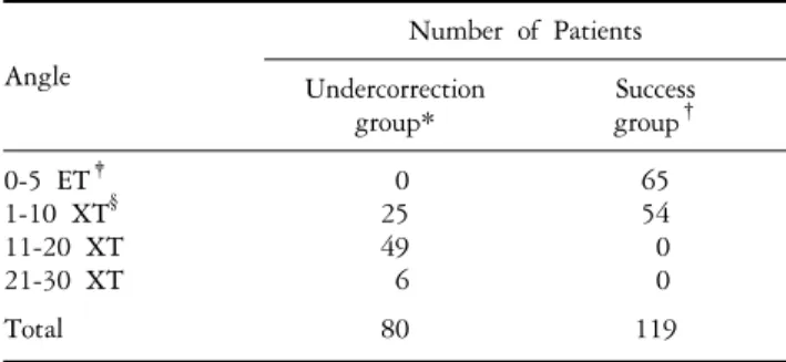
At the last follow-up, the undercorrection group showed an average 14.4 PD and 14.3 PD deviation angle at near and far distance, respectively, and average of 0.88 weeks were required to turn the postoperative overcorrection into the orthophoria. The undercorrection group showed that an average of 36.3 weeks was required to achieve exotropia of more than 9 PD from the orthophoria and an average of 36.9 weeks were needed to achieve exotropia of less than 9 PD.
Tables 8 and 9 show the deviation angle distribution at near
and far distance in the undercorrection group and success
group at the last follow-up.
Factor Number of patients
Undercorrection group* Success group
†P value
Amblyopia P
‡(n=32)
A
§(n=167)
18 62
14 105
P=0.058
Vertical deviation P (n=76) A (n=123)
28 52
48 71
P=0.447
Anisometropia P (n=6)
A (n=193)
3 77
3 116
P=0.619
D.V.D P (n=2)
A (n=197)
2 78
0 119
P=0.083
A-V type P (n=2)
A (n=197) 1
79 1
118 P=0.776
P value >0.05, Chi-Sqaure test.
*: The group whose near and far angle of deviation 1 year after surgery was 9 or more.
†: The group whose near and far angle of deviation was 8 or below or who had orthophoria.
‡: presence,
§: absence.
Table 7. Surgical outcome according to the preoperative factors
Angle
Number of Patients Undercorrection
group*
Success group
†0-5 ET
‡1-10 XT
§11-20 XT 21-30 XT
0 25 49 6
65 54 0 0
Total 80 119
P value=0.000, 2 sample t-test.
*: The group whose near and far angle of deviation 1 year after surgery was 9 or more.
†: The group whose near and far angle of deviation was 8 or below or who had orthophoria.
‡: esodeviation,
§
: exodeviation.
Table 8. Distribution according to the deviation angle at near distance on final visit
Angle
Number of Patients Undercorrection
group*
Success group
†0-5 ET
‡1-10 XT
§11-20 XT 21-30 XT
0 27 48 5
74 45 0 0
Total 80 119
P value=0.000, 2 sample t-test.
*: The group whose near and far angle of deviation 1 year after surgery was 9 or more.
†: The group whose near and far angle of deviation was 8 or below or who had orthophoria.
‡: esodeviation,
§