Influence of Tibial Rotation on EMG Activities of Medial and Lateral Hamstrings During Maximal Isometric Knee Flexion
Woo-taek Lim
1,2, PhD, PT
11
1Dept. of Physical Therapy, College of Health and Welfare, Woosong University
2Advanced Institute of Convergence Sport Rehabilitation, Woosong University
Abstract 1)
Background: The hamstring muscles in the lower extremity are highly important for knee joint stability and can be classified into medial and lateral hamstrings according to the anatomical position, which have some different functions. To measure the strength of the individual hamstring muscles, manual muscle testing is clinically performed by dividing rotation postures into internal and external postures. However, this has no sufficient scientific background.
Objects: This study aimed to test the difference that the tibial rotation would cause in the muscle activity of the medial and lateral hamstrings.
Methods: The muscle activities of the biceps femoris, semitendinosus, and semimembranosus were measured in a total of three different postures (neutral position and internal and external rotations) with 3 replications. During the maximal isometric contraction, resistance was constantly provided by the string attached to the strap, not by manual resistance of the examiner. Before and after electromyography measurements, the participants underwent hamstring flexibility measurement using the active knee extension test in the supine position on the treatment table.
Results: The semitendinosus had a 12.56% reduction in muscle activity in external rotation as compared with that in neutral position. The biceps femoris and semimembranosus showed reduced muscle activities in both external and internal rotations as compared with those in neutral position. Only the women showed significant decreases in the comparison between pre and post-active knee extension.
Conclusion: Only the semitendinosus muscle was consistent with the anatomical speculation. However, the reduction in the muscle activity of the semitendinosus as compared with that in neutral position was only 12.56%, the clinical value of which may be difficult to justify.
Key Words: Electromyography; Hamstrings; Isometric contraction; Tibial rotation.
Introduction
The lower extremities are more developed than the rest of the body parts to support the entire body against gravity and to maintain the standing position.
In the lower extremities, the hamstring muscles par- ticularly maintain the stability of the knee joints during movement, enabling smooth motions. Thus, in case of anterior cruciate ligament injuries, strength- ening the hamstring muscles is considered the most
important factor in the rehabilitation of the lower extremities (Li et al, 1999; Pandy and Shelburne, 1997; Wilk and Andrews, 1992). The hamstring mus- cles are composed of four muscles, namely the bi- ceps long and short heads, semitendinosus (ST), and semimembranosus (SM). While simultaneous con- tractions of all the hamstring muscles produce flex- ion of the knee joint, each muscle has a different orientation from the origin to the insertion, which, accordingly, makes a difference in individual func- Corresponding author: Woo-taek Lim [email protected]
The research was supported by 2018 Woosong University Academic Research Funding and Basic Science Research
Program through the National Research Foundation of Korea (NRF) funded by the Ministry of Education
(NRF-2017R1C1B5076885)
tions (Lewek et al, 2004; Lynn and Costigan, 2009).
In detail, the hamstring muscles are largely divided into the medial hamstring (MH) and lateral ham- string (LH) based on the anatomical location. The biceps femoris originates from the ischial tuberosity and inserts in the lateral side of the head of the fib- ula, whereas the ST and SM originate from the is- chial tuberosity surface and attached to the medial tibia and medial tibial condyle, respectively. Owing to the attached site, the biceps femoris rotates laterally during flexion of the knee joint, while the ST and SM medially rotate on the femur.
To configure clinical rehabilitation programs ap- propriate for individual patients, the extent of the injury and the resulting muscle strength deficits must be accurately assessed. Currently, muscle strength is measured in various ways, using a wide range of measuring devices, from specialized devices such as Biodex, which has high validity and reli- ability for measurement, to portable devices such as a handheld dynamometer. However, manual muscle testing (MMT), in which therapists manually apply resistance for measurement, is used widely in clin- ical practices owing to its convenience for measure- ment and affordability. MMT is a highly valuable method that can easily measure the force generated in the contractile unit of the muscle. It has been in use since the early 1900 and is now widely applied in clinical practice (Martin and Lovett, 1915). In general, in MMT, measurements are performed in a single posture predetermined for each muscle, except for the hamstring muscles, which are sometimes measured in external or internal rotation owing to their anatomical characteristics.
For MMT measurement, many clinicians include Daniels and Worthingham’s Muscle Testing and Muscles: Testing and Function, with Posture and Pain (Avers and Brown, 2018; Kendall et al, 2005).
For measurement of the biceps femoris, the authors described that the knee joint needs to be externally rotated so that the toes face outside while bending the knee <90˚, and then, the subject should induce
contraction of the LH to isolate it with downward resistance in inward directions. At this point, the biceps femoris must be maintained with the foot ends facing outward during knee flexion. By con- trast, the ST and SM are measured while applying downward resistance in outward directions after in- ternal rotation of the knee joint. Although the two references are widely used in clinical practice and cited in many papers, they do not describe the sci- entific evidence for these measurements. Wide clin- ical use cannot be used as a logical basis. In other words, whether different amounts of muscle activity are actually generated in the MH and LH according to tibial rotation must be quantitatively measured.
The present study aimed to investigate if the me- dial and lateral compartments of the hamstring muscles have differences in quantitative contractions in tibial torsion, for which an electromyography de- vice was used.
Methods
Subjects
Thirty-five young and healthy college students participated in the study (Table 1). They had no in- jury or disease in the hip and knee joints and were given a full explanation about the study. They pro- vided informed consent prior to participation in the study. This study was approved and monitored by the Institutional Review Board of Woosong University (approval number: 1041549-180419-SB-59).
Instrumentation
Surface electromyography (EMG) signal was col-
lected using a Trigono™ Wireless EMG system
(Delsys Inc., Boston, MA, USA). The EMG data
were collected from the selected Hamstrings. Delsys
EMGworks® Acquisition and Analysis software was
used for the analysis. The raw EMG signals were
sampled 2,000 ㎐ and were processed into a root
mean square with a window of 125 ms. A band pass
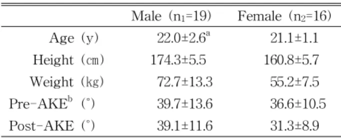
Male (n
1=19) Female (n
2=16) Age (y) 22.0±2.6
a21.1±1.1 Height (㎝) 174.3±5.5 160.8±5.7 Weight (㎏) 72.7±13.3 55.2±7.5 Pre-AKE
b(˚) 39.7±13.6 36.6±10.5 Post-AKE (˚) 39.1±11.6 31.3±8.9
a
mean±standard deviation,
bactive knee extension.
Table 1. Subjects characteristics and changes in
hamstring flexibility (N=35)
fileter of 20-450 ㎐ was used together with notch filters at 60 ㎐.
Procedure
The participants comfortably lied down in the prone position on the treatment table and bent the knee joint to 60˚ (Albertus-Kajee et al, 2011; Fauth et al, 2010; Onishi et al, 2002). The pelvis and con- tralateral lower extremity were fastened to the table with a belt. As for the long head of biceps femoris (BF), the electrodes of the EMG sensor were placed at 50% on the line between the ischial tuberosity and the lateral epicondyle of the tibia. For the ST, the electrodes must be placed 50% on the line be- tween the ischial tuberosity and medial epicondyle of the tibia. For the SM, the electrodes were attached laterally to the ST tendon in the apex of the “V”
between ST tendon and the biceps femoris. Sensor was placed in accordance with SENIAM (surface EMG for non-invasive assessment of muscles) guide- lines and the text book of Anatomical guide for the electromyopgapher (Perotto, 2011). Before EMG at- tachment, the skin was fully shaved. During meas- urement of the maximal isometric contraction of the hamstring muscles, resistance was constantly pro- vided by the string attached to the strap, not by manual resistance of the examiner. One end of the strap was tied at 3 ㎝ below the lateral malleolus, and the other end was tied to a fixed post to pre- vent movement during contraction. The string con- nected to the strap was made perpendicular to the ankle. The three measurement postures were neu-
tral position and internal and external rotations.
Each subject was measured in a random order of the postures. The value measured in neutral posi- tion was set as the maximum voluntary isometric contraction (MVIC), and values obtained in internal and external rotations were calculated as the
%MVIC relative to that in neutral position. In flex- ion, the subjects were instructed to maximally con- tract the leg after internal or external rotation, in which resistance was applied in the opposite direc- tion to the contraction. The MVIC was measured 3 times per posture (5 sec/time, 15 sec in total), and the mean value was used. Before and after the EMG measurements, the participants were also subjected to the hamstring flexibility measurement in the supine position on the treatment table using the active knee extension test (AKE). The AKE defined by the longitudinal axes of the tibia and femur was recorded at maximal knee extension and was subtracted from 180˚.
Data Analyses
All the statistical analyses were performed us- ing IBM SPSS Statistics ver. 23 experimental data (IBM Corp., Armonk, NY, USA). One-sample t-test was performed to test the difference in %MVIC de- pending on the tibial torsion within individual mus- cles (internal or external rotation), and the Bonferroni correction was applied. Pre- and post-AKE were compared using the paired sample t-test. Statistical significance was set at p values of <.025. All the re- sults were expressed as mean ± standard deviation.
Results
When the %MVIC of the hamstring muscles in
the knee joint was examined during rotation in com-
parison with that in neutral position, statistically sig-
nificant differences were found in the BF and ST,
but not in the SM. First, the BF showed a sig-
nificant decrease in %MVIC in both external (p=.006)
Biceps femoris Semitendinosus Semimembranosus
External rotation 92.13±15.84
a* 87.44±17.47* 91.81±18.20*
Internal rotation 88.94±14.85* 106.77±18.06 95.86±16.58
a