Corresponding author: Hae-Gyeong Baek
Department of Laboratory Medicine, Kwangju Christian Hospital, 37 Yangnim-ro, Nam-gu, Gwangju 61661, Korea
E-mail: [email protected]
ORCID: https://orcid.org/0000-0003-3414-8604
ORIGINAL ARTICLE
Effective Identification of Ochrobactrum anthropi Isolated from Clinical Specimens
Hyun-Mi Ko
1, Jun-Hyeon Jo
2, Hae-Gyeong Baek
21Dental Science Research Institute, Department of Oral Anatomy, School of Dentistry, Chonnam National University, Gwangju, Korea
2Department of Laboratory Medicine, Kwangju Christian Hospital, Gwangju, Korea
임상검체에서 분리된
Ochrobactrum anthropi
의 효과적인 동정고현미
1,조준현
2,백해경
21전남대학교 치과대학 치의학연구소 구강해부학교실, 2광주기독병원 진단검사의학과
ARTICLE INFO ABSTRACT
Received July 29, 2020 Revised 1st August 10, 2020 Revised 2nd September 3, 2020 Accepted September 3, 2020
Ochrobactrum anthropi is a non-fermentative oxidative gram-negative bacillus that produces oxidase. Distinguishing a mixed culture with non-fermenting bacteria having a similar appearance and oxidase-positive is difficult, and there is a limit to accurate identification with a biochemical identification system. This paper proposes that the Matrix-Assisted Laser Desorption/Ionization Time-of-Flight Mass Spectrometry Platform (MALDI-TOF) method is useful for classifying bacteria that are difficult to identify using biochemical testing methods. As a result of analyzing five cases of O. anthropi examined using MicroScan, it took 6.5 days to the final report, which was 3.5 days more than the 3.0 days of E. coli. The pus sample in patient 5 was a mixed infection with Achromobacter xylosoxidans, and it took 11.3 days because of multiple subculture and retests. Four patients were over 60 years old with an underlying disease, and the possibility of opportunistic and nosocomial infections could not be excluded. Among them, samples collected after 92 days of hospitalization were resistant to imipenem and meropenem. Therefore, an examination using the MALDI-TOF method will be useful for the rapid and adequate treatment of patients with difficult identification, such as O. anthropi.
Copyright Ⓒ 2020 The Korean Society for Clinical Laboratory Science. All rights reserved.
Key words Achromobacter MALDI-TOF MicroScanWalkAway Ochrobactrum anthropi
서 론
Ochrobactrum anthropi는 oxidase 양성, 포도당과 유당 을 산화적으로 분해하는 비 발효 산소성 그람음성 막대 균으로 urease가 양성 또는 음성이고 주모성 편모가 있어 운동성을 가 지며 맥컨키한천배지에서 연한 분홍색의 점액성 집락을 형성한 다[1]. O. anthropi는 집락의 특징과 색소 생성능, 냄새 등으로 Pseudomonas species와는 구별이 가능함에도 불구하고 생
화학적 성상이 Pseudomonas를 비롯한 다양한 세균과 비슷하 여 정확한 동정이 어렵고 종종 다른 세균으로 보고되고 있다[2].
비 발효 산소성 그람음성 막대균인 Pseudomonas aeru- ginosa와 Acinetobacter baumannii는 기회감염과 원내감 염원으로서 MRAB (multidrug-resistance Acinetobacter baumannii)나 MRPA (multidrug-resistance Pseudomonas aeruginosa)는 지정 감염병으로 지정되었고 MRAB의 폐렴으 로 인한 사망률은 40%에 이른다[3]. O. anthropi 또한 1980년 인체감염 첫 사례 발표 이후 꾸준히 감염사례가 증가되고 있으 며[4-8], 임상검체뿐 아니라 병원 환경에서도 검출되며 다양한 서식환경에 존재하는 것으로 보고되고 있다[9, 10]. 입원 중 카 테터 등에 의한 원내감염으로 인해서 균혈증 유발이 보고[5-8]
Korean Society for Clinical Laboratory Science
되었고 면역기능저하나 기저질환이 있는 환자에게서 인체감염 보고가 점차 증가하고 있으며[11-13], 우리나라의 경우 2009 년 meropenem내성 O. anthropi에 의한 균혈증이 보고된 바 있다[14]. 하지만 O. anthropi의 정확한 병인성과 항생제 내성 기전이 아직까지 보고된 바 없어 내성기전이 밝혀진 다른 비 발 효 세균들에 비해 중요하게 인식되지 않고 있다.
비 발효 세균 중 O. anthropi와 같이 동정이 까다로운 세균 은 계대배양과 재검사 이외에 추가검사가 쉽지 않아 세균동정보 다 항균제 검사에 치중한 보고를 하고 있다. 하지만 기저질환을 동반한 장기 입원 환자에게서 내성 균의 기회감염과 원내감염이 환자 사망과 직결된다면 신속하고 정확한 동정검사결과를 임상 에 보고해야 한다. MicroScan (Beckman Coulter, Brea, CA, USA)와 VITEK 2 system (bioMérieux, Marcy l’Étoile, France)는 신속하게 세균의 생화학적 특성을 이용해 균을 동정 하는 동시에 항균제 감수성검사가 가능해 지금까지 임상미생물 검사실에서 보편적으로 사용하고 있으나[15, 16], 생화학적 반 응을 보기 위해 기질에 사용되는 충분한 양의 세균과 반응시간 이 요구되므로 배양이 까다로운 세균이나 혼합감염이 된 세균들 을 동정하는 경우 잘못된 결과를 보이기도 한다.
따라서 본 연구는 O. anthropi의 생화학적 동정방법과 Matrix- Assisted Laser Desorption/Ionization Time-of-Flight Mass Spectrometry Platform (MALDI-TOF)법을 이용한 동 정을 통해 생화학적 동정 장비 검사가 주는 한계를 확인하고 O.
anthropi로 보고된 사례를 수집해 기저질환 유무와 검체 채취 까지 입원기간, 검사에 소요된 기간, 연령 및 항균제 감수성을 분 석하여 원내감염 가능성을 예측하고 신속한 환자 치료를 위해 MALDI-TOF를 이용한 동정검사가 유용함을 제안하고자 한다.
재료 및 방법
1. 연구대상
2016년 1월부터 2020년 5월까지 광주광역시 소재 500병상 규모의 K종합병원 미생물검사실로 배양 의뢰된 164,841건 중 에서 생화학적 동정 장비인 MicroScan (Beckman Coulter, Brea, CA, USA)로 검사하여 90% 이상의 확률로 동정 보고된 O. anthropi 7례 중 동일 환자에서 반복 동정된 2례를 제외하 고 5례를 대상으로 분석하였다(Table 1). 최종동정 보고까지 소 요된 시간 분석에 2020년 4월 MicroScan에서 E. coli로 보고 된 49검체를 분석하였다. 본 연구는 광주기독병원 생명윤리위 원회의 승인을 얻어 실시하였다.
2. 배양
혈액배양은 자동혈액배양기인 BacT/Alert 3D (BioMérieux, Durham, NC, USA)를 이용하여 호기성(Bact/Alert 3D stan- dard aerobic)과 혐기성(Bact/Alert 3D standard aerobic) 을 동시에 진행하였다. 증식된 균을 다시 혈액한천배지와 맥컨 키한천배지에 5% CO2 , 35°C 환경에서 5시간 이상 배양하였 다. 소변, 고름, 객담, 비강 검체는 혈액한천배지와 맥컨키한천 배지에 접종하여 5% CO2 , 35°C 환경에서 20시간 이상 배양하 였고 필요 시 같은 환경에서 48시간 배양하였다.
3. 세균 동정
생화학적 동정장비인 MicroScan을 이용하여 NC63 panel (Beckman Coulter, West Sacramento, CA, USA)에 접종하 여 동정하였다. 5번 환자 검체는 생화학적 동정장비 VITEK 2 system (bioMérieux, Marcy l’Étoile, France)에서 GN (bioMérieux, Marcy l’Étoile, France)카드로 재검사하였으 며 MALDI-TOF법을 이용한 검사장비 MALDI-TOF MS system, the brukerBiotyper MS (BrukerDaltonics, Bremen, Germany)와 ASTA MicroIDSys system (ASTA, Suwon, Korea)을 이용해 교차 동정하였다. 그람염색은 Gram-Hucker’s Stain Solution (Muto pure chemicals LTD, Tokyo, Japan) 을 이용하여 solution I, 1분; solution II, 2분; 95% ethanol, 10초; solution III, 30초 시행하여 광학현미경(Olympus Corp.,Tokyo, Japan)으로 확인하였다.
4. 항균제 감수성검사
MicroScan의 NC63 panel을 이용해 최소발육억제농도 (MIC) 결과 값은 CLSI 지침에 따라 감수성(susceptible), 중등 도 내성(intermediate) 또는 내성(resistant)로 분류하였다.
결 과
1. 환자 검체에서O. anthropi의 분리 동정
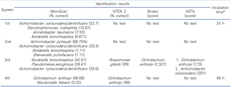
Table 1은 다양한 임상 검체에서 O. anthropi가 검출됨을 보여주고 있으며, 5번 환자 고름 검체 동정을 위해 맥컨키한천 배지에서 24시간 배양한 집락을 MicroScan에서 첫 번째 동정 결과 85% 이하의 낮은 동정률을 보였다(Table 2). 고름 검체 채 취 과정에서 오염이 의심되어 단일 집락을 계대 배양 후 재검사 하였으나 1차 검사와는 다른 종류의 세균들로 낮은 동정율을 보 였다. 육안으로 확인되진 않지만 집락 내 여러 세균이 섞여있을
Table 1. Clinical features of patients with Ochrobactrum anthropi reported
No. Sex/age Source of isolate Isolation date Underlying disease DHC* Number of days reported Outcome
1 M/4 Urine 2016. 01. 08 Pneumonia 0 6.1 Recovered
2 M/62 Nasal swab 2018. 04. 30 Heart failure
Renal failure
0 5.3 Died
3 F/86 Blood 2018. 07. 13 Parkinsonism
Hypertension
6 4.7 Recovered
4 F/60 Sputum 2018. 07. 21 Cerebral hemorrhage
Epilepsy
92 5.0 Died
5 M/86 Pus 2020. 04. 23 Diabetes mellitus
Alzheimer’s disease
29 11.3 Recovered
Abbreviation: *, date from hospitalization to collection.
Table 2. Comparision of the results for pus of No.5 patient from four systems
System
Identification results
Incubation time*
MicroScan (% correct)
VITEK 2 (% correct)
Bruker (score)
ASTA (score) 1st Achromobacter xylosoxidans/denitrificans (22.7)
Stenotrophomonas maltophilia (15.67) Acinetobacter baumannii (7.53) Bordetella bronchiseptica (0.871)
No test No test No test 24 h
2nd Achromobacter pichaudii (68.75%) Achromobacter xylosoxidans/denitrificans (29.3)
Bordetella bronchiseptica (1.11) Shewanella putrefaciens (1.11)
No test No test No test
3rd Bordetella bronchiseptica (42.51) Pseudomanas.aeruginosa (39.41) Achromobacter xylosoxidans/denitrificans (29.0)
Roseomonas gilardii (99)
Ochrobactrum anthropi (2.327)
1. Ochrobactrum anthropi (173) 2. Achromobacter xylosoxidans (201) 4th Ochrobactrum anthropi (96.68)
Wautersiella falsenii (3.32) Ochrobactrum anthropi (90)
No test No test 48 h
*, at 5% CO2, 35°C.
가능성을 배제할 수 없으므로 두 개의 집락을 각각 배양하여 3차 검사를 진행한 결과, 두 집락에서 동정 결과는 동일하였으나, 1, 2차 검사와는 다른 Bordetella bronchiseptica (42.51%)로 낮은 동정률의 결과를 확인할 수 있었다. 오염가능성을 없애면 서 진행한 반복된 검사에도 생화학적 반응을 보는 검사로는 정 확한 결과를 확인하기 어려움이 있으므로 높은 동정률 획득을 위해 맥컨키한천배지에서 24시간 자란 집락을 MALDI Biotyper (Bruker Daltonics)로 동정한 결과 O. anthropi (2.327)로 동정되었고, MicroIDSys (ASTA)에서는 한 배지에서 두 가지 집락을 각각 검사한 결과 O. anthropi (173)와 Achromo- bacter xylosoxidans (201)로 동정되었다(Table 2). 이후 하 나의 집락 만으로 동정이 가능할 만큼 48시간 집락을 배양한 후 MicroScan과 VITEK 2로 검사한 결과 동일하게 O. anthropi 로 동정되었다.
2. O. anthropi의 형태학적 및 생화학적 성상
Table 1의 No.5 환자로부터 분리된 O. anthropi를 혈액한 천배지에 접종하여 5% CO2 , 35°C 에서 24시간 배양하였을 때 1 mm 이하의 점액성인 회백색 단일 집락을 형성하였으며 (Figure 1A), 48시간 이상 배양하였을 때는 Figure 1C 에서 보 는 바와 같이 약 2 mm 정도의 점액성의 우윳빛 집락을 형성하 며, 용혈현상은 관찰되지 않았다. O. anthropi를 맥컨키한천 배지에서 배양 시 혈액한천배지에서 배양할 때와 생장속도가 비 슷하나 점액성인 연한 분홍색 집락이 관찰 되었으며(Figure 1B), 혈액한천배지에서 24시간 자란 집락은 oxidase 양성을 나 타낸 반면 맥컨키한천배지에서 24시간 자란 집락은 oxidase 음성을 나타내었고 48시간 배양 후 양성을 나타냈다(Table 2).
그람염색 결과 O. anthropi는 붉은 색의 전형적인 그람음성 막 대균 형태가 관찰되었다(Figure 2).
Figure 1. Macroscopic appearance of O. anthropi (A) Growth on Blood agar plate as non-hemolytic, milky pinpoint colonies after 24 h of incubation at 5% CO2, 35°C in an aerobic environment. (B) Growth on MacConkey agar as pink colonies with mucus after 24 h of incubation at 5% CO2, 35°C in an aerobic environment. (C) Growth on Blood agar plate as non-hemolytic, about 2mm colonies after 48 h of incubation at 5% CO2, 35°C in an aerobic environment. (D) Growth on MacConkey agar as pink colonies with mucus about 2mm after 48 h of incubation at 5% CO2, 35°C in an aerobic environment.
The specimen was isolated from No. 5 patient of Table 1.
Figure 2. Gram stain microscopic photo of O. anthropi isolated from No. 5 patient (1000×).
Figure 3. Comparision of the MicroScan and the MALDI-TOF for reporting periods of E. coli and O. anthropi in clinical specimens.
3. O. anthropi의 기회감염 및 동정에 소요된 시간
Table 1에서 보는 바와 같이 2016년 1월부터 2020년 5월까 지 MicroScan에서 O. anthropi로 보고된 5례는 60대 이상이
80.0%이며 모두 심부전, 고혈압, 당뇨, 뇌출혈등 기저질환을 가 지고 있으며 입원부터 검체를 채취하기까지 경과된 기간은 최저 0일에서 최고 92일이었다. 임상검체채취 후 O. anthropi의 최 종보고까지 걸린 시간을 다른 균의 경우와 비교를 위해 2020년 4월 같은 장비 MicroScan에서 E. coli로 보고된 49검체의 동 정보고에 소요된 시간을 분석한 결과 O. anthropi 동정보고에 걸린 시간은 평균 6.5일이며, 이는 2020년 4월 E. coli의 동정 보고시간 평균 3.0일보다 3.5일이 더 소요되었다(Figure 3).
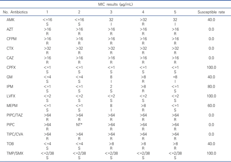
4. O. anthropi의 항균제 감수성
Table 3에서는 O. anthropi의 항균제 감수성결과로 총 보 고된 횟수와 감수성으로 보고된 횟수와의 비를 제시하였다. 환 자의 검체에서 분리된 O. anthropi는 aztreonam과 cefepime, cefotaxim, ceftazidim에 모두 내성이었고, levofloxacin (100%), ciprofloxacin (100%), trimethoprime/sulfame- thoxazole (100%), imipenem (80.0%), meropenem (60.0%)에 감수성을 나타냈다. 4번 환자의 객담 검체에서 분리 된 O. anthropi의 경우 imipenem과 meropenem에 모두 내 성을 나타냈다.
고 찰
본 연구에서는 O. anthropi의 전통적인 생화학적 동정방법 을 이용한 검사에서 단시간 배양에서 얻어진 작은 집락을 이용 할 때 여러 번 반복검사를 통해 오랜 시간이 걸려 동정되는 과정 을 보여 주었다. 특히 O. anthropi는 초기에 Achromobacter
Table 3. MIC results of each antibiotic by MicroScan
MIC results (μg/mL)
No. Antibiotics 1 2 3 4 5 Susceptible rate
AMK <=16
S
<=16 S
32 I
>32 R
32 I
40.0
AZT >16
R
>16 R
>16 R
>16 R
>16 R
0.0
CFPM >16
R
>16 R
>16 R
>16 R
>16 R
0.0
CTX >32
R
>32 R
>32 R
>32 R
>32 R
0.0
CAZ >16
R
>16 R
>16 R
>16 R
>16 R
0.0
CPFX <=1
S
<=1 S
<=1 S
<=1 S
<=1 S
100.0
GM <=4
S
<=4 S
8 I
>8 R
=8 I
40.0
IPM <=1
S
<=1 S
2 S
>8 R
<=1 S
80.0
LVFX <=2
S
<=2 S
<=2 S
<=2 S
<=2 S
100.0
MEPM <=1
S
<=1 S
8 I
>8 R
<=1 S
60.0
PIPC/TAZ >64
R
>64 R
>64 R
>64 R
>64 R
0.0
PIPC >64
R
NT* >64
R
>64 R
>64 R
0.0
TIPC/CVA >64
R
>64 R
>64 R
>64 R
>64 R
0.0
TOB <=4
S
<=4 S
>8 R
>8 R
>8 R
40.0
TMP/SMX <=2/38
S
<=2/38 S
<=2/38 S
<=2/38 S
<=2/38 S
100.0
Abbreviations: R, resistant; I, intermediate; S, susceptible; AMK, amikacin; AZT, aztreonam CFPM, cefepime; CTX, cefotaxime; CAZ, ceftazidim; CPFX, ciprofloxacin; GM, gentamicin; IMP, imipenem; LVFX, levofloxacin; MEPM, meropenem; PIPC/TAZ, piperacillin/tazobactam;
PIPC, piperacillin; TIPC/CVA, ticacillin/clavulanic acid; TOB, tobramycin; TMP/SMX, trimethoprime/sulfamethoxazole.
*, not tested.
그룹과의 유사성 때문에 Achromobacter species biotype Vd로 분류되었으나, 1988년 Holmes 등이 DNA-DNA hy- bridization 법으로 새로운 균임을 밝혀 O. anthropi로 새롭 게 명명되었다[17]. Table 2에 제시된 5번째 환자검체의 동정 과정에서는 Achromobacter와 혼합 배양되었음에도 외관상 구별이 어려웠으며, 수 차례 계대배양 과정에서 오염되었는지 처음부터 혼합감염 또는 채취과정에서의 오염인지 알 수 없으나 한 배지에서 자란 oxidase 양성인 두 세균은 MALDI-TOF법을 이용한 검사장비 Micro ID Sys로 검사했을 때 O. anthropi와 A. xylosoxidans로 각각 동정되었다(Table 2). 24시간 배양한 O. anthropi 집락 크기는 1 mm 이하이므로 외관상 구별이 힘 든 세균과의 혼합배양 시 여러 개의 집락을 사용해 검사하여야 하는 생화학적 동정장비 MicroScan이나 VITEK 2에서는 정확 한 동정이 이루어질 수 없었다. 또한 MALDI-TOF를 이용한 E.
coli의 최종 보고까지 소요된 시간은 항균제감수성검사로 인해 MicroScan에서 16시간 이상 배양해야 하는 이유로 단축되지 않지만 Table 2의 4번째 동정에서 볼 수 있듯이 O. anthropi를 MALDI-TOF로 동정한다면 항균제검사에 이용할만한 충분한 크기의 단일집락 형성을 위해 48시간을 배양한다 하여도 최종 보고까지 2.5일을 단축할 수 있다(Figure 3).
최근 기저질환을 동반한 장기 입원사례가 늘어 MRAB, MRPA, CRE를 포함해 병원환경에 존재하는 미생물에 의한 감 염을 주의해야 하며 신속하고 정확한 균 동정은 환자치료와 동 시에 감염관리를 위해 반드시 필요하다. 원내감염 혹은 2차감염 원인 균으로 MRAB와 MRPA는 질병관리본부의 지침에 의해 격 리 등의 적절한 관리가 의무화되었지만, O. anthropi는 병원환 경에 존재하면서 카테터를 통한 균혈증 야기 등 원내감염이 보 고[11-13]되고 있으며 Table 1에서 보는 바와 같이 입원부터
검체 채취까지 경과된 기간이 최저 0일에서 92일로 면역이 저하 된 환자들에게 기회감염 또는 원내감염 가능성을 배제할 수 없 음에도 동정이 까다로워 신뢰할만한 통계보고조차 없는 것이 현 실이다. 게다가 본 연구에 의하면 O. anthropi는 평균 6일 이상 의 동정기간이 소요되었고, 혼합 배양된 경우는 더욱 많은 시간 이 필요하여 5번 환자의 경우 보고까지 시간이 11.3일이 소요되 어 치료가 늦어졌고 한달 동안 O. anthropi가 검출되었다. 따 라서 O. anthropi의 경우 현재까지 발표된 사례보다 훨씬 더 많은 감염가능성을 배제할 수 없으며, 균 동정에 소요되는 시간 이 길므로 신속하고 정확한 치료가 어렵고 더불어 O. anthropi 의 항균제 내성률을 높이는 원인이 될 수 있다. 최근에는 O.
anthropi의 carbapenem계열 항균제 내성이 보고[2]되고 있 으며 Table 1에서 보는 바와 같이 4번째 환자의 경우, 지주막하 출혈로 인한 뇌간 기능부전으로 입원 92일 만에 채취한 객담에 서 O. anthropi가 검출될 시 위 결과에 표기되진 않았지만 같 은 검체에서 MRAB와 동시에 검출되어 지침에 따라 격리상태였 고, 환자는 사망했는데 imipenem과 meropenem의 carbape- nem 계열 항균제 및 모든 베타락탐계 항생제에는 내성을 가지 고 있는 것으로 보아(Table 3) O. anthropi의 신속하고 정확한 동정 및 병인성과 항생제 내성 기전에 관한 연구는 필수불가결 한 실정이다.
기회감염은 고령이거나 기저질환에 의한 면역이 약화된 환자 에서 주로 발생한다. Table 1에서 보는 바와 같이 O. anthropi 가 검출된 연령대는 60대 이상이 80%를 차지하였다. 이 결과만 으로 O. anthropi의 검출과 연령과의 상관관계를 확언할 수는 없지만, 4세의 소아 1명과 암 또는 기저질환이 있는 4명의 환자 에서 O. anthropi가 검출된 것은 면역이 약하거나 기저질환의 유무가 이 세균의 감염과 관련 있다는 다른 그룹의 연구결과들 과 일치하는 결과이다[18-20].
MALDI-TOF법은 분자량이 비교적 큰 시료와 매트리스가 혼 합된 결정체에 레이저를 조사하여 이온화시킨 후 전하를 띤 이 온들을 질량분석기에 통과시켜 검출기까지의 도달시간을 측정 하여 분자량을 분석해 이미 구축된 각 균종에 대한 정보와 비교 분석하여 균종을 동정한다[21]. MALDI-TOF MS system을 이 용한 세균동정 시간은 균주 당 평균 6분 정도 소요된다는 보고가 있으며[22], 이는 세균동정시간을 단축시키는 획기적인 방법으 로[23] 전통적인 생화학 동정 장비를 이용한 동정검사와 비교한 연구에서 그 유용성이 보고되었고[24], 특히 혈액배양시 그람양 성막대균 동정에 대한 유용성과 산소성세균 동정에 대한 유용성 이 보고된 바 있다[25, 26]. 그러나 MALDI-TOF를 이용해 신속 한 동정이 가능해도 항균제감수성 검사가 MicroScan이나
VITEK 2 등의 MIC 결과에 의존하고 있어 최종보고에 이르는 시간을 단축하기에는 한계가 있다. 하지만 O. anthropi와 같이 동정과정에서 많은 시간을 소비하는 균들은 MALDI-TOF를 이 용해 동정시간이 단축되면 항균제감수성검사를 위해 48시간 배 양하더라도 최종보고까지 4일이면 충분하며(Figure 3), 최근에 는 내성유전자를 보유하고 있는 균을 MALDI-TOF를 이용해 검 출하는 방법이 계속 연구되고[27-29] 있으므로 향후 내성균들 에 대한 정확하고 빠른 보고가 가능하리라 예상한다.
현재 MALDI-TOF법은 많은 대형 병원에서 미생물동정검사 에 이용 중이다. 하지만 아직까지 전통적인 생화학적 동정방법 에 의존하고 있는 중소병원에서도 집락외관이나 생화학적 특징 의 유사성과 느린 생장으로 인해 검출동정이 까다로워 반복검사 를 할 수 밖에 없는 O. anthropi와 같은세균 동정을 위해 적은 양의 세균으로도 빠른 동정이 가능한 MALDI-TOF법을 도입한 다면 환자치료와 감염관리에 매우 유용할 것으로 사료된다.
요 약
Ochrobactrum anthropi는 oxidase를 생산하는 비발효 산소성 그람음성 막대균으로 외관이 비슷하고 oxidase가 양성 인 비발효 세균과 혼합배양 시 구분이 힘들고 생화학적 동정 장 비로는 정확한 동정에 한계가 있다. 따라서 본 연구에서는 생화 학적 검사 방법으로 동정이 힘든 세균동정의 Matrix-Assisted Laser Desorption/Ionization Time-of-Flight Mass Spec- trometry Platform (MALDI-TOF) 법의 유용성을 제시하고 자 하였다. MicroScan을 이용해 검사했던 O. anthropi 5례를 분석한 결과, 최종보고까지 6.2일이 소요되었으며 E. coli의 3.0일에 비해 3.5일이 더 소요되었다. 5번 환자 고름 검체는 Achromobater xylosoxidans와 혼합감염으로 여러 번의 계 대 배양과 재검사로 인해 11.3일이 소요되었는데, MALDI- TOF법으로 검사한 경우 한 번에 동정되었다. 4명의 환자는 기 저질환이 있는 60세 이상이었고 기회감염과 원내감염의 가능성 을 배제할 수 없었으며, 그 중 92일 만에 채취된 검체는 imipenem 과 meropenem에 내성이었다. 따라서 O. anthropi처럼 동정 이 까다로운 세균은 신속하고 적절한 환자 치료를 위해 MALDI- TOF법을 이용한 검사가 매우 유용할 것으로 사료된다.
Acknowledgements: None Conflict of interest: None
Author’s information (Position): Ko HM1, Adjunct professor; Jo JH2, M.T.; Baek HG2, M.T.
REFERENCES
1. Cieslak TJ, Robb ML, Drabick CJ, Fischer GW. Catheter-asso- ciated sepsis caused by
Ochrobactrum anthropi
: report of a case and review of related nonfermentative bacteria. Clin Infect Dis.1992;14:902-907.
2. Kim GM, Jin SJ, Yoo JS, Kim CO, Choi JY, Kim JM, et al. A case of meropenem-resistant
Ochrobactrum anthropi
bacteremia. Infect Chemother. 2009;41:62-64. https://doi.org/10.3947/ic.2009.41.1.62
3. Nasir N, Mahmood SF. Mortality in patients with respiratory and non respiratory carbapenem resistant-multidrug resistant
Acine- tobacter
infections. J Ayub Med Coll Abbottabad. 2017;29:511- 513.4. Appelbaum PC, Campbell DB. Pancreatic abscess associated with
Achromobacter
group Vd biovar1. J Clin Microbiol. 1980;12:282-283. https://doi.org/10.1128/JCM.12.2.282-283.1980 5. Jimenez G. Antony S.
Ochrobactrum anthropi
– an unusual causeof line related sepsis. current knowledge of the epidemiology and clinical features of this pathogen. Br J Med Med Res. 2016;18:1-7.
https://doi.org/10.9734/BJMMR/2016/29365
6. Aguilera-Arreola MG, Ostria-Hernández ML, Albarrán-Fernández E, Juárez-Enriquez SR, Majalca-Martínez C, Rico-Verdín B, et al.
Correct identification of
Ochrobactrum anthropi
from blood cul- ture using 16rRNA sequencing: a first case report in an im- munocompromised patient in Mexico. Front Med(Lausanne).2018;5:205. https://doi.org/10.3389/fmed.2018.00205
7. Mastroiani A, Cancellieri C, Montini G.
Ochrobactrum anthropi
bacteremia: case report and review of the literature. Clin Microbiol Infect. 1999;5:570-573. https://doi.org/10.1111/j.1469- 0691.1999.tb00437.x8. Gransden WR, Eykyn SJ. Seven cases of bacteremia due to
Ochrobactrum anthropi
. Clin Infect Dis. 1992;15:1068-1069.https://doi.org/10.1093/clind/15.6.1068
9. Babic I, Fischer-Le Saux M, Giraud E, Boemare N. Occurrence of natural dixenic association between the symbiont Photorhabdus luminescens and bacteria related to
Ochrobactrum
spp. in trop- ical entomopathogenic Heterorhabditis spp. (Nematoda, Rha- bditida). Microbiology. 2000;146:709-718. https://doi.org/10.1099/00221287-146-3-709
10. Shilton CM, Brown GP, Benedict S, Shine R. Spinal arthropathy associated with
Ochrobactrum anthropi
in free-ranging cane toads (Chaunus [Bufo] marinus) in Australia. Vet Pathol. 2008;45:85-94. https://doi.org/10.1354/vp.45-1-85
11. SitiRohani AH, TzarMN.
Ochrobactrum anthropi
catheter-related bloodstream infection: the first case report in Malaysia. Med J Malaysia. 2013;68:267–268.12. Menezes FG, Abreu MG, Kawagoe JY, Warth AN, Deutsch AD, Dornaus MF, et al.
Ochrobactrum anthropi
bacteremia in a pre- term infant with cystic fibrosis. Braz J Microbiol. 2014;45:559–561. https://doi.org/10.1590/S1517-83822014005000043 13. Mrozek S, Dupuy M, Hoarau L, Lourtet J, Martin-Blondel G,
Geeraerts T. Brain empyema due to
Ochrobactrum anthropi
. Med Mal Infect. 2014;44:128–129. https://doi.org/10.1016/j.medmal.2014.01.003
14. Stager CE, Davis JR. Automated systems for identification of
microorganisms. Clin Microbiol Rev. 1992;5:302–327. https://doi.
org/10.1128/cmr.5.3.302
15. Funke G, Monnet D, deBernardis C, von Graevenitz A, Freney J.
Evaluation of the VITEK 2 system for rapid identification of med- ically relevant gram-negative rods. J Clin Microbiol. 1998;36:
1948-1952. https://doi.org/10.1128/JCM.36.7.1948-1952.1998 16. Garcia-Garrote F, Cercenado E, Bouza E. Evaluation of a news-
ystem, VITEK 2, for identification and antimicrobial suscepti- bility testing of Enterococci. J Clin Microbiol. 2000;38:2108–2111.
https://doi.org/10.1128/JCM.38.6.2108-2111.2000
17. Velasco J, Romero C, López-Goñi I, Leiva J, Díaz R, Moriyón I.
Evaluation of the relatedness of Brucella spp. and
Ochrobactrum anthropi
and description ofOchrobactrum intermedium sp
. nov., a new species with a closer relationship toBrucella spp
. Int J Syst Bacteriol. 1998;48:759–768. https://doi.org/10.1099/00207713- 48-3-75918. Gupta A, Chauhan K, Pandey A. Neonatal Septicaemia by
Ochrobactrum anthropi
: A missed pathogen. Int J Curr Microbiol App Sci. 2018;7:1651-1654. https://doi.org/10.20546/ijcmas.2018.705.195
19. Berman AJ, Del Priore LV, Fischer CK. Endogenous
Ochrobac- trum anthropi
endophthalmitis. Am J Ophthalmol. 1997;123:560-562. https://doi.org/10.1016/s0002-9394(14)70190-4 20. Haditsch M, Binder L, Tschurtschenthaler G, Watschinger R,
Zauner G, Mittermayer H. Bacteremia caused by
Ochrobactrum anthropi
in an immunocompromised child. Infection. 1994;22:291-292. https://doi.org/10.1007/BF01739922
21. Murray PR. What is new in clinical microbiology-microbial iden- tification by MALDI-TOF mass spectrometry: a paper from the 2011 William Beaumont Hospital Symposium on molecular pathology. J Mol Diagn. 2012;14:419-423. https://doi.org/10.
1016/j.jmoldx.2012.03.007
22. Seng P, Drancourt M, Gouriet F, La Scola B, Fournier PE, Rolain JM, et al. Ongoing revolution in bacteriology: routine identi- fication of bacteria by matrix-assisted laser desorption ioniza- tion time-of-flight mass spectrometry. Clin Infect Dis. 2009;49:
543-551. https://doi.org/10.1086/600885
23. Kim TS, Lee K, Hong YJ, Hwang SM, Park JS, Park KU, et al.
MALDI-TOF MS: Its application in the clinical laboratory and a paradigm shift in clinical microbiology. Lab Med Online. 2015;
5:176-187. http://doi.org/10.3343/lmo.2015.5.4.176
24. Guo L, Ye L, Zhao Q, Ma Y, Yang J, Luo Y. Comparative study of MALDI-TOF MS and VITEK 2 in bacteria identification. J Thorac Dis. 2014;6:534-538. http://doi.org/10.3978/j.issn.2072-1439.
2014.02.18
25. Choi JU, Yu YB, Kim SH, Won S, Kim YK. Two years quaternary isolation of Gram-positive bacilli using MALDI-TOF MS in pos- itive blood culture of a university hospital. Korean J Clin Lab Sci.
2018;50:414-421. https://doi.org/10.15324/kjcls.2018.50.4.414 26. Kim M, Kwon MJ, Chung HS, Lee Y, Yong D, Jeong SH, et al.
Evaluation of matrix-assisted laser desorption ionization-time of flight mass spectrometry for identification of aerobic bacteria in a clinical microbiology laboratory. Korean J Clin Microbiol.
2012;15:60-66. https://doi.org/10.5145/KJCM.2012.15.2.60 27. Camoez M, Sierra JM, Dominguez MA, Ferrer-Navarro M, Vila J,
Roca I. Automated categorization of methicillin-resistant
Sta-
phylococcus aureus
clinical isolates into different clonal com-plexes by MALDI-TOF mass spectrometry. Clinl Microbiol Infect.
2016;22:161.e1-161.e7. https://doi.org/10.1016/j.cmi.2015.10.009 28. Rapp E, Samuelsen Ø, Sundqvist M. Detection of carbapenemases
with a newly developed commercial assay using matrix assisted laser desorption ionization-time of flight. J Micorobiol Methods.
2018;146:37-39. https://doi.org/10.1016/j.mimet.2018.01.008
29. Kim YA, Yong D, In YH, Park HS, Lee K. Application of ma- trix-assisted laser desorption ionization time-of-flight mass spectrometry to screen the extended-spectrum β-lactamase-pro- ducing ST131
Escherichia coli
strains. Ann Clin Microbiol.2016;19:65-69. https://doi.org/10.5145/ACM.2016.19.3.65