IEG 환경지질연구정보센터
9
0
0
전체 글
(2) 416. Yul Roh, Chul-Min Chon and Ji-Won Moon. an electron acceptor and formed magnetite, siderite, green rust, and vivianite [Fe3(PO4)2·8H2O] crystals (Zhang et al., 1997; Roh et al., 2001, 2002, 2003, 2006; Fredrickson et al., 2001; Zachara et al., 2002). Magnetite, siderite, and vivianite are common mineral products and ubiquitous in natural environments (Emerson, 1976; Emerson and Widmer, 1978; Pye et al., 1990). Although a direct link can not be established between these laboratory-produced biogenic minerals and those observed in nature, several lines of evidence suggest that at least some proportion of these minerals present in natural environments are biogenic (Pye et al., 1990). Little is known about metal reduction and iron biomineralization under extreme conditions, and only recently has bacterial Fe3+ reduction been demonstrated under thermophilic (Liu et al., 1997; Roh et al., 2002), psychrophilic (Zhang et al., 1999; Roh et al., 2002), or acidic conditions (Kusel et al., 1999). Sites contaminated with toxic metals can have drastically different environmental conditions, and the biological reduction of most metals has commonly been studied at circum neutral pH values (Strab et al., 2001). Microbial metal reduction and iron biomineralization under alkaliphilic growth conditions has not been demonstrated well. A novel metal-reducing, alkaliphilic species named as Alkaliphilus metalliredigenes sp. nov. was proposed by Ye et al. (2004) and they studied genomic characteristics and physiology of the microorganism in detail. Therefore, the objectives of this study were to further examine metal reduction and iron mineral formation in alkaline conditions as well as to perform mineralogical characterization of the precipitates formed via microbial reduction of Fe(III)-citrate and Fe(III)-EDTA using an alkaliphilic bacterium, Alkaliphilus metalliredigens (QYMF), isolated from a boron containing pond. 2. MATERIALS AND METHODS 2.1. Source of Microorganisms In this study, we examined the microbial formation of iron minerals and metal reductions using an alkaliphilic bacterium (QYMF, Alkaliphilus metalliredigens sp. nov.) isolated from boron leachate ponds at the U.S. Borax Mines in Boron, CA (Ye et al., 2004). The hydrogeochemical analysis of water samples from the boron leachate ponds indicated that Na, Si, and K were the major elements. Sodium concentration is 11,800 mg/L. Boron and Fe concentrations were about 2000 mg/L and 4.9 mg/L, respectively. Cr and Ba concentrations were 0.2 mg/L and 0.4 mg/L, respectively. U concentrations ranged from 0.2 mg/L to 0.4 mg/L, and arsenic concentration was 126 mg/L. Phylogenetic analysis of the Small subunit rDNA indicated the bacterium, QYMF, was a low G+C Gram-positive microorganism in the Clostridiaceae, and had 96% and 92% nucleotide identity with. Fig. 1. An SEM image of endspores produced by isolate QYMF grown with Fe -EDTA as an electron acceptor and lactate as an electron donor. 3+. Alkaliphilus transvaalensis and Alkaliphilus crotonoxidans, respectively (Ye et al., 2004). The isolate might represent a novel metal-reducing, alkaliphilic species and the name Alkaliphilus metalliredigenes sp. nov. was proposed (Ye et al., 2004). Bacterial cell morphology examined using scanning electron microscopy (SEM) showed that QYMF cells were straight with some cells slightly curved, and a mean length of 3 to 6 mm and an approximate width of 0.5 mm (Ye al., 2004). As the cells entered stationary phase growth, terminal endospores were observed (Fig. 1). The temperature range for growth was between 4 °C (lowest temperature attempted) and 45 °C (Ye et al., 2004), The maximum Fe(III)-citrate reduction rates and bacterial growth rates were observed at 35 °C. When the Alkaliphilus metalliredigens sp. (QYMF) was grown in the presence of Fe(III)citrate at 25 °C, the pH range for growth was between 7.0 and 11.0, growth below pH 7.5 or above pH 11.0 was negligible, and the pH optimum was approximately 9.5. By definition, true alkaliphiles should have an optimum pH above 9.0, and display little growth at or below pH 7.0. Microbial Fe(III) reduction by Alkaliphilus metalliredigens sp. (QYMF) occurred at a salinity range of 0–80 g/L NaCl at 25 °C using lactate (10 mM) as an electron donor and Fe(III)-citrate as an electron acceptor. Sodium chloride was not essential for growth, but the optimal concentration appeared to be approximately 20 g/l. The isolate could grow and reduce Fe(III) in the presence of up to 1.5% borax (Na2B4O7). 2.2. Growth Conditions Two types of growth media including sodium carbonate (NaHCO3) buffer medium and tris(hydroxymethyl)amino.
(3) Metal reduction and biomineralization by a metal reducer. 417. Table 1. Media composition for microbial reduction of metals NaCO -buffer medium. CaCl MgCl (NH ) SO 100X Trace Mineral Solution NaCl Na CO tris(hydroxymethyl)amino methane (TRIS) Yeast extract Na B O K HPO Headspace 2. 2. 4 2. 2. 2. 2. 4. 3. 4. 7. 4. TRIS-buffer medium. 3. Component. g/L 1.2 10 ml 20 3.0 0.25 2 1 N (100%) or N -CO (80%:20% v:v) 2. 2. methane (TRIS) buffer medium were prepared anaerobically and used for microbial metal reduction and biomineralization (Table 1). The trace mineral solution contained 1.5 g of nitrilotriacetic acid, 3 g of MgSO4, 0.5 g of MnSO4, 1.0 g of NaCl, 0.1 g of FeSO4, 0.1 g of CaCl2, 0.1 g of CoCl2, 0.154 g of ZnSO4, 0.01 g of CuSO4, 0.01 g of AlK(SO4), 0.01 g of H3BO3, 0.025 g of Na2MoO4, 0.1 g of NiCl2 per liter of deionized water. The medium was prepared under N2 or N2-CO2 (80% N2-20% CO2) and had a final medium pH of 9.5. To assess the capability of metal reduction and mineral formation by QYMF (Alkaliphilus metalliredigenes sp. nov.), various electron acceptors such as Fe(III)-citrate (10-20 mM), Fe(III)-EDTA (10 mM), cobalt(III)-EDTA (5 mM), potassium chromate (100 µM), Mn(IV)-oxide (10 mM), iron oxyhydroxide [akaganeite, β-FeOOH, ~70 mM]], selenate (5 mM), and uranyl carbonate (1 mM) were examined at 25 °C with lactate (20 mM) or acetate (20 mM) as an electron donor. Iron reduction and biomineralization were also examined by QYMF with Fe(III)-citrate (15 mM) in the presence of arsenate (5 mM) using lactate (10 mM) or acetate (10 mM) as an electron donor. The Mn(IV) oxide was prepared as described by Roh et al. (2002) and uranyl carbonate [UO2(CO3)22-] was prepared by mixing uranyl-nitrate [UO2 (NO3)22-] solution with sodium bicarbonate (Roh et al., 2000). Analysis of X-ray diffraction showed that the Mn oxide was amorphous. The iron oxyhydroxide was prepared by neutralizing a solution of 0.4 M FeCl3 with 10M NaOH as described previously (Roh et al., 2003). Analysis of X-ray diffraction showed that the iron oxyhydroxide was poorly crystalline akaganeite (βFeOOH). 2.3. Metal Reduction Studies To examine the chemical conditions of metal reduction and mineral formation by the alkaliphilic Fe(III)-reducing bacteria, subsamples (1 ml) of bacterial cultures and abiotic. 2. 5.545 9.5 1.2 10 ml 20 6.15 0.25 2 1 N (100%) or N -CO (80%:20% v:v) 2. 2. 2. controls were taken from the culture bottles at different times. Fe(II) and Cr(VI) concentrations (0.5 N HCl soluble) were determined by measuring the absorbance at 562 nm (for Fe2+) and at 540 nm (for Cr6+) on a Hewlett Packard 8453 spectrophotometer using the ferrozine method with anaerobic water for sample dilution (Stookey, 1970; Zhang et al., 1996). 2.4. Mineralogical Characterization Precipitates in each sample were collected on a 0.22 μm Millipore filter (Millipore Corporation, Bedford, MA) and washed three times with deionized water (Roh et al., 2003). The filtered precipitates were dried under an N2 atmosphere to prevent oxidation of the precipitated minerals. The filtered and dried precipitates were carbon coated with a Ted Pella carbon sputter coater and immediately examined. The morphology, mineralogy, and chemistry of precipitated phases during microbial metal precipitation of iron were determined with scanning electron microscope (SEM) (XL30FEG, Phillips, Netherlands) with energy dispersive X-ray (EDX) analysis. X-ray diffraction analysis (a Scintag XDS 2000, Scintag, Sunnyvale, CA) was used to monitor mineralogical characteristics with operating at a scan rate of 2° 2θ min-1. Cobalt K-α radiation (40 kV, 35 mA) was used to minimize fluorescence of Fe-rich minerals. Laser induced plasma spectroscopy (LIPS) was used to detect boron in the precipitate formed by the bacterium (Martin et al., 2002). 3. RESULTS AND DISCUSSION 3.1. Metal Reduction Fe(II) concentrations were monitored at 25 °C in a time course experiments using Na2CO3 buffered medium containing Fe(III)-citrate (20 mM) as an electron acceptor and acetate (20 mM) as an electron donor. A dramatic increase in Fe(II) concentration was observed within four days of.
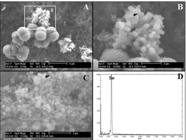
(4) 418. Yul Roh, Chul-Min Chon and Ji-Won Moon. Fig. 2. (A) Time course experiments of Fe (III) reduction using the Na CO buffered medium. (B) Time course experiments of K CrO (100 µM) reduction. The results are the means of duplicate experiments. 2. 3. 2. 4. incubation at 25 °C. The bacteria reduced the Fe(III)-citrate and changed Fe(II) concentration from 1 mM to 8.8 mM Fe(II) within 100 hours of bacterial incubation (Fig. 2a). The color of the culture solution also changed from yellowish brown (ferric citrate) to colorless at 25 °C. Fe(II) concentration ranged from 0.7 to 2.2 mM in the abiotic controls, which may result from abiotic reduction of Fe(III) by organic nutrients such as yeast extract in the medium. However, the abiotic controls did not show any color change in the time course experiment. The bacterium was also able to reduce Fe(III)-EDTA (10 mM) as an electron acceptors using acetate or lactate as an electron donor. Cr(VI) concentrations with time were also examined using Na2CO3 buffered medium containing ~100 µM K2CrO4 and 20 mM acetate at 25 °C. A dramatic decrease in Cr(VI) concentration was observed within 5 days of incubation at 25 °C and reached 30 µM Cr(VI) (Fig. 2b) and showed the color change of the culture solution from yellow (K2CrO4) to colorless at 25 °C. The bacteria reduced the Cr(VI) and changed Cr(VI) concentration from 100 µM up to below detection limit within 150 hours of bacterial incubation (Fig. 2b). Cr(VI) concentration decreased slightly from 102 to 83 µM in the abiotic controls, which may result from abiotic reduction of Cr(VI) by organic nutrients such as yeast. extract in the medium. However, the abiotic controls did not show any color change in the time course experiment. The ability of the Alkaliphilus metalliredigens sp (QYMF) to reduce metals besides Fe(III) and Cr(VI) was also examined using Na2CO3- or TRIS-buffered medium. The bacterium was able to reduce Co(III) [Co(III)-EDTA, 5 mM] to Co(II) and U(VI) (UO2CO32-, 1 mM) to U(IV) with lactate (10 mM) as an electron donor, as indicated by the color change of the culture solution from purple [Co(III)-EDTA] and yellow (uranyl carbonate) to colorless at 25 °C. The reduced cobalt, Co(II), is colorless and the reduced U(VI) is precipitated as UO2 in the media (Roh et al., 2002). The bacterium was also able to reduce selenate [Se(VI), 5 mM] and precipitated reddish brown phases. An SEM analysis showed that the bacterium precipitated trigonal crystals or ball-like selenium (0) particles during selenate reduction using lactate as an electron donor at pH=10 (Fig. 3). The precipitated phases mainly consisted of Se, thereby demonstrating the reduction of Se(VI) to Se(0) (Fig. 3). The reduction of the oxyanions, arsenate and selenate, is a common biogeochemical process in other alkaliphiles such as Bacillus selenitrireducens and Bacillus arsenicoselenatis (Stoltz and Oremland, 1999). These results indicated that the Alkaliphilus metalliredigens sp. (QYMF) can reduce several other transition metals beside iron as long as their concentrations are held below the toxic levels. The bacterium was also able to reduce Mn(IV)-oxide (10 mM) in Na2CO3 buffer medium and to produce white precipitates using lactate (10 mM) as an electron donor. The XRD analysis showed that the precipitated phase formed by reduction of Mn(IV)-oxide has 1.3 nm and 0.7 nm peaks (Fig. 4d). It is an unknown mineral and further study is required for proper identification. The SEM with EDX analyses showed that this crystalline phase contained significant quantities of Mn, Ca, and P (Fig. 4b) and had a leaf-shaped morphology (Fig. 4a). But the Alkaliphilus metalliredigens sp (QYMF) could not reduce akaganeite (~70 mM) as an electron acceptor. The Alkaliphilus metalliredigens sp (QYMF) used acetate (10 mM), glucose (10 mM), lactate (10 mM), pyruvate (10 mM), and H2 (20% H2-80% N2) as an electron donor. The Alkaliphilus metalliredigens sp (QYMF) could not use fumurate (10 mM) as an electron donor to reduce Fe(III)-citrate. This study showed that metal reduction by microbial Fe(III) reduction can be an important process for organic matter oxidation in anaerobic alkaline subsurface environments. Previous studies have shown that organic compounds such as lactate, formate, acetate, and pyruvate are potentially available in terrestrial subsurface environments (Lovley, 1991). The oxidation of organic compounds coupled with the reduction of Fe(III) oxides can be expected to release Fe(II) ions in subsurface environments (Lovley, 1993; Fredrickson et al., 1998). Hydrogen gas is generated from anaerobic decomposition of organic matter as well as from.
(5) Metal reduction and biomineralization by a metal reducer. 419. Fig. 3. Scanning electron photomicrographs of precipitates formed by the selenate reduction with QYMF. Extracellular trigonal (A and B) or ball-like (C) selenium particles are elemental selenium as determined by the EDX analysis (D).. Fig. 4. SEM photomicrographs together with EDX and XRD analyses of minerals formed by the microbial reduction of manganese oxide using alkaliphilic Fe(III)-reducing bacteria, QYMF..
(6) 420. Yul Roh, Chul-Min Chon and Ji-Won Moon. geochemical processes in subsurface environments and probably constitutes a sustainable source of energy for subsurface biosphere ecosystems. 3.2. Biomineralization of Vivianite No magnetite was formed with the microbial reduction of Fe(III)-citrate and Fe(III)-EDTA. During the course of Fe(III) reduction, color of Fe(III)-citrate and Fe(III)-EDTA changed from amber to green. Approximately 3 days after reduction of the Fe(III)-citrate and Fe(III)-EDTA, a white or brown mineral precipitates were produced when QYMF cells were grown in bicarbonate or TRIS buffered medium in the presence of lactate or acetate as an electron donor at pH 7.5-11.0. Detailed mineralogical characterization of the white precipitate or greenish brown precipitates formed during microbial reduction of Fe(III)-citrate and Fe(III)-EDTA were performed. No precipitates were formed in control tubes without cells. Analysis with SEM and EDX analysis showed that the precipitated phase mainly contained Fe and P with keg- or barrel-shape morphology and a pronounced platy habit (Fig. 5). The XRD analysis of the precipitate formed by reduction of Fe(III)-citrate displayed sharp reflection patterns and all peaks were attributable to vivianite [Fe3(PO4)2·8H2O]. The. precipitate formed by reduction of Fe(III)-citrate displayed a broader background than that of precipitate formed by Fe(III)-EDTA reduction (Fig. 6). The broadness may be attributable to the poor crystallinity of the former vivianite crystals. On the other hand, the latter vivianite crystals showed two sharp and intense XRD peaks, attesting the highly crystalline nature of the precipitate. The presence of two vivianite peaks only could be attributed to flattened {010} growth of the precipitates (Fig. 6). The crystals can be found in encrusting masses with a divergent bladed structure. They occur as relatively well-crystallized secondary minerals in natural environment (Deer et al., 1966). Vivianite can be found in sedimentary deposits where it is often associated with bone, decayed wood, and other remains. Microbes play a role in the authigenic or diagenetic formation of the phosphate minerals such as vivianite, strengite (FePO4·2H2O), and variscite (AlPO4·2H2O) (Ehrlich, 1990). Vivianite has been observed in the upper iron reduction zone in anaerobic lake sediments (Emerson and Widmer, 1978), freshwater swamps (Postma, 1981) and in river sediments (Hearn et al., 1983). Vivianite also forms in iron rich sediments within or below the sulfate reduction zone or the methanogenic zone. Vivianite has been detected in anoxic river sediments receiving the treated sewage, although little. Fig. 5. SEM photomicrographs and EDX results of microbially precipitated phases using Fe(III)-citrate and acetate (A, C) and for those using Fe(III)-EDTA and lactate(C, D)..
(7) Metal reduction and biomineralization by a metal reducer. 421. Fig. 6. X-ray diffraction patterns of precipitates from the reduction of (A) Fe(III)-citrate and (B) Fe(III)-EDTA.. Fig. 7. An SEM photomicrograph (A) and EDX result (B) of microbially precipitated phases using Fe(III)-citrate (15 mM) with arsenate (5 mM) and lactate (10 mM) and an SEM photomicrograph (C) and laser induced spectra (D) of the precipitated phases using Fe(III)citrate and acetate.. is known about the condition of formation and the frequency of occurrence for vivianite in sediments (House, 2003). A previous study by Zhang et al. (1996) showed that no crystalline minerals precipitated when Fe(III)-citrate or Co(III)EDTA were used as the electron acceptor, which is probably due to chelation of Fe(II) with dissolved citrate or EDTA. in solution. Citrate acts as a chelating agent in decontamination operations, forming highly recalcitrant and mobile metal-citrate (Jardine et al., 2002). An industrial chelating agent EDTA also forms very strong complexes with divalent and trivalent metal ion (Jardine et al., 2002). Newly isolated cultures of Alkaliphilus metalliredigens sp (QYMF) was, however, able to grow using Fe(III)-citrate and Fe(III)-.
(8) 422. Yul Roh, Chul-Min Chon and Ji-Won Moon. EDTA as an electron acceptor, while iron mineral precipitation was promoted by the addition of inorganic phosphate. Borate was present in the culture medium (2.0 g/l), and the SEM and LIPS results (Fig. 7c,d) indicated that boron was incorporated into the crystalline iron-precipitate, vivianite. Naturally occurring bacteria from boron-containing sites may prove beneficial in minimizing the effects of boron toxicity from fly ash ponds. The alkaliphilic Fe(III)reducing bacteria (QYMF) reduced Fe(III)-citrate and precipitated vivianite in the presence of up to 5 mM arsenate. The SEM-EDX analysis suggested that arsenic can be reduced and co-precipitated with vivianite. The formation of sparingly-soluble vivianite precipitates, mediated by the alkaliphilic Fe(III)-reducing bacterium, may sequester iron, phosphate, and arsenic into more stable and less toxic forms of iron phosphate minerals. Naturally-occurring bacteria from boron-containing sites (e.g. the U.S. Borax mine, Boron, California) may not only prove beneficial in reducing the effects of boron toxicity of fly ash ponds but also potentially assist the remediation of boron contaminated soils and sediments through bacterially-facilitated precipitation; i.e., by complexing boron into mineral forms that are both sparingly soluble and unavailable for bio-uptake. These results indicate that metal-reduction can occur in alkaline habitats, and iron mineral precipitation may vary according to the dynamics of chemical environments. Future work is needed to fully understand the role of metal-reducing microorganisms in alkaline environments and to assess the potential applicability for immobilization of boron via iron biomineralization. 4. CONCLUSIONS Alkaliphilus metalliredigens sp (QYMF), isolated from a leachate-pond containing high levels of salt (Na concentration = 440–12,100 mg/L) and boron (2000–3000 mg/L) at pH 9.0-10.0, was able to use lactate, acetate and hydrogen as alternative electron donors and Fe(III)-citrate, Fe(III)EDTA, Cr(VI), Co(III)-EDTA, and Mn(IV) as electron acceptors. The reduction of Fe(III)-citrate and Fe(III)-EDTA in the presence of K2HPO4 and boron resulted in the precipitation of vivianite [Fe3(PO4)2·8H2O]. The variation in chemical milieu altered the size and morphology of vivianite precipitates. The formation of sparingly-soluble iron phosphate, mediated by the alkaliphilic Fe(III)-reducing bacterium, may sequester iron, phosphate, boron and other metals (e.g. As and Se) into more stable and less toxic forms. These results suggest that bioremediation of metal-contaminated alkaline environments should be feasible, and that the process of metal-reduction occurs in alkaline habitats. Natural bacteria from boron-containing sites may prove beneficial for immobilizing boron in subsurface environments.. ACKNOWLEDGEMENTS: We thank M. Z. Martin at Oak Ridge National Laboratory for her help with laser induced spectroscopic analyses. This study was supported by the Basic Research Project (073211) of the Korea Institute of Geoscience and Mineral Resources, funded by the Ministry of Science and Technology of Korea. We are grateful for the constructive and helpful review of anonymous reviewers. We are grateful to Mr. B. Park and Ms. K. Song for SEM analysis in KBSI.. REFERENCES Deer, W. A., Howie, R. A. and J. Zussman, 1966, An Introduction to the Rock Forming Minerals. Longman Group Limited, London. 528p. Ehrlich, H.L., 2002, Geomicrobiology. 4th Ed. Rev. and Expanded. Marcel Dekker, Inc., New York, USA, 768 p. Emerson, S., 1976, Early diagenesis in anaerobic lake sediments: chemical equilibria in interstitial waters. Geochimica et Cosmochimica Acta, 40, 925–934. Emerson, S. and Widmer, G., 1978, Early diagenesis in anaerobic lake sediments: II. Thermodynamic and kinetic factors controlling the formation of iron phosphate. Geochimica et Cosmochimica Acta, 42, 1307–1316. Francis, A.J., Dodge, C.J. and Meinken, G.E., 2002, Biotransformation of pretechnetate by Clostridia. Radichimica Acta, 90, 791− 797. Fredrickson, J.K., Zachara, J.M., Kennedy, D.W., Dong, H., Onstott, T.C., Hinman, N.W. and Li, S., 1998, Biogenic iron mineralization accompanying the dissimilatory reduction of hydrous ferric oxide by a groundwater bacterium. Geochimica et Cosmochimica Acta, 62, 3239−3257. Fredrickson, J.K., Zachara, J.M., Kukkadau, R.K., Gorby, Y.A., Smith, S.C. and Brown, C.F., 2001, Biotransformation of Ni-substituted hydrous ferric oxide by a Fe(III)-reducing bacterium. Environmental Science & Technology, 35, 703−712. Hearn, P. P., Parkhurst, D. L. and Callender, E., 1983, Authigenic vivianite in Potomac river sediments: control by ferric oxyhydroxide. J. Sedimentary Petrology, 53, 165−177. House, W.A., 2003, Geochemical cycling of phosphorous in rivers. Applied Geochemistry, 18 (5), 739−748. Jardine, P.M., Mehlhorn, T.L., Larsen, I.L., Bailey, W.B., Brooks, S.C., Roh, Y. and Gwo. J.P., 2002, Influence of hydrological and geochemical processes on the transport of chelated metals and chromate in fractured shale bedrock. Journal of Contaminant Hydrology, 55, 137−159. Kusel, K., Dorsch, T., Acker, G. and Stackebrandt. E., 1999, Microbial reduction of Fe(III) in acidic sediments: isolation of Acidphilum cryptu JF-5 capable of coupling the reduction of Fe(III) to the oxidation of glucose. Applied and Environmental Microbiology, 65, 3633−3640. Liu, S.V., Zhou, J., Zhang, C., Cole, D. R., Gajdarziska-Josifovska, M. and Phelps, T.J. 1997, Thermophilic Fe(III)-reducing bacteria from the deep subsurface: the evolutionary implications. Science, 277, 1106−1109. Lovley, D.R., 1991, Dissimilatory Fe(III) and Mn(IV) reduction. Microbiology Reviews, 55, 259−287. Lovley, D.R., 1993. Dissimilatory metal reduction. Annual Review of Microbiology, 47, 263−290. Martin, M., Wullschleger, S. and Garten Jr., C., 2002, Laser-induced breakdown spectroscopy for environmental monitoring of soil carbon and nitrogen. Proceedings of SPIE, Eds. Tuan Vo-Dinh.
(9) Metal reduction and biomineralization by a metal reducer. and Stephanus Buttgenbach, 4576, 188−195. Nealson, K.H. and Cox. B.L., 2002, Microbial metal-ion reduction and Mars: extraterrestrial expectations? Current Opinion in Microbiology, 5, 296−300. Postma, D., 1981, Formation of siderite and vivianite and the pore water composition of a recent bog sediment in Denmark. Chemical Geology, 31, 225−244. Pye, K., Dickson, J.A.D., Schiavon, N., Coleman, M.L. and Cox, M., 1990, Formation of siderite-Mg–calcite-iron sulphide concretions in intertidal marsh and sandflat sediments, north Norfolk, England. Sedimentology, 37, 325–343. Roh, Y., Lee, S.Y., Elless, M.P. and Foss. J.E., 2000, Incorporation of radioactive contaminants into pyroaurite minerals by electrochemical synthesis. Clays and Clay Minerals, 48(2), 266−271. Roh, Y., Lauf, R. J., McMillan, A.D., Zhang, C., Rawn, C.J., Bai, J. and Phelps. T.J., 2001, Microbial synthesis and the characterization of some metal-doped magnetite. Solid State Communications, 118(10), 529−534. Roh, Y., Liu, S., Li, G., Huang, H., Phelps, T.J. and Zhou, J., 2002, Isolation and characterization of metal-reducing Thermoanaerobacter strains from deep subsurface environments. Applied and Environmental Microbiology, 68, 6103−6020. Roh, Y., Zhang, C.-L., Vali, H., Lauf, R.J., Zhou, J. and Phelps, T.J., 2003, Biogeochemical and environmental factors on iron biomineralization: magnetite and siderite formation. Clays and Clay Minerals, 51(1), 83-95. Roh, Y, Gao, H., Vali, H., Gao, W., Kennedy, D.W., Yang, Z., Gao, W., Dohnalkova, A.C., Stapleton, R.D., Moon, J.-W., Phelps, T.J., Fredrickson, J.K. and Zhou. J., 2006, Metal Reduction and Iron Biomineralization by a Psychrotolerant Fe(III)-Reducing Bacterium Shewanella sp. PV-4. Applied and Environmental Microbiology, 72, 3236−3244. Stapleton, R.D. Jr., Sabree, Z.L., Palumbo, A.V., Moyer, C., Devol, A., Roh, Y. and Zhou, J., 2005, Metal reduction at cold temperatures by Shewanella isolates from various marine environments. Aquatic Microbial Ecology, 38, 81−91.. 423. Stoltz, J.F. and Oremland, R.S., 1999, Bacterial respiration of arsenic and selenium. FEMS Microbiology Review, 23, 615−627. Stookey, L.L., 1970, Ferrozne-a new spectrometric reagent for iron. Analytical Chemistry, 42, 779−781. Straub, K.L., Benz, M. and Schink, B., 2001, Iron metabolism in anoxic environments at near neutral pH. FEMS Microbiology Review, 34, 181−186. Weigel, J. and Hanel, J., 2002, Chemolithoautotrophic thermophilic iron (III) reducer. In L. Ljungdahl, M.W.W. Adams, M. Johnson, and T.L. Baxton (eds.), Biochemistry and physiology of anaerobic bacteria. Springer-Verlag, New York, N.Y. p. 235-251. Ye, Q, Roh, Y., Carroll, S.L., Blair, B., Zhou, J., Zhang, C. and Fields. M. W., 2004, Alkaline anaerobic respiration: Isolation and characterization of a novel alkaliphilic and metal-reducing bacterium. Applied and Environmental Microbiology, 70(9), 5595−5602. Zachara, J.M., Kukkadapu, R.K., Fredrickson, J.K., Gorby, Y.A. and Smith, S.C., 2002, Biomineralization of poorly crystalline Fe(III) oxides by dissimilatory metal reducing bacteria (DMRB). Geomicrobiology Journal, 19, 179−207. Zhang, C., Liu, S.V., Logan, J., Mazumder, R. and Phelps, T.J., 1996, Enhancement of Fe(III), Co(III), and Cr(VI) reduction at elevated temperatures and by a thermophilic bacterium. Applied Biochemistry and Biotechnology, 57/58, 923−932. Zhang, C., Liu, S., Phelps, T.J., Cole, D.R., Horita, J., Fortier, S.M., Elless, M. and Valley, J.W., 1997, Physiochemical, mineralogical, and isotopic characterization of magnetite rich iron oxides formed by thermophilic bacteria. Geochimica et Cosmochimica Acta, 61, 4621−4632. Zhang, C., Stapleton, R.D., Zhou, J., Palumbo, A.V. and Phelps. T.J., 1999, Iron reduction by psychrotrophic enrichment cultures, FEMS Microbiol Ecology. 30, 367−371. Manuscript received October 19, 2006 Manuscript accepted December 17, 2007.
(10)
수치


관련 문서
CMP process is effectively used to flat dielectric layer such as IMD, ILD, PMD and metal layer such as W, Al, Cu in order to perform multi layer
This study, made up of 6 chapters in all, aims to handle the details and problems of the spouse inheritance system under existing law, as well as to review the draft of
As a result, this study suggests some desirable teaching methods to gain knowledge of traditional Korean music in a familiar way, as well as a simple and effective teaching
Objective : The study aim was to estimate the prevalence of sleep disturbance and depressive symptoms as well as to examine the moderating effect of
Therefore, in order to help educational instruction in the educational field, this study aims to examine whether in families of middle school students
Reduction (환원) : reduction of metal oxides using gases such as.. Reduction (환원) : reduction of metal oxides using gases such as hydrogen
Therefore this study is to examine various characteristics of Doseon, extract the truth of tale inherent in the tale and show the aspects of Doseon tale
The present study, according to the sports instructor introduction of satisfaction and to perform sustained exercise classes.. Effects on the purpose of this
