520
Copyright © 2017 The Korean Society of Fisheries and Aquatic Science pISSN:0374-8111, eISSN:2287-8815
서 론
뱀장어(Anguilla japonica)는뱀장어목(order Anguilliformes), 뱀장어과(family Anguillidae), 뱀장어속(genus Anguilla)에속 하는어류로써뱀장어속어류는전세계적으로 19종이알려져 있으며, 이중우리나라에는동북아에주로출현하는뱀장어(A.
japonica)와열대성으로적대지역에주로서식하며, 한반도남 부가분포지의 북한계인무태장어 A. marmorata 2종이분포 한다(Kim and Park, 2007). 뱀장어(Anguilla japonica)는다른 어종에비해서단백질, 지방, 무기질, 비타민등이풍부하게함 유되어있는대표적인담수어종으로한국, 일본, 중국등동남 아시아에서는기호식품으로오래전부터이용되고있다(Cho
et al., 2011) 또한 뱀장어에는 기능성 저분자 디펩타이드인
Carnosine이많이함유되어져있는것으로알려져있으며, 뱀 장어유래 carnosine의추출방법및항산화효과와같은기능 성에관한연구결과가보고되고 있다(Song et al., 2006; Lee et al., 2007; Song et al., 2009). 러시아과학자인 Gluevitch와 Amiradgibi (1900)에의해처음알려진 Carnosine은 histidine
과 β-alanine이펩타이드결합한디펩타이드화합물로서초기
Decker (1992)와 Chan et al. (1993)은 carnosine의자유라디 칼및금속이온소거능과항산화능에대해보고한바있고, 최근 의연구결과에따르면기타항산화제와는달리노화및질병과 관련된반응에직간접적으로관여할것이라는연구결과가보 고되고있다(Lee et al., 1999; Salah et al., 2000; Hipkiss et al., 2001; Kang et al., 2002).
또한 carnosine이 항염증작용 및 동맥경화와 당뇨병치료
뱀장어( Anguilla japonica) 추출 Carnosine이 과산화수소로 유도된 인체 백혈구의 DNA 손상과 Repair에 미치는 효과
송호수*
영산대학교 서양조리학과
The Effect of Carnosine Extracted from Eels Anguilla japonica on Oxidative DNA Damage Induced by Hydrogen Peroxide and the DNA Repair Capacity of Human Leukocytes
Ho-Su Song
Department of Western Cuisine & Culinary Arts, Youngsan University, Busan 48015, korea
Carnosine was recently reported to protect against the DNA damage induced by oxidative stress. In this study, we investigated the protective effect of eel Anguilla japonica carnosine extracts prepared using different methods (heat treatment extracts, HTEs; ion exchange chromatography, IEC; ultrafiltration permeation, UFP) on leukocyte DNA damage using the comet assay. Human leukocytes were incubated with extracts of eel carnosine at concentrations (of 10, 50, 100 μg/mL), and then subjected to an oxidative stimulus [200 μM hydrogen peroxide (H2O2)].
Pretreatment of the cells for 30 min with carnosine significantly reduced the genotoxicity of H2O2 measured as DNA strand breaks. The protective effects of the three types of extract (HTE, IEC, and UFP) increased with concentration.
At the highest concentration (100 g/mL). there were no statistical differences in oxidative damage between each ex- tract treatment and PBS-treated negative controls. When leukocytes were incubated with carnosine for 30 min after exposure to H2O2. the protective ability of each extract changed. Therefore, eel carnosine inhibits the H2O2 induced damage to cellular DNA in human leukocytes, supporting the protective effect of this compound against oxidative damage.
Key words: Eel, Anguilla japonica, Carnosine, Genotoxic effect, DNA damage
This is an Open Access article distributed under the terms of the Creative Commons Attribution Non-Commercial Licens (http://creativecommons.org/licenses/by-nc/3.0/) which permits unrestricted non-commercial use, distribution, and reproduction in any medium, provided the original work is properly cited.
https://doi.org/10.5657/KFAS.2017.0520 Korean J Fish Aquat Sci 50(5) 520-526, October 2017
Received 24 July 2017; Revised 21 August 2017; Accepted 16 October 2017
*Corresponding author: Tel: +82. 51. 540. 7142 Fax: +82. 51. 540. 7137 E-mail address: [email protected]
효과등이 알려져있다(Chasovnikova, 1990; Bucala, 1995;
Gayiva, 1999).
일반적으로알려진질병및노화의원인으로는반응성이매 우큰활성산소(active oxygen)가세포구성성분들인지질, 단 백질, 당, DNA 등에대해비선택적, 비가역적인파괴작용을 하게되며(Adelson et al., 1988; Escalante et al., 2001) 식품에 있어서도색, 향, 조직및영양적가치에좋지않은영양을미치 게된다. 또한, 이와같은반응으로부터야기된독성을가진물 질은암과노화를비롯하여알츠하이머병, 뇌졸중, 면역질환, 동맥경화등의다양한질병을야기시키는것으로보고되고있 다. (Decker et al., 1995; Kansci et al., 1997; Zhou et al., 1999).
단백질에산화적손상이야기되면열적불안정성(heat labil- ity), 효소활성, 단백질분해효소에대한항원성과반응성이변 화하게된다. 산화적손상에의한단백질의변화는단백질의 3 차원적구조가변하게되며, 이로인해노화된단백질의교체가 지연되며세포내에축적되어노화가진행됨에따라반응성이 풍부한카르보닐(carbonyl)기의함량이증가하게되는것이다.
혈액과세포내당질은단백질이나 DNA와결합하게되는데 이것을당화(glycation)라고하며당산화(glycoxidation) 반응 을거쳐당과단백질이결합된최종당화산물(advanced glyca- tion endproducts, AGE)을형성하게된다(Bucala et al., 1995).
정상적인 상태에서 단백질은 비 효소적으로 당과 반응하는 maillard 반응을일으키며, 초기반응산물인 schiff base를형성 한후재배열되어아마도리(amadori)형의조기당화산물이생 성되며, 이단계까지의반응은가역적으로일어나농도가조절 되나일정농도이상혈당에서는아마도리산물이재배열되어 단백질과교차결합되어비가역적인최종당화산물(AGE)이생 성되며조직에축적되게되며, 또한, 이것은혈당이정상으로조 절되어도분해되지않으며, 단백질생존기간동안단백질조직 의구조와기능성을비정상적으로변화시킨다(Nagasaya et al., 2001; Yokozaya et al., 2001). Carnosine은이와같은산화적 스트레스로야기된최종당화산물에대한저항성을증대및단 백질기능성저하를억제시키는능력을가지고있는것으로알 려져있다.
현재까지보고된바에따르면 carnosine의생리적기능은분 자내이미다졸고리(imidazole ring)에의한완충작용(Harris et al., 1990), 금속킬레이트능(Quinn et al., 1992) 및자유라디칼 (Boldyrev et al., 1995)과활성당분자의소거능에기인하는것 으로추정되고있다(Lee et al., 1999). 또한, 천연물에서추출 한 carnosine이합성 carnosine에비해상대적으로높은항산화 능및단백질당화억제능력을가지고있는것으로알려져있다 (Lee et al., 2007; Song et al., 2009). 이에본연구는뱀장어로 부터추출한천연물유래 carnosine을이용하여, 천연 carnosine
의기능특성규명을위해인체 DNA 손상억제및회복에미치
는영향등에대해조사하였다.
재료 및 방법
시료 및 시약
본 연구에 사용한 뱀장어(Anguilla japonica)는 부산광역 시 남천동에 소재한 남천 해변시장에서 체장 40-70 cm, 평
균체중 200-400 g의뱀장어를구매하여살아있는상태로실
험실로운반해각종실험에사용하였다. Comet assay에사용 된 histopaque 1077, low melting point agaroses, hydrogen peroxide, normal melting point agaroses Triton X-100, di- sodium salt ethylenediamine-tetraacetic, sodium hydrogen phosphate,potassium phosphate 등의시약은 Sigma chemical Co., (St. Louis, MO, USA)에서구입하였으며, 그외의모든시약 은특급시약을사용하였다.
시료 전처리
뱀장어로부터 Carnosine 추출을위해수욕상에서 3시간정 도 방치시킨후즉살시켜두부와껍질, 뼈, 내장을제거한후 carnosine 추출용시료로사용하였다.
뱀장어 Carnosine 추출
Carnosine 추출은 Song et al. (2006)의방법에따라 3가지방 법을이용하여추출하였으며, 가열추출방법, 한외여과처리방 법과이온교환처리방법을병행하여수행하였다.
가열추출
뱀장어육약 100 g을잘게썰어 2배의탈이온수(4℃)를가하 여마쇄(60초, 2회)한뱀장어육을 homoginizer (PH-91, SMT Co., Japan)를이용하여균질화(8,000 g, 2분, 4회) 시킨후원심 분리(8,000 g, 30분, 4℃)시켜상등액을취한후 Whatman No.
4 필터(Whatman, London, England)를이용하여여과한액을 80℃에서 10분간가열처리후다시 8,000 g에서 15분간원심 분리시켜침전물을제거한후 Whatman No. 4 필터로여과한 후실험용시료로사용하였다.
이온교환처리
뱀장어육에 10배가량의 1% picric acid를가해마쇄한뱀장 어육을균질화한후 8,000 g에서 30분간원심분리시켜침전물 을제거한상등액을 Dowex-2 chloride column (2.5×30 cm) 을이용하여단백질잔여물과 picric acid를제거하여실험용시 료로사용하였다
한외여과처리
한외여과처리는 Amicon사의 stirred cell 한외여과장치(Am- icon Co., Beverly, MA, USA)를이용하였다. 즉, 저분자펩타 이드인 carnosine 분리를위해서가열처리및이온교환처리한 시료를 membrane filter (YM50, YM30, YM10, YM3, YM1,
YC05)로걸러서분자량을최종 500 Da이하까지조절하였으
며여과액을 -50℃ 이하로냉동시킨후동결건조하여실험용
시료로사용하였다.
혈액 내 백혈구 세포 분리
24세와 25세의건강한비흡연성인남성 2명으로부터채혈한 신선한전혈 5 mL를 Hisopaque 1077을이용해백혈구만을분 리해낸후본실험에사용하였다.
산화적 스트레스 유발
산화적스트레스유발은 3가지농도의뱀장어추출 carnosine 을이용하여 2가지방법으로산화적스트레스를유발하여실험 하였으며그방법은다음과같다.
(1) 준비된백혈구세포에다양한농도의뱀장어추출 carno- sine을 10, 50, 100 μg /mL의농도로처리하여 37℃에서 30분 간반응시켰다. 반응이끝난후에는백혈구를 PBS로세척한후 인위적으로산화적스트레스를유발하기위해 200 μM H2O2 를백혈구에처리하여 4℃에 5분간반응시킨다음다시 PBS로 세척하였다.
(2) 200 μM H2O2를백혈구에처리하여 4℃에 5분간반응 시킨다음뱀장어추출 carnosine을 10, 50, 100 μg/mL의농도 로처리하여 37℃에서 30분간반응시킨후다시 PBS로세척 하였다. Positive control을위해뱀장어추출시료대신용매인 DMSO를처리한후 200 μM H2O2 해를처리하였고, negative control인용매(DMSO)처리세포에는 H2O2를처리하지않았 다.
Comet assay법을 이용한 DNA의 산화적 손상 측정 인체백혈구 DNA를대상으로 H2O2로부터야기된유전자독 성(genotoxic)에대한뱀장어유래 carnosine의보호효과에대 해알아보기위해활성산소에의한산화적스트레스유발시세 포내의 DNA의손상정도를직접확인할수있는유용한지표 로사용되고있는 comet assay (single-cell gel electrophoresis) 법을사용하였으며, 이에뱀장어에서추출한 carnosine 첨가에 따른 DNA 손상억제도를확인하기위해 Singh et al. (1988)의 분석법을약간의수정을가한후 comet assay를실시하였다.
24세와 25세의건강한비흡연남성에서채취한세포현탁액 에 0.5% low melting agarose (LMA) 75 μL를혼합하고 0.5%
normal melting agarose 150 μL가 precoating 된 slide 위로백 혈구와 LMA 현탁액이골고루분산시킨후 cover slip으로덮어 4℃에서 10분간냉장보관하였다. Gel이응고된후 cover slip 을제거한후다시한번 0.7% LMA 용액 75 μL를 slide 위에 첨가한후다시 cover slip을덮어 gel이응고될때까지냉장보 관하였다. Gel이굳은후 cover slip을제거하고 slide를차가운 alkali lysis buffer (2.5M NaCl, 100 mM Na2 EDTA, 10 mM Tris, 1% Triton X-100, 10% DMSO, 1% laurosylsarcosinate, pH 10)에담가 60분간 4℃의암실에두면서세포단백질을제 거하였다.
Lysis가끝난 slide는 alkaline 용액(10mM Na2 EDTA, 300
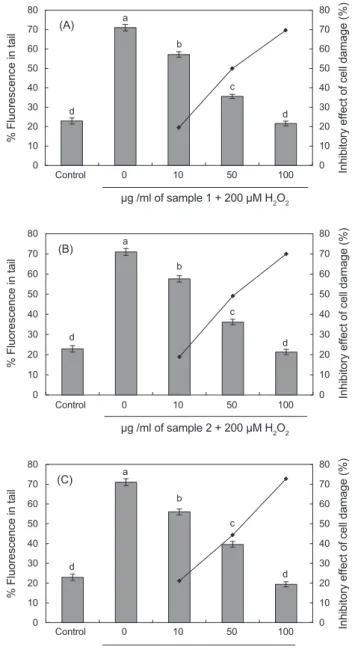
Fig. 1. The preventive effect of supplementation in vitro with different concentration of eel Anguilla japonica carnosine (A, Heat treatment extracts; B, Ion exchange chromatography treated; C, Ultrafiltration permeated) on 200 μM H2O2-induced human leuko- cytes DNA damage. Control, PBS treated normal control. Values are mean with standard error of duplicate experiments with leu- kocytes from each of two different donors. Values not sharing the same letter are significantly different from one another (P<0.05).
0 10 20 30 40 50 60 70 80
0 10 20 30 40 50 60 70 80
Inhibitory effect of cell damage (%)
% Fluorescence in tail
d d
c b
a
Control 0 10 50 100
μg /ml of sample 1 + 200 μM H2O2
Control 0 10 50 100
μg /ml of sample 3 + 200 μM H2O2
Control 0 10 50 100
200 μM H2O2 + μg /ml of sample 1
Control 0 10 50 100
200 μM H2O2 + μg /ml of sample 3 (A)
Control 0 10 50 100
μg /ml of sample 2 + 200 μM H2O2
0 10 20 30 40 50 60 70 80
0 10 20 30 40 50 60 70 80
Inhibitory effect of cell damage (%)
% Fluorescence in tail
d d
c b
(B) a
0 10 20 30 40 50 60 70 80
0 10 20 30 40 50 60 70 80
Inhibitory effect of cell damage (%)
% Fluorescence in tail
d d
c b
(C) a
0 10 20 30 40 50 60 70 80
0 10 20 30 40 50 60 70 80
Inhibitory effect of cell damage (%)
% Fluorescence in tail d d
c b
(A) a
Control 0 10 50 100
200 μM H2O2 + μg /ml of sample 2 0 10 20 30 40 50 60 70 80
0 10 20 30 40 50 60 70 80
Inhibitory effect of cell damage (%)
% Fluorescence in tail d d
c b
(B) a
0 10 20 30 40 50 60 70 80
0 10 20 30 40 50 60 70 80
Inhibitory effect of cell damage (%)
% Fluorescence in tail d
cd c
b (C) a
mM NaOH, pH 13)이포함된전기영동기에넣고 4℃에서 40 분간 unwinding 시킨후 25 V, 300 mA에서 25분간전기영동을 실시하였다. 전기영동한슬라이드글라스를중성화시키기위해 0.4 M Tris (pH 7.5)용액으로 5분간격으로 2회세척하였으며,
LC/MS를이용하여표품 carnosine과본연구에서추출한뱀장 어추출 carnosine 모두동일한분자량(Mw 226)을가진물질 임을확인한후(결과미제시) 3가지방법으로추출한 carnosine 시료를이용하여 0 μg/mL, 10 μg/mL, 50 μg/mL, 100 μg/mL 의함량으로첨가시킨후 30분후에 200 μM H2O2 첨가하여 인체백혈구 DNA의손상도를측정한결과를 Fig. 1A-1C에제 시하였다.
무첨가구의경우 H2O2에의해 DNA가손상되어이에따
른 % fluorescence in tail이 71%를나타낸반면에가열처리추 출 carnosine을첨가한경우는 DNA tail %가 각각 57%, 35%, 12%를나타내었으며, 세포손상억제효과는각각 19%, 49%, 69%이었다(Fig. 1A). 이온교환처리 carnosine은 10 μg/mL, 50 μg/mL, 100 μg/mL 첨가한경우각각 57%, 36%, 21%를나 타내었으며, 이에따른세포손상억제효과는각각 18%, 49%, 69%로나타났다(Fig. 1B). 한외여과처리 carnosine은 10 μg/
mL, 50 μg/mL, 100 μg/mL 첨가한경우각각 56%, 39%, 19%
를나타내었으며이에따른세포손상억제효과는각각 21%, 44%, 72%로나타났다(Fig. 1C).
또한, 각각다른농도의 carnosine을 100 μg/mL로첨가한경 우 H2O2에의해손상되지않은 DNA와동일한보호효과가있 는것으로나타났으며, DNA 손상에따른 Comet image 분석결 과(Fig. 2) H2O2에손상되지않은 DNA에비해무첨가구의경
우 DNA 꼬리가길게생성된반면뱀장어에서추출한한외여과
처리 carnosine을첨가한경우첨가량의증대에따라서손상된 꼬리가짧아지는결과를나타내었다. 특히 100 μg/mL 의함량 으로첨가한실험결과손상된꼬리가없는구의형태를나타내 Fig. 2. Comet images of human leukocytes. A, negative control; B, leukocytes treated with 200 μM H2O2 ; C, leukocytes treated with 10 μg/ml eel Anguilla japonica carnosine (UFP, Ultrafiltration permeated)+200 μM H2O2 ; D, leukocytes treated with 50 μg/ml eel carnosine (UFP, Ultrafiltration permeated)+200 μM H2O2 ; E, leukocytes treated with 100 μg/ml eel carnosine (UFP, Ultrafiltration permeated)+200 μM H2O2.
ethanol 3 mL로 3분간건조시킨후 comet image 분석을위 해 20 μL농도의 ethidium bromide용액으로핵을염색시켜형 광현미경(Leica MZ16 FA, Germany)으로관찰하였다. 그후 CCD camera (Nicon, Japan)를통해얻은세포핵 image는이미 지자동분석소프트웨어(Kormet 4.0, Kinetic Imaging, UK)을 이용하여분석하였다. Comet assay에의한백혈구 DNA 손상 도의측정은핵으로부터이동한 DNA 파편의거리(tail length, TL) 또는 tail length에 tail 내함유된 DNA%를곱해준 tail mo-
ment (TM)값을측정하여백혈구에서 DNA 손상도를측정하
였다. 통계처리
모든자료의통계처리는 SPSS-PC+통계 package를이용하였 으며, 각항목에따라백분율과평균치±표준오차(SE)를구하 고, 각물질의 DAN 손상억제정도를비교하기위해 ANOVA test를통해 F값을구하고 Duncan’s multiple range test를이용 하여유의성을나타내었으며, 통계적유의성은 5% 수준에서 평가하였다.
결과 및 고찰
DNA의 손상에 미치는 영향 보호 효과
인체백혈구 DNA를대상으로 H2O2로부터야기된유전자
독성(genotoxic)에대한뱀장어유래 carnosine의보호및재생 효과에대해알아보기위해 Song et al. (2006)이밝힌바와같이
524 송호수
DNA의손상을효과적으로억제하는것으로나타났다. 재생 효과
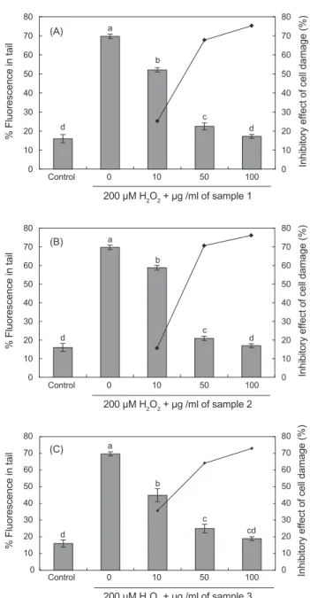
H2O2에의해손상된 DNA의재생효과를살펴보기위해 200 μM H2O2를백혈구에처리하여 4℃에 5분간반응시킨후뱀장 어추출 carnosine을농도별로 0 μg/mL, 10 μg/mL, 50 μg/mL, 100 μg/mL의함량으로첨가한후 37℃ 에서 30분간반응시킨 실험결과는 Fig. 3A-3C에서보는바와같다.
H2O2 무첨가구의경우 DNA가손상되어이에따른 DNA tail %가 69%를나타낸반면가열처리추출 carnosine 첨가시 DNA tail %가각각 52%, 22%, 17%, 손상된세포의재생효 과는각각 25%, 67%, 75%였다(Fig. 3A). 이온교환처리 car- nosine을첨가한경우에는각각 58%, 20%, 16%를나타내었 으며, 이에따른손상된세포의재생효과는각각 15%, 70%, 76%로나타났고(Fig. 3B), 한외여과처리뱀장어추출 carno- sine을 10 μg/mL, 50 μg/mL, 100 μg/mL로각각첨가한경우 44%, 24%, 18%를나타내었으며, 이에따른각각의세포재생 효과는 35%, 64%, 72%로나타났다(Fig. 3C). 특히 carnosine 100 μg/mL 첨가하였을경우 H2O2에의해손상된 DNA를손 상되지않은 DNA와동일하게재생시키는결과를나타내었으 며, 추출방법에따른영향은크지않은것으로나타났다. Song et al. (2006)과 Min et al. (2017)은수산어류로부터 carnosine
및 anserine과같은저분자펩타이드의추출에있어서가열처
리, 이온교환, 한외여과추출방법을이용한결과유리철과철 단백질과같은산화촉진물의함량은효과적으로감소되었으나 각추출방법에따른저분자펩타이드의함량변화는크지않았 다고보고하였으나, 향후이에대한추가적인연구가필요할것 으로판단된다.
이상의실험결과를종합해보면뱀장어에서추출한 carno- sine은산화적스트레스유발물질인 H2O2에의해유도된백혈 구 DNA의손상에효과가있는것을확인하였으며, 이에본연 구에서는뱀장어로부터추출한 carnosine을이용하여, 천연물 유래 carnosine의기능특성규명을위해인체 DNA 손상억 제및회복에미치는영향등에대해조사함으로써향후산업 적및의학적으로중요한기능성소재로써활용이가능할것으 로판단된다.
사 사
이연구는 2017년영산대학교교내연구비의지원을받아수행
되었음.
References
Adelson R, Saul RL and Ames BN. 1988. Oxidative damage to DNA: Relation to species metabolic and life span. Proc Natl Acad Sci USA 85, 2706-2708. https://doi.org/10.1073/
pnas.85.8.2706.
Fig. 3. The recovery effect of supplementation in vitro with different concentration of eel Anguilla japonica carnosine (A, Heat treatment extracts; B, Ion exchange chromatography treated; C, Ultrafiltration permeated) on 200 μM H2O2-induced human leuko- cytes DNA damage. Control, PBSO treated normal control. Values are mean with standard error of duplicate experiments with leu- kocytes from each of two different donors. Values not sharing the same letter are significantly different from one another (P<0.05).
0 10 20 30 40 50
0 10 20 30 40 50
Inhibitory effect of cell damage (%)
% Fluorescence in tail
d d
c
Control 0 10 50 100
μg /ml of sample 1 + 200 μM H2O2
Control 0 10 50 100
μg /ml of sample 3 + 200 μM H2O2
Control 0 10 50 100
200 μM H2O2 + μg /ml of sample 1
Control 0 10 50 100
200 μM H2O2 + μg /ml of sample 3
Control 0 10 50 100
μg /ml of sample 2 + 200 μM H2O2 0 10 20 30 40 50 60 70 80
0 10 20 30 40 50 60 70 80
Inhibitory effect of cell damage (%)
% Fluorescence in tail
d d
c b
(B) a
0 10 20 30 40 50 60 70 80
0 10 20 30 40 50 60 70 80
Inhibitory effect of cell damage (%)
% Fluorescence in tail
d d
c b
(C) a
0 10 20 30 40 50 60 70 80
0 10 20 30 40 50 60 70 80
Inhibitory effect of cell damage (%)
% Fluorescence in tail d d
c b
(A) a
Control 0 10 50 100
200 μM H2O2 + μg /ml of sample 2 0 10 20 30 40 50 60 70 80
0 10 20 30 40 50 60 70 80
Inhibitory effect of cell damage (%)
% Fluorescence in tail d d
c b
(B) a
0 10 20 30 40 50 60 70 80
0 10 20 30 40 50 60 70 80
Inhibitory effect of cell damage (%)
% Fluorescence in tail d
cd c
b (C) a
었다. 이러한결과는 ferritin과 H2O2로인한산화적 DNA 손상 에있어서합성 carnosine과 homocarnosine 첨가를통해 DNA 의손상을억제시켰다는 Kang (2010)의연구결과와같이뱀 장어에서추출한한외여과처리 canosine 또한, H2O2로인한
buffering capacity and dipeptide content in the throughbred horse, greyhound dog and man. Comp Biochem Physiol 97, 249-251. https://doi.org/10.1016/0300-9629(90)90180-z.
Hipkiss AR, Brownson C and Carrier MJ. 2001. Carnosine, the anti-ageing, anti-oxidant dipeptide, may react with protein carbonyl groups. Mech Ageing Dev 122, 1431-1445. https://
doi.org/10.1016/s0047-6374(01)00272-x.
Kang JH. 2010. Protective effects of carnosine and homocarno- sine on ferritin and hydrogen peroxide-mediated DNA dam- age. Kor Soc Biochem Mol Biol 43, 683-687. https://doi.
org/10.3858/bmbrep.2010.43.10.683.
Kang JH, Kim KS, Choi SY, Kwon HY, Won MH, and Kang TC.
2002. Protective effects of carnosine, homocarnosine and anserine against peroxyl radial-mediated Cu, Zn-superoxide dismutase modification. Biochimica et Biophysica Acta 1570, 89-96. https://doi.org/10.1016/s0304-4165(02)00158- Kanner J, Harel S and Jaffe R. 1991. Lipid peroxidation of mus-7.
cle food as affected by sodium chloride. J Agric Food Chem 39, 1017-1024. https://doi.org/10.1021/jf00006a002.
Kansci G, Genot C, Meynier A and Gandemer G. 1997. The an- tioxidant activity of carnosine and its consequences on the volatile profiles of liposomes during iron/ascorbate induced phospholipid oxidation. Food Chem 60, 165-175. https://
doi.org/10.1016/s0308-8146(95)00257-x.
Kim IS and Park JY. 2007. Freshwater fishes of Korea. Kyo- haksa, Seoul, Korea, 467.
Lee BJ, Park JH, Lee YS, Cho MH, Kim YC and Hendricks DG. 1999. Effect of Carnosine and Related Compounds on Glucose Oxidation and Protein Glycation In Vitro. J Bio- chem Mol Biol 32, 370-378.
Lee KT, Song HS and Prak SM. 2007. Antioxidant Effect of carnosine Extracted from the eel Anguilla japonica extracts.
Korean J Fish Sic 40, 193-200. https://doi.org/10.5657/
kfas.2007.40.4.193
Min HO, Park IM and Song HS. 2017. Effect of extrac- tion method on anserine, protein, and iron contents of salmon(onchorhynchus) Extracts. J Korean Soc Food Sci Nutr 46, 220-228. https://doi.org/10.3746/
jkfn.2017.46.2.220.
Nagasawa T, Yokozawa T and Terassawa K. 2001. A study of kampo medicines in a diabetic nephrophathy model. J Trad Med 19, 161-168.
Quinn PR, Boldrev AA and Formazuyk VE. 1992. Carnosine : its properties, functions and potential therapeutic ap- plications. Mol Aspects Med 13, 379-444. https://doi.
org/10.1016/0098-2997(92)90006-l.
Salah E, Gariballa, Alan J and Sinclair. 2000. Carnosine: physi- ological properties and therapeutic potential. Age Ageing 29, 207-210. https://doi.org/10.1093/ageing/29.3.207.
Song HS, Lee KT and Kang OK. 2006. Effect of extraction method on the carnosine, protein, and iron contents of Boldyrev A, Abe H, Stvolinsky S and Tyulina O. 1995. Effects of
carnosine and related compounds on generation of free oxy- gen species: a comparative study. Comp Biochem Physiol 12, 481-485. https://doi.org/10.1016/0305-0491(95)00084- Bucala R, Cerami A and Vlassara H. 1995. Advanced glycosyl-4.
ation end products in diabetic complications. Diabetes Rev 3, 258-268. https://doi.org/10.14310/horm.2002.11140.
Chan KM, Decker EA and Means WJ. 1993. Extraction and activity of carnosine, a naturally occuring antioxidant in beef muscle. J Food Sci 58, 1-7. https://doi.org/10.1111/
j.1365-2621.1993.tb03199.x.
Chan KM, Decker EA and Means WJ. 1993. Extraction and activity of carnosine, a naturally occuring anti- oxidant in beef muscle. J Food Sci 58, 1-7. https://doi.
org/10.1111/j.1365-2621.1993.tb03199.x.
Chasovnikova LV, Formazyuk VE, Sergienko VI, Boldyrev AA and Severine SE. 1990. The antioxidative properties of carnosine and other deuge. Biochem Int 20, 1097-1103.
Cho HS, Choi JH and Ko HB. 2011. Evaluation of major nu- trients of domestic farmed eels Anguilla japonica. Ko- rean J Fish Aquat Sci 44, 237-24. http//doi.org/10.5657/
KFAS.2011.0237.
Crush KG. 1970. Carnosine and related substances in ani- mal tissues. Comp Biochem Physiol 34, 3-30. https://doi.
org/10.1016/0010-406x(70)90049-6.
Decker EA and Crum AD. 1993. Antioxidant activity of carno- sine in cooked ground pork. Meat sci 34, 245-253. https://
doi.org/10.1016/0309-1740(93)90031-c.
Decker EA, Chan KM, Livisay SA, Butterfield DA and Faust- man C. 1995. Interactions between carnosine and the differ- ent redox states of myoglobin. J Food Sci 60, 1201-1204.
https://doi.org/10.1111/j.1365-2621.1995.tb04555.x.
Decker EA, Crum AD and Calvert JT. 1992. Differences in the Antioxidant Mechanism of Carnosine in the Presence of Cooper and Iron. J Agric Food Chem 40, 756-759. https://
doi.org/10.1021/jf00017a009.
Escalante AS, Djenane D, Torescano, Beltran JA and Roncales P. 2001. The effects of ascorbic acid, taurine, carnosine and rosemary powder on colour and lipid stability of beef patties packaged in modified atmosphere. Meat Sci 58, 421-429.
https://doi.org/10.1016/s0309-1740(01)00045-6.
Gayiva EI. Kron V, Pavlisak M, Fedurco and Novakova B.
1999. carnosine in patients with diabetes mellitus type I.
Bratisl Lek Listy 100, 500-502.
Gopalakrishnan J, Decker EA and Means WJ. 1999. Antioxidant activity of mechanically separated pork extracts. Meat Sci 2, 101-110. https://doi.org/10.1016/s0309-1740(98)00154-5.
Gulevitch VS and Amiradgibi S. 1900. Uber das carnosine, eine neueorganiche base des fleischextraktes. ber Dtsch Chem Ges 33, 1902.
Harris RC, Marlin DJ, Snow DH and Hultman E. 1990. Muscle
eel(anguilla japonica) extracts. Korean J Fish Sic 39, 384- 390. https://doi.org/10.5657/kfas.2006.39.5.384.
Song HS, Lee KT and Kang OK. 2009. Inhibitory Effects of eel(Anguilla japonica) extracted carnosine on Protein Glyca- tion. Korean J Fish Sic 42, 104-108. https://doi.org/10.5657/
kfas.2009.42.2.104.
Yokozawa T, Nakagawa T and Terasawa K. 2001. Effects of oritental medicines on the production of advanced glycation products. J Trad Med 18, 107-112
Zho S and Decker EA. 1999. Ability of carnosine and other skeletal muscle components to quench unsaturated aldehy- dic lipid oxidation products. J Agric Food Chem 47, 51-55.
https://doi.org/10.1021/jf980780j.