강원대학교 의과대학 생화학교실, 강원대학교병원 임상의학연구소, 강원대학교 의과대학 내과학교실 성지현1, 이미은2, 한선숙3, 이승준3, 하권수1, 김우진3
Gene Expression Profile of Lung Cancer Cells Following Photo- dynamic Therapy
Ji Hyun Sung, M.D.1, Mi-Eun Lee, M.D.2, Seon-Sook Han, M.D.3, Seung-Joon Lee, M.D.3, Kwon-Soo Ha, M.D.1, Woo Jin Kim, M.D.3
1Department of Molecular and Cellular Biochemistry, College of Medicine, Kangwon National University, and 2Clinical Research Institute of Kangwon National University Hospital, 3Department of Internal Medicine, College of Medicine, Kangwon National University, Chunchon, Korea
Background: Photodynamic therapy is a viable option for lung cancer treatment, and many studies have shown that it is capable of inducing cell death in lung cancer cells. However, the precise mechanism of this cell death has not been fully elucidated. To investigate the early changes in cancer cell transcription, we treated A549 cells with the photosensitizer DH-I-180-3 and then we illuminated the cells.
Methods: We investigated the gene expression profiles of the the A549 lung cancer cell line, using a DEG kit, following photodynamic therapy and we evaluated the cell viability by performing flow cytometry. We identified the genes that were significantly changed following photodynamic therapy by performing DNA sequencing.
Results: The FACS data showed that the cell death of the lung cancer cells was mainly caused by necrosis. We found nine genes that were significantly changed and we identified eight of these genes. We evaluated the expression of two genes, 3-phosphoglycerate dehydrogenase and ribosomal protein S29. The expressed level of carbonic anhydrase XII, clusterin, MRP3s1 protein, complement 3, membrane cofactor protein and integrin beta 1 were decreased.
Conclusion: Many of the gene products are membrane-associated proteins. The main mechanism of photodynamic therapy with using the photosensitizing agent DH-I-180-3 appears to be necrosis and this may be associated with the altered production of membrane proteins. (Tuberc Respir Dis 2007; 63: 52-58)
Key Words: Gene expression profiling, Lung neoplasms, Photosensitizing agents.
This work was supported in part by a grant of the Korea Health 21 R & D Project, Ministry of Health & Welfare, Republic of Korea (A030003)
Address for correspondence: Woo Jin Kim, M.D.
Department of Internal Medicine, Kangwon National University Hospital, 17-1 Hyoja-3 dong, Chunchon, Kangwon-do, 200-947, Korea
Phone: 82-33-258-2377, Fax: 82-33-258-2455 E-mail: [email protected]
Received: Apr. 5. 2007 Accepted: Jun. 7. 2007
서 론
폐암은 수년간의 연구에도 불구하고 세계적으로 매 년 100만명 이상이 사망에 이르고 있으며1, 전반적으 로 5년 이상 생존율이 15% 정도에 못 미치는 심각한 질병이다2. 그러므로, 새로운 치료방식의 개발 및 도 입으로 폐암 치료의 결과가 개선되어야 할 필요성이 있다. 최근 보고에 의하면 기관지내시경을 이용한 광
역학 치료가 조기 폐암의 치료와 진행한 폐암의 고식 적 치료에서 효과적 치료 방법으로서 역할을 할 수 있 을 것으로 제시되고 있다3.
광역학 치료는 체내의 산소와 빛에 대해 예민한 반 응을 보이는 광과민성 물질(photosensitizer)을 투여 하고, 광과민성 물질이 활성화되는 파장의 빛을 조사 하여 암세포를 특이적으로 사멸시키는 치료법이다.
광역학 치료의 주요기전은 활성 산소와 이에 의하여 유발되는 자유 라디칼(free radical)에 의해 암세포가 선택적으로 파괴된다는 것이다. 그러나 광역학 치료 에 의한 세포자멸사(apoptosis)나 세포괴사(necrosis) 가 일어나는 정확한 기전은 확실히 밝혀지지 않은 상 태이다4.
현재 일부 광과민성 물질에 대한 사용허가가 많은 나라에서 이루어져 있으며 새로운 광과민성 물질의 개발이 진행되고 있다. 최근 연구에 의하면 염소계 (chlorine) 광과민성 물질인 DH-I-180-3이 폐암 세포
의 시험관내(in vitro) 실험과 생체내(in vivo) 실험에 서 효과가 있는 것으로 보고되어 있다5,6. DH-I-180-3 은 bacteriochlorin의 유도체인 pheophorbide계통의 광과민성 물질이라는 것은 알려져 있지만 DH-I- 180-3이 종양세포를 파괴하는 기전은 많이 연구되어 있지 않다.
이에 우리는 광역학 치료법에 의해 세포사멸이 일 어나는 기전을 알아보기 위하여 FACS(fluorescence- activated cell sorting)을 시행하고, 유전자 발현의 변 화를 알아보고자 하였다. 임의의 특정 프라이머 (primer)들을 이용하여, 발현의 차이를 보이는 유전자 들을 발굴하는 원리로 개발된 DEG(differentially expressed genes) kit를 이용하여 광역학 치료에 따른 폐암 세포의 유전자 발현의 변화를 알아보았다7,8.
방 법 1. 암세포주 배양
본 연구에 사용하는 세포주로는 American Type Culture Collection (ATCC)으로부터 구입한 인간 폐 암 세포주 A549를 사용하였으며, 10% fetal bovine serum(FBS: Hyclone Laboratory, Logan, UT, USA) 및 항생제가 포함되어 있는 RPMI-1640 배양액을 사 용하여 35-mm 페트리접시에 5% CO2, 37℃의 조건으 로 배양하였다.
2. 광역학 치료와 세포처리
광역학 치료에 광과민성 물질로 DH-I-180-3을 사 용하였다. DH-I-180-3을 디메칠설폭사이드(dimethyl sulfoxide, DMSO: Sigma Chemical Co., St. Louis, MO, USA)에 녹여 사용하였으며, 보관시에는 -20℃
에 보관하였다. 각각의 실험을 위해 DMSO에 녹인 DH-I-180-3용액을 배양액에 희석하여 최종농도가 1
㎍/ml이 되도록 하여 12시간동안 5% CO2, 37℃의 조 건으로 배양하였다.
광역학 치료 실험을 위해 세포주를 35 mm 페트리 접시에 80% 이상 배양시켰고, 레드 필터가 장착된 할
로겐 램프를 이용하여 670 nm 이상 파장의 빛을 0.8 J/cm2의 세기로 5분간 조사하였다. 빛을 조사한 뒤 20 분 후에 세포로부터 DH-I-180-3용액을 제거하고 PBS(phosphate buffered saline)로 세척한 뒤 RNeasy Mini kit(Qiagen GmbH, Hilden, Germany)를 이용하 여 RNA를 추출하였다.
3. Fluorescence-activated cell sorting (FACS)
광역학 치료 후, 35 mm 페트리접시에 있는 세포에 트립신(trypsin)-EDTA (0.05% trypsin, 0.53% EDTA) 를 처리하여 세포를 떼어낸 후 PBS로 트립신- EDTA를 제거하였다.
세포(2x10⁵ cells)에 5 ㎕ 아넥신(Annexin) V- FITC(fluorescein isothiocyanate)와 5 ㎕ propidium iodide(PI)를 300 ㎕ 아넥신 V 결합 완충제(binding buffer)와 함께 넣어준 후 4℃의 암실 조건에서 15분 간 배양하였다. 형광물질이 염색된 세포의 FACS는 FACSCalibur(Becton Dickinson, Franklin Lakes, NJ, USA)에 의해 488 nm의 아르곤 레이저로 분석을 하 였으며, CellQuest Software program(Becton Dic- kinson)을 이용하여 세포사멸의 정도를 분석하였다.
4. cDNA 합성을 위한 역전사
역전사를 위하여 RNA 3 μg, 5 x reaction buffer (Promega, Madison, WI, USA) 4 ㎕, dNTPs 5 ㎕, 10 mM dT-ACP1 2 ㎕(5'-CTGTGAATGCTGCGACT ACGATIIIIIT(18)-3'), RNasin RNase inhibitor (40 U/㎕) 0.5 ㎕, Moloney murine leukemia virus reverse transcriptase(200 U/ ㎕; Promega) 1 ㎕ 을 넣어준 후 최종 양이 20 ㎕가 되도록 하여 42℃에서 역전사 반응을 시켰다.
5. ACP(annealing control primer)-based Gene- Fishing PCR(polymerase chain reaction)
발현상의 차이가 있는 유전자는 GeneFishing DEG kit를 이용하여 ACP-based PCR 방법에 의해 조사하
Figure. 1. Quantitative analysis of the apoptotic and necrotic cells. A549 cells were analyzed 1, 2, 3, and 12h following PDT. While no increase in apoptotic or necrotic cells was seen with the photosensitizer only, about 50%
were necrotic till 3h and about 30% were necrotic by 12h. FACS data showed that the mechanism of cell death is mainly necrosis.
였다(Seegene, Seoul, South Korea). ACP는 시동체 의 구조가 3부분으로 되어 있어, 높은 붙임 온도에서 도 재현성있게 긴 PCR 산물을 얻을 수 있도록 고안된 방법이다7,8.
우선 PCR 조건을 94℃ 1분, 50℃ 30분, 72℃ 1분간 1 사이클(cycle)로 수행하여 second-strand cDNA를 합성하였고, 두번째 단계로 94℃ 40초, 65℃ 40초, 7 2℃ 40초로 40사이클과 72℃에서 5분간으로 PCR 증 폭단계를 수행하였다. PCR 결과물들은 2% 우무겔 (agarose gel)에서 분석하였다.
6. 염기서열분석
발현상의 차이를 보이는 밴드를 GENCLEAN II kit(Q-BIO Gene, Carlsbad, CA, USA)를 이용하여 추 출하였고, ABI Prism 3100-Avant genetic analyzer
(Applied Biosystems, Foster City, CA, USA)를 이용 하여 염기서열분석을 하였다.
결 과 1. 광역학 치료에 의한 세포사멸
광역학 치료에 의해 A549 세포에서 세포사멸이 일 어남을 알 수 있었다. 세포사멸이 세포괴사(necrosis) 에 의한 것인지 세포자멸사에 의한 것인지에 대한 정 량분석은 아넥신 V와 PI로 수행되었다. 광역학 치료 후 2시간 경과 시 세포의 약 50%는 사멸하였고, 세포 자멸사는 거의 관찰되지 않았다. 광역학 치료 후 12시 간 경과 시점까지 세포사멸의 비율은 유지되었다 (Figure 1). 3회의 반복 실험에서 비슷한 결과를 얻었 다.
NumberName GenBank Acc. No. Homology(%)
1 3-PGDH AF006043 526/528(99)
2 ribosomal protein
S29 BC035313.1 217/217(100)
3 CA-XII NM206925.1 271/271(100)
4 MRP3 AF154001 520/520(100)
5 betal intefrin BC020057.1 264/265(99)
6 MCP BC030594.2 383/383(100)
7 complement 3 K02765.1 333/334(99) 8 clusterin BC019588.2 580/580(100) PGDH: phosphoglycerate dehydrogenase; CA:carbonic anhydtase; MRP: multidrug resistance-associated protein.
Table 1. Eight genes that were dignificantly changed following PDT using DH-I-180-3. The mRNA levels of first two genes increased and the expression of the rest genes decreased.
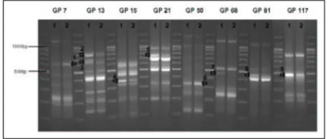
Figure. 2. Differential expression of genes in agarose gel with or without PDT. Two bands increased (arrow 1,6) and seven decreased following PDT (arrow 2-5,7-9). (1; pre-PDT, 2; post-PDT, GP; GeneFishing primer)
2. DEG (Differentially expressed genes)
ACP(annealing control primer) 기법을 이용한 PCR 방법으로 광역학 처리를 한 것과 광역학 처리를 하지않은 두 조건의 세포들의 mRNA의 발현차이를 비교하였다. 120가지 임의의 특정 프라이머들과 2개 의 oligo(dT) 프라이머들에 의해 광역학 치료 전, 후 의 발현상의 차이가 나는 유전자 밴드들을 찾은 결과, 9가지의 밴드가 확인되었다(Figure 2).
9개의 밴드 중에서 광역학 치료 후 발현이 증가된 것은 2개, 발현이 감소된 것은 7개였다.
3. Sequencing
9개의 밴드 중 발현의 증감이 확실히 확인된 8개의 밴드를 선택하여 염기서열분석을 하였고, 그 결과 BLAST(Basic Local Alignment Search Tool)에서 상동성이 있는 유전자를 찾았다,
발현이 증가된 2개의 유전자는 3-phosphoglycerate dehydrogenase(PGDH) 와 ribosomal protein S29 이 었고, 발현이 감소된 6개의 유전자는 carbonic anhy- drase XII, clusterin, MRP(multidrug resistance- associated protein) 3s1 protein, complement 3, membrane cofactor protein 그리고 β-1 integrin이었 다(Table 1).
고 찰
본 연구는 DH-I-180-3을 사용한 광역학 치료에 의 해 폐암 세포가 세포사멸단계에 이르게 되는 결과를 확인하였으며, 이 과정에서 발현변화를 보이는 9개의 유전자를 확인하였고, 이 중 8개의 유전자를 밝혀내었 다.
먼저 광역학 치료에 의해 폐암 세포가 세포사멸에 이르게 되는 것이 세포괴사(necrosis)에 의한 것인지 세포자멸사에 의한 것인지에 대해서는 세포주의 종 류, 광과민성 물질의 종류, 광역학 치료의 조건 등에 의해 의존적으로 달라질 수 있다9,10. 본 연구에서는 DNA와 RNA의 이중 사슬(base pair)에 결합하게 되 는 PI(Propidium iodide)와 세포자살 과정에서 세포 내막이 바깥쪽으로 노출 되었을 때 phosphatidyl serine에 결합하게 되는 아넥신 V를 염색하여11 FACS로 분석하였다. 이를 통해 염소계의 광과민성 물질인 DH-I-180-3에 의해서는 주로 세포괴사로 인 하여 종양세포가 사멸에 이르는 것을 확인하였다. 최 근 다른 연구에서도 광과민성 물질에 의한 항종양의 주요 기전이 원형질 막의 붕괴로 인해 일어나는 세포 괴사임이 확인되었다12.
본 실험에서 광역학 치료 후에 여섯개의 유전자 발
현이 감소되는 것으로 확인이 되었는데 이 중 많은 유 전자들이 막 단백질과 관련되고 이중 일부는 최근 종 양치료의 표적이 되는 유전자이다. 탄산탈수소효소 XII(carbonic anhydrase XII)는 막당단백질(trans- membrane glycoprotein)로서 폐암, 위암, 신장 암 등 에서 발현이 증가되며13,14 종양치료 연구의 표지가 될 가능성을 가지고 있는 유전자로 최근 확인되었다15.
MRP3는 폐의 정상세포와 폐암 세포의 막 부분에 위치하고 있고16, 이는 본질적으로 폐암 세포의 생성 을 억제하기위해 내재되어있는 유전자이다17. 본 연구 에서 MRP3의 RNA발현은 광역학 치료 후 초기에 감 소되는 것으로 나타났다.
β-1 integrin은 세포와 세포외 간질(extracellular matrix)의 상호작용에 관여하는 이질이량체 막수용체 (heterodimeric transmembrane receptor) 이며, 최근 폐암에서 나쁜 예후와 관련이 있다는 보고가 있었다
18. 이온화 방사선에 의해서는 β-1 integrin이 증가된 다고 보고되어 있으나19, 본 실험에서는 광역학 치료 후에 β-1 integrin의 mRNA발현이 감소되는 것으로 나타났다. 또한, β-1 integrin은 일부 암세포에서 치료 의 가능성 있는 표지인자로 연구되고 있다20. 막 보조 단백질(membrane cofactor protein)은 세포 손상을 보호하는 단일사슬의 제 1형 막당단백질이다21. 이는 면역저하와 종양의 전이 그리고 유방암에서의 좋지 않은 예후와 관련이 있는 것으로 알려져 있다22.
C3 mRNA도 광역학 치료 후 감소한 유전자인데, 이전의 보고에 의하면 폐암세포에서 photofrin에 의해 광역학 치료를 하였을 때 C3 mRNA의 발현이 증가되 었으며23 photofrin계열 광과민성 물질에 의해 종양세 포가 파괴되었다고 보고된 바가 있다. 이것은 광과민 성 물질의 종류에 따라서 빛에 의한 화학적 반응이 일 어나는 기전이 조금씩 다른 이유와 관련될 지도 모른 다. 본 실험은 mRNA수준에서 확인을 한 것이기 때문 에 제한이 있고, 명확한 결론에 이르기 위해서는 보체 (complement)의 정확한 농도를 직접 측정해야 할 것 으로 사료된다.
Clusterin은 당화 단백질(glycosylated protein)로서 인간의 여러 가지 악성종양에서 증가되는 유전자이다
24. 이것은 발암과 종양성장에 중요한 단백질이고 칼
슘에 의해 영향을 받는 proapoptotic와 antiapoptotic 양쪽 모두와 연관이 있다25. 최근 clusterin은 폐암 환 자에서 치료의 표적으로 연구되고 있다26. 이 유전인 자의 발현은 광역학 치료에 의하여 감소되었고 따라 서 광역학 치료에서 중요한 표적으로 이용될 수 있을 것이다.
다음의 2개의 유전자는 광역학 치료 후에 발현이 증가되었다. 3-PGDH는 그 조절 기전이 잘 알려져 있 지 않으나, 세린(serine) 생합성에 중요하며27, 암세포 에서 발현이 증가되는 것으로 알려져 있다. Riboso- mal protein S29는 세포자살을 조절하는 단백질이며28 폐암 세포에서 적은 농도로 존재하고29, 이 유전자가 트랜스펙션(transfection) 될 경우 폐암 세포에서 세 포자멸사를 유도하기도 하고 화학요법에서 세포들을 민감하게 만들기도 한다30.
이전의 다른 광과민성 물질에 의해 광역학 치료 후 RNA발현을 조사한 결과 caspase-8과 phosphatase 1 등을 포함하는 40개의 유전자가 있음이 보고된 바 있 다31. 이러한 유전학적 조사의 결과가 다양하게 나오 는 것은 세포주의 종류, 광과민성 물질의 종류, 광역 학 치료의 조건 등과 연관이 있을 것이다.
본 연구에서 광역학 치료 후 발현이 감소된 유전자 들 중 많은 부분이 막과 결합되어 있었고, 다른 연구 에서도 광과민성 물질에 의한 항종양의 주요 기전이 원형질 막의 붕괴로 인해 일어나는 세포괴사로 보여 진다는 보고가 있었다12.
광역학 치료에 의해 감소된 유전자들 중 일부가 최 근 치료 표적으로 연구되고 있다. 이 유전자중 일부는 종양괴사의 중요한 표적이거나 또는 그 상위단계에 해당되는 표적일수도 있다. 일부 약제내성 악성세포 에 대해서 광역학 치료가 효과적일 수 있는데4 이는 MRP3를 포함한 광역학 치료 후 발현이 감소된 유전 자에 의해서 설명이 될 수도 있다. 좀 더 자세한 유전 자 작용 기전을 알아보기 위해서는 광역학 치료의 주 요 표적 유전자가 어떤 것인지 밝혀져야 할 것이다.
결론적으로, 본 연구에서는 DH-I-180-3를 사용하 여 광역학 치료를 한 결과 암세포의 사멸 기전이 주로 세포괴사에 의해 이루어진다는 것을 시사하였고, 광 역학 치료 후에 8개의 유전자 발현이 영향을 받았는
데, 2개는 발현이 증가되었고 6개는 발현이 감소되었 으며, 발현이 감소된 유전자들의 많은 부분들이 막 결 합 또는 발암과 관련이 있었다. 광역학 치료는 폐암에 대한 효과적인 새로운 치료법이 될 수 있다고 사료된다.
요 약
연구배경: 광역학 치료는 폐암 치료에 실질적으로 이용 가능하며, 많은 연구들에서 폐암 세포에서 세포 사멸을 일으킨다는 것이 이미 알려져 있다. 그러나 이 세포사멸의 기전은 아직 정확히 알려져 있지 않으며, 이에 암세포의 전사에서 초기 변화가 어떻게 일어나 는 지를 알아보기 위하여 실험을 수행하였다.
방 법: 광과민성 물질인 DH-I-180-3으로 A549 세포에 처리를 하고 광역학 치료를 한 후 관찰하였다.
광역학 치료 후 DEG kit를 이용하여 폐암 세포주에서 의 유전자 발현을 보았으며, 유세포 분석기를 이용하 여 세포 사멸을 측정하였다. 광역학 치료 후 의미있는 변화를 보인 유전자는 염기서열분석으로 확인하였다.
결 과: 유세포분석 결과 폐암세포주는 대부분 세 포괴사에 의하여 사멸되었다.광역학 치료 후, 9개의 유전자에서 명확한 변화가 있음을 발견했으며 이 중 8개의 유전자를 밝혀내었다. 3-phosphoglycerate dehydrogenase와 리보솜 단백질 S29의 유전자 발현 이 증가되어 있었으며, carbonic anhydrase XII, clusterin, MRP3s1 protein, complement 3, membrane cofactor protein, β-1 integrin의 유전자 발현은 감소 되어 있었다.
결 론: 본 연구는 광과민성 물질인 DH-I-180-3을 이용한 광역학 치료에서 폐암 세포의 세포사멸의 주 된 기전이 세포괴사에 의해 이루어 진 것임을 밝혀냈 으며, 이와 관련된 유전자들 대부분이 막단백의 변화 를 통해 이루어짐을 알 수 있었다.
참 고 문 헌
1. Spiro SG, Porter JC. Lung cancer--where are we today? Current advances in staging and nonsurgical treatment. Am J Respir Crit Care Med 2002;
166:1166-96.
2. Spira A, Ettinger DS. Multidisciplinary management of lung cancer. N Engl J Med 2004;350:379-92.
3. Moghissi K, Dixon K. Is bronchoscopic photodynamic therapy a therapeutic option in lung cancer? Eur Respir J 2003;22:535-41.
4. Dougherty TJ, Gomer CJ, Henderson BW, Jori G, Kessel D, Korbelik M, et al. Photodynamic therapy. J Natl Cancer Inst 1998;90:889-905.
5. Kim CS, Lee CH, Lee PH, Han S. Inactivation of mitochondrial electron transport by photosensitization of a pheophorbide a derivative. Mol Cells 2004;
17:347-52.
6. Lim DS, Ko SH, Won DH, Lee CH, Lee WY.
Photodynamic anti-tumor activity of a new chlorine- based photosensitizer against Lewis lung carcinoma cells in vitro and in vivo. J Porphyrins Phthalo- cyanines 2003;7:155-61.
7. Kim YJ, Kwak CI, Gu YY, Hwang IT, Chun JY.
Annealing control primer system for identification of differentially expressed genes on agarose gels. Biote- chniques 2004;36:424-30.
8. Cui XS, Shin MR, Lee KA, Kim NH. Identification of differentially expressed genes in murine embryos at the blastocyst stage using annealing control primer system. Mol Reprod Dev 2005;70:278-87.
9. Fabris C, Valduga G, Miotto G, Borsetto L, Jori G, Garbisa S, et al. Photosensitization with zinc (II) phthalocyanine as a switch in the decision between apoptosis and necrosis. Cancer Res 2001;61:7495-500.
10. Dellinger M. Apoptosis or necrosis following photofrin photosensitization: influence of the incubation protocol. Photochem Photobiol 1996;64:182-7.
11. Vermes I, Haanen C, Steffens-Nakken H, Reute- lingsperger C. A novel assay for apoptosis. Flow cytometric detection of phosphatidylserine expression on early apoptotic cells using fluorescein labeled Annexin V. J Immunol Methods 1995;184:39-51.
12. Ahn WS, Bae SM, Huh SW, Lee JM, Namkoong SE, Han SJ, et al. Necrosis-like death with plasma membrane damage against cervical cancer cells by photodynamic therapy. Int J Gynecol Cancer 2004;
14:475-82.
13. Ulmasov B, Waheed A, Shah GN, Grubb JH, Sly WS, Tu C, et al. Purification and kinetic analysis of recombinant CA XII, a membrane carbonic anhydrase overexpressed in certain cancers. Proc Natl Acad Sci USA 2000;97:14212-7.
14. Leppilampi M, Saarnio J, Karttunen TJ, Kivela J, Pastorekova S, Pastorek J, et al. Carbonic anhydrase isoenzymes IX and XII in gastric tumors. World J Gastroenterol 2003;9:1398-403.
15. Supuran CT, Scozzafava A, Casini A. Carbonic anhydrase inhibitors. Med Res Rev 2003;23:146-89.
16. Torky AR, Stehfest E, Viehweger K, Taege C, Foth H.
Immuno-histochemical detection of MRPs in human lung cells in culture. Toxicology 2005;207:437-50.
17. Young LC, Campling BG, Cole SP, Deeley RG, Gerlach JH. Multidrug resistance proteins MRP3, MRP1, and MRP2 in lung cancer: correlation of protein levels with drug response and messenger RNA levels. Clin Cancer Res 2001;7:1798-804.
18. Oshita F, Kameda Y, Ikehara M, Tanaka G, Yamada K, Nomura I, et al. Increased expression of integrin beta 1 is poor prognostic factor in small-cell lung cancer. Anticancer Res 2002;22;1065-70.
19. Cordes N, Blaese MA, Meineke V, Van Beuningen D.
Ionizing radiation induces up-regulation of functional beta1-integrin in human lung tumour cell lines in vitro. Int J Radiat Biol 2002;78:347-57.
20. Park CC, Zhang H, Pallavicini M, Gray JW, Baehner F, Park CJ, et al. Beta1 integrin inhibitory antibody induced apoptosis of breast cancer cells, inhibits growth, and distinguished malignant from normal phenotype in three dimensional cultures and in vivo.
Cancer Res 2006;66:1526-35.
21. Hakulinen J, Junnikkala S, Sorsa T, Meri S.
Complement inhibitor membrane cofactor protein is constitutively shed from cancer cell membrane in vesicles and converted by a metalloproteinase to a functionally active soluble form. Eur J Immunol 2004;34:2620-9.
22. Madjd Z, Durrant LG, Pinder SE, Ellis IO, Ronan J, Lewis S, et al. Do poor-prognosis breast tumours express membrane cofactor proteins (CD46)? Cancer Immunol Immunother 2005;54:149-56.
23. Cecic I, Serrano K, Gyongyossy-Issa M, Korbelik M.
Characteristics of complement activation in mice bearing Lewis lung carcinomas treated by photo-
dynamic therapy. Cancer Lett 2005;225:215-23.
24. Shannan B, Seifert M, Leskov K, Willis J, Boothman D, Tilgen W, et al. Challenge and promise: roles for clusterin in pathogenesis, progression and therapy of cancer. Cell Death Differ 2006;13:12-9.
25. Pajak B, Orzechowski A. Clusterin: the missing link in the calcium-dependent resistance of cancer cells to apoptogenic stimuli. Postepy Hig Med Dosw 2006;60:45-51.
26. July LV, Beraldi E, So A, Fazli L, Evans K, English JC, et al. Nucleotide-based therapies targeting clusterin chemosensitize human lung adenocarcinoma cells both in vitro and in vivo. Mol Cancer Ther 2004;3:223-32.
27. Cho HM, Jun DY, Bae MA, Ahn JD, Kim YH.
Nucleotide sequence and differential expression of the human 3-phosphoglycerate dehydrogenase gene. Gene 2000;245:193-201.
28. Khanna N, Reddy VG, Tuteja N, Singh N. Differential gene expression in apoptosis: identification of ribo- somal protein S29 as an apoptotic inducer. Biochem Biophys Res Commun 2000;277:476-86.
29. Zhou ZD, Bao L, Liu DG, Li MQ, Ge YZ, Huang YL, et al. Low content of protein S29 in ribosomes of human lung cancer cell line a549: detected by two- dimensional electrophoresis. Protein Pept Lett 2003;10:91-7.
30. Khanna N, Sen S, Sharma H, Singh N. S29 ribosomal protein induces apoptosis in H520 cells and sensitizes them to chemotherapy. Biochem Biophys Res Commun 2003;304:26-35.
31. Wild PJ, Krieg RC, Seidl J, Stoehr R, Reher K, Hofmann C, et al. RNA expression profiling of normal and tumor cells following photodynamic therapy with 5-aminolevulinic acid-induced protoporphyrin IX in vitro. Mol Cancer Ther 2005;4:516-28.