In Vitro
항산화능 측정법에 대한 특징 분석과 채소․과일 시료에 대한 적용 사례 고찰⁃
총 설⁃
김민정․박은주†
경남대학교 식품영양학과
Feature Analysis of Different In Vitro Antioxidant Capacity Assays and Their Application to Fruit and Vegetable Samples
Min-Jung Kim and Eunju Park†
Dept. of Food and Nutrition, Kyungnam University, Gyeongnam 631-701, Korea
Abstract
Reactive oxygen species (ROS), including singlet oxygen (O
21), superoxide anion radical (O
2․
-), hydroxyl radi- cal (HO․), peroxyl radical (ROO․), hydrogen peroxide (H
2O
2), and hypochlorous (HOCl), are generated as by- products of normal cellular metabolism. ROS induce damage to many biological molecules, such as lipids, proteins, carbohydrates, and DNA. It is widely believed that some degenerative diseases caused by ROS can be prevented by the high intake of fruits and vegetables due to their antioxidant activities. Recently, research on natural antioxidants has become increasingly active in various fields. Several assays have been developed to measure the total antioxidant capacity of antioxidants in fruits and vegetables in vitro. These assays include those for DPPH radical scavenging activity, SOD-like activity, total polyphenol content, oxygen radical absorbance ca- pacity, reducing power, trolox equivalent antioxidant capacity (ABTS assay), single-cell gel electrophoresis (comet assay), and a cellular antioxidant activity assay. Because different antioxidant compounds may act through different mechanisms in vitro, no single assay can fully evaluate the total antioxidant capacity of foods.
Due to the complexity of the composition of foods, it is important to be able to measure antioxidant activity using biologically relevant assays. In this review, recently used assays were selected for extended discussion, including a comparison of the advantages and disadvantages of each assay and their application to fruits and vegetables.
Key words: antioxidant capacity assay, reactive oxygen species, fruit, vegetable
†
Corresponding author. E-mail: [email protected]
†
Phone: 82-55-249-2218, Fax: 82-505-999-2139
서 론
인체의 생명유지에 필수적인 호기성 에너지 대사에는 반 드시 산소의 이용이 필요하다. 그러나 호흡과정에서 체내로 공급된 산소 중 일부분은 정상세포 대사의 부산물인 활성 산 소종(reactive oxygen species, ROS)으로 전환된다. Singlet oxygen(O
21) 및 superoxide anion radical(O
2․
-), hydroxyl radical(HO․), peroxyl radical(ROO․), hydrogen peroxide (H
2O
2), hypochlorous(HOCl) 등의 활성 산소종은 세포에 산 화적 스트레스를 유발하는 것으로 알려져 있다(1). 심각한 산화적 스트레스는 지질 및 단백질, 탄수화물, DNA와 같은 여러 가지 생물학적 분자들에 손상을 일으켜 암과 관상동맥 질환 같은 만성적 질병과 노화의 원인이 된다(2).
폴리페놀 화합물과 비타민 E, C, 카로티노이드, 플라보노 이드 등과 같은 식이성 항산화제는 비효소적 항산화제로 산
화적 스트레스와 관련 있는 질병의 예방에 효과가 있는 것으
로 알려져 있으며, 이러한 식이성 항산화 물질은 특히 채소
와 과일에 많이 함유되어 있다(3). 채소와 과일이 풍부한 식
이의 섭취는 인체의 항산화 수준에 긍정적인 영향을 미치는
것으로 보고되고 있다. Superoxide dismutase(SOD) 및
catalase, glutathione peroxidase(GSH-Px) 등의 효소는 생
체 내에 존재하는 항산화물질로 활성 산소종의 유리기를 제
거함으로써 산화적 스트레스로부터 인체를 보호함으로써
항산화 상태를 유지한다(4). 생체 내 항산화 상태는 노화와
관련된 질병 예방에 관여하기 때문에 식이성 항산화제나 항
산화물질이 함유된 식품의 섭취가 생체 내 항산화 상태에
실질적으로 기여하는지에 대한 평가는 반드시 이루어져야
한다(5). 특히 두드러진 항산화 활성으로 채소와 과일의 섭
취가 세계적으로 권장되고 있는 가운데 지난 10년간 항산화
제와 산화적 스트레스에 대한 연구 발표가 4배 가까이 증가
되었다(6).
총 항산화능을 빠르고 정확히 다각적으로 측정할 수 있는 편리한 측정법에는 SOD-like activity(SOD assay) 및 DPPH radical scavenging activity(DPPH assay), oxygen radical absorbance capacity(ORAC) assay, total polyphenol con- tent(TPC assay), reducing power(RP), trolox equivalent antioxidant capacity(TEAC assay, ABTS assay), single cell gel electrophoresis(Comet assay), cellular antioxidant activity assay(CAA assay) 등이 있으며, 이들 방법은 생체 외(
in vitro) 실험으로 채소와 과일을 포함한 다양한 시료의 항산화능을 screening 하는데 널리 이용되고 있다. 그러나 식품의 구성성분은 식품 내에서 복합체를 이루고 있고 각각 의 항산화제는 단일 시스템에서 다수의 메카니즘에 의해 작 용한다(7). 따라서 이러한 식품 구성 성분의 복합성으로 인 해 각각의 항산화 성분에 대한 연구는 비용이 많이 들며 비 효율적이다(8). 그러므로 각각의 시료에 따라 적절한 측정법 의 선택과 사용이 더욱 중요시된다고 사료된다. 이에 본 총 설의 목적은 현재 채소와 과일을 포함하여 다양한 시료 추출 물에 폭 넓게 사용되고 있는 생체 외(
in vitro) 항산화능 측정 법의 특징을 분석하고 선행연구에서의 적용사례를 고찰함 으로써 다양한 시료에 따른 적합한 측정법의 활용에 기여를 하고자 하였다.
In Vitro
항산화능 측정법에 대한 특징 분석DPPH radical scavenging activity(DPPH assay)
DPPH(2,2-diphenyl-1-picrylhydrazyl) assay는 빠르고 간단하게 이용할 수 있는 측정법으로 식물이나 식품 추출물 또는 단일 화합물의 항산화능을 측정하기 위해 널리 사용되 는 방법이다(9). DPPH는 짙은 보라색을 나타내는 organic nitrogen radical로 에탄올 용액 상태에서 525 nm 파장대에 서 최대의 흡광도를 보인다(10). 항산화 활성이 있는 물질과 만나면 매우 빠른 속도로 hydrogen radical의 전자를 받아들 이면서 환원되어 안정한 화합물인 2,2-diphenyl-1-picryl hydrazine으로 비가역적으로 전환되며 짙은 보라색이 엷어 지는 특징을 가진다(11). 색깔이 엷어지는 정도는 추출물이 나 화합물의 DPPH radical 소거능이 큰 것을 의미하며(12), 일반적으로 free radical과의 반응력이 큰 분자일수록 DPPH radical을 효과적으로 소거한다. DPPH radical과 antioxi- dants의 반응 과정을 간단히 나타내면 아래와 같다(13).
DPPH․+AH(antioxidants) → DPPH-H+A․
DPPH assay는 각 실험실에서 Mensor 등(14)의 방법과 Schwarz 등(15)의 방법, Jeong 등(16)의 방법 외에 여러 가 지 측정법을 약간 변형하여 이용하고 있다. DPPH를 녹이는 용매는 ethanol이나 methanol을 주로 사용하고 흡광도는 분 광광도계(spectrophotometer)나 ELISA로 측정 가능하다.
DPPH radical 소거능(%)은 아래의 식으로 구할 수 있다.
Radical scavenging activity (RSA, %)=(1-A/B)×100 A: 시료 첨가구의 흡광도, B: 무처리구의 흡광도
DPPH assay는 시료에 포함되어 있는 항산화 물질의 농 도에 비례하므로 반응 초기의 DPPH 농도를 50%로 감소시 키는 항산화 물질의 농도를 EC
50(half maximal effective concentration; μg/mL), 또는 IC
50(half maximal inhibitory concentration; μg/mL)으로 규정하여 나타낼 수도 있다.
Standard 대조구로 α-tocopherol이나 L-ascorbic acid를 사 용하여 시료의 활성과 비교한다.
DPPH assay는 간단한 실험 방법이나, 항산화 물질과 peroxyl radical과의 반응에서 일어나는 수소 분자 전이 (hydrogen atom transfer)의 기계론적(mechanistic) 차이점 으로 인해 실험의 적용에 한계가 있다. 색이 진한 시료의 경우 시료의 색에 의해 흡광도의 측정에 방해를 받을 수 있 으므로 시료 자체 색에 대한 보정이 반드시 필요하다. DPPH 는 오래 지속되는 nitrogen radical로 지질 과산화반응에서 생성되는 일시적이고 반응성이 높은 peroxyl radical와의 반 응에는 유사성을 보이지 않는다(8). 또한 단백질은 알코올성 용매의 조건에서 침전하므로 plasma 등의 항산화능 측정에 DPPH assay를 사용하지 않는다(17).
Total polyphenol content(TPC assay)
TPC 측정법은 시료의 항산화능을 측정하는 방법은 아니 지만, 시료에 함유되어 있는 폴리페놀 화합물의 양을 측정함 으로써 항산화능을 예측할 수 있으므로 항산화 연구에 폭넓 게 이용되는 방법이다. 대표적인 총 폴리페놀 화합물 측정법 은 Folin-Ciocalteu(FC) 시약을 사용하는 Folin-Denis 방법 (18)이다. TPC assay는 간단하고 민감하고 정확한 방법으 로 오랜 기간 동안 천연 물질의 총 폴리페놀 화합물을 측정 하는데 사용되어져 왔다(19). 이 측정법의 기본 메카니즘은 산화 환원 반응으로 다른 여러 측정법들과 마찬가지로 항산 화 측정법으로 인식되어진다(19). 천연에 존재하는 주요한 폴리페놀 화합물에는 flavone 및 isoflavone, flavonone, anthocyanin, catechin 등이 있으며, 이러한 화합물들은 radical 소거능과 강력한 항산화능을 포함한 폭 넓은 생물학 적 활성을 가지는 것으로 알려져 있다(20). 폴리페놀 화합물 의 벤젠 고리에 치환되어 있는 여러 개의 수산기가 free radical과의 환원 반응에 참여함으로써 항산화 활성이 나타 나므로, 일반적으로 시료에 포함되어 있는 총 폴리페놀 함량 이 증가할수록 항산화 활성이 증가한다(21). 채소와 과일을 대상으로 실시한 많은 선행연구에서 총 폴리페놀 화합물과 항산화 활성의 양의 상관관계를 보고하였다(22,23).
TPC assay는 발색을 이용하는 측정법으로 반응용액은
765 nm 파장대에서 높은 흡광도를 가진다(19). 측정된 흡광
도는 gallic acid나 tannin acid, caffeic acid 등을 사용한 표준
검량곡선을 이용하여 시료의 총 폴리페놀 화합물의 함량을
mg/100 g(equivalent), mg/g(equivalent) 또는 μM(equiv-
alent)로 나타낸다.
TPC assay와 ORAC assay와의 상관관계는 대체로 뚜렷 하게 나타나고(19), 총 폴리페놀 함량은 DPPH assay의 활성 에 중요한 역할을 한다고 보고되어진다(24). 그러나 TPC assay는 표준화가 되어 있지 않기 때문에 동일한 천연 식품 이나 시료의 폴리페놀 측정값에 차이를 보인다. 따라서 FC 측정법의 표준화에 대한 노력과 연구가 계속되어져야 한다 고 사료된다. 또한 FC 측정법은 당 및 방향족 아민, ascorbic acid, 유기산 등의 물질에 의해 영향을 받으므로 이들 물질에 대한 보정이 요구된다(19).
Oxygen radical absorbance capacity(ORAC) assay
ORAC assay는 2004년 7월에 플로리다주의 Olando에서 열린 제1회 International Congress on Antioxidant Method 에서 승인받은 방법으로 Delange와 Glazer(25)의 초기 연구 보고에 기반을 두고 있고, Cao 등(26)에 의해 더욱 발전되었 다. ORAC assay는 산화 물질(peroxyl radical generator)에 서 유도된 peroxyl radical에 대한 항산화 물질의 저해능을 측정하는 방법으로 수소 원자의 전이에 의해 나타나는 항산 화능을 반영한다(27). ORAC assay에서 peroxyl radical은 형광 probe와 반응하여 비형광의 생성물을 형성하는데, 항 산화 활성은 시간에 따른 비형광 생성물의 양과 감소율을 평가하여 항산화 활성을 측정한다(19). 초기 ORAC assay를 이용한 연구에서는 형광물질로 적색의 B-phycoerythrin (B-PE)을 사용했으나 몇몇 단점으로 인해 현재는 녹색의 fluorecsein(FL; -dihydroxyspiro[isobenzofuran-1[3H], 9[9H]- xanthen]-3-one)을 사용하며(27), peroxyl radical gen- erator로는 2,2'-azobis(2-amidinopropane) dihydrochloride (AAPH)를 사용한다. 반응과정을 간단히 나타내면 아래와 같다(19).
R-N=N-R → N
2+2ROO (28): peroxyl radical generate ROO․+probe (fluorescent) →
ROOH+oxidized probe (loss of fluorescence) ROO․+AH → ROOH+A․
ROO․+A․→ ROOA
형광 probe와 peroxyl radical의 반응은 96 well plate와 fluorescence reader를 사용하여 37
oC, pH 7.4의 조건에서 매분 또는 2분마다 30분∼1시간 동안 측정하며, 이때 파장은 485 nm(excitation)와 535 nm(emission)를 사용한다(29). 항 산화 물질의 방어 효과 계산은 형광이 감소하는 곡선 아래 부분의 총 면적(net area under the curve: net AUC=
AUC
sample-AUC
blank)으로 산출한다. ORAC value는 시료 1
g 또는 1 L에 대하여 1 μM trolox equivalents(TE)로 나타낸 다(27,30).
ORAC assay는 multi-functional plate reader를 이용하 여 손쉽게 자동화하여 측정 가능하고, 재현성이 좋으며 측정 법의 원리는 다른 radical을 활용하는 것에도 적용 가능하다 (31). 예를 들면, radical generator로 copper(II)-H
2O
2를 이
용하는 방법(ORAC
HO)과 금속 산화제로 copper(I)를 이용하 는 방법(ORAC
Cu)이 있다(17). ORAC 반응은 온도에 민감하 므로 온도의 조절이 필수적이며(30), ORAC assay를 이용하 여 동물의 조직이나 plasma, 채소, 과일 등 다양한 시료의 총 항산화 상태를 평가할 수 있으나(17), plasma나 단백질 함량이 높은 시료일 경우 단백질의 thiol group에 의해 peroxyl radical 소거가 방해 받게 된다(32).
Reducing power(RP)
환원력은 빠르고 간단하며 항산화 활성과 밀접한 관련이 있는 항산화능 측정법으로 항산화 활성은 환원력에 의한 free radical 소거능으로 나타내어진다(24). 항산화 물질의 환원제는 수소 원자를 제공하여 free radical chain reaction 을 중단시킴으로써 환원력을 제공한다. 환원제는 또한 peroxide의 전구체와 반응하여 peroxide의 형성을 방지한다 고 보고되었다(33). 식물 추출물에서 나타나는 환원력은 항 산화 활성과 밀접한 관계를 나타낸다(34). Chou 등(35)의 방 법은 potassium ferricyanide를 이용하여 전자 공여능을 측 정하는 방법으로 항산화 물질은 ferric ion/ferricyanide 복합 체를 ferrous form인 Perl’s Prussian blue complex로 환원시 킨다. 그러므로 환원력은 잠재적 항산화 활성의 중요한 지표 를 제공한다(36). Perl’s Prussian blue complex는 700 nm에 서 최대의 흡광도를 가지며 흡광도로서 항산화 활성을 측정 한다. 흡광도로서 환원력을 나타내기도 하며, 시료 무처리구 의 흡광도와 비교하여 IC
50(μg/mL)으로 나타내기도 한다.
SOD(superoxide dismutase)-like activity(SOD assay)
세포의 호흡작용에 의해 생성되는 활성 산소종 중 super- oxide에 존재하는 free radical anion은 세포에 독성을 일으 키므로 재빨리 제거되어야 한다. 세포질이나 미토콘드리아 에 존재하는 SOD는 superoxide를 산소와 과산화수소로 바 꾸어주는 반응을 촉매하는 효소이다. SOD의 작용으로 세포 를 독성으로부터 방어하게 되므로 인체 내에 없어서는 안 될 중요한 효소 중의 하나이다. SOD가 촉매하는 반응을 간 단히 나타내면 아래와 같다.
O
2․
-+O
2․
-+H
2---→ O SOD
2+H
2O
2SOD assay는 이러한 SOD의 작용과 유사한 활성을 평가 하는 실험 방법이다. Xanthine-xanthine oxidase를 이용하 는 방법 등은 감도가 높아 단일물질의 활성을 분석하기에는 적합하나 식물 추출물과 같은 혼합물의 분석에는 간섭이 많 아 새로운 항산화 물질의 개발 연구에는 부적절하다(37).
Marklund와 Marklund(38)의 방법은 superoxide와 반응하
여 갈변물질을 생성하는 pyrogallol 자동산화 반응을 측정하
는 것으로 항산화제의 superoxide 산화 억제 작용을 알아보
기 위한 실험이다. 이 방법은 감도는 낮지만 천연의 원료로
부터 새로운 항산화 물질을 찾을 때 연구 초기 단계의 탐색
방법으로 유용하게 사용된다. 갈변물질 생성이 적어 흡광도
가 낮을수록 항산화효과가 뛰어난 것을 나타내나, 시료에 따 라 pyrogallol 자동산화를 촉진시키는 경우도 있다(37). 흡광 도는 분광광도계(spectrophotometer)나 ELISA로 420 nm 파장으로 측정한다. SOD-like activity(%)는 아래의 식으로 구할 수 있다.
SOD-like activity (%)=(1-A/B)×100
A: 시료 첨가구의 흡광도, B: 무처리구의 흡광도 SOD-like activity는 무처리구의 흡광도를 50%로 감소시 키는 항산화 물질의 농도를 EC
50(μg/mL), 또는 IC
50(μg/mL) 으로 규정하여 나타내고 standard 대조구로 α-tocopherol이 나 L-ascorbic acid를 사용하여 시료의 활성과 비교한다.
DPPH assay와 마찬가지로 색이 진한 시료의 경우 시료의 색에 의해 흡광도의 측정에 방해를 받을 수 있으므로 시료 자체 색에 대한 보정이 반드시 필요하다. 자동산화를 일으키 는 pyrogallol은 매 실험마다 신선하게 만들어 사용해야하며 pyrogallol 산화는 pH에 의해 크게 영향을 받으므로 시료용 액의 pH는 반드시 8.2로 보정하여야 한다(37).
ABTS cation radical scavenging activity(ABTS; TEAC assay)
ABTS assay 또는 TEAC assay는 Miller 등(39) 및 Rice- Evans와 Miller(40)에 의해 처음 보고된 측정법으로 2,2- azinobis-(3-ethylbenzothiazoline-6-sulphnate) 양이온 (ABTS․
+)에 대한 항산화제의 소거능을 측정하는 방법이다.
표준물질로 trolox를 사용하므로 Trolox Equivalent Anti- oxidant Capacity(TEAC assay)라고 한다. 실험 방법이 간 단하여 항산화능 측정에 널리 사용되는 방법 중 하나로(19) 여러 가지 시료에 대한 TEAC 값이 보고되고 있다. ABTS는 peroxyl radical이나 산화제에 의해 양이온 radical로 산화되 며, ABTS radical은 상당히 안정하므로 산화촉진제를 필요 로 하지 않는다(41). ABTS radical은 DPPH radical와 마찬 가지로 합성된 radical이나 물과 유기용매 모두에 용해되므 로 극성 시료뿐만 아니라 비극성 시료의 항산화능 측정에 모두 사용 가능하다(42). 그러므로 DPPH radical에 비해 다 양한 시료의 항산화능 측정에 사용된다. ABTS assay에서 ABTS는 potassium persulfate와 반응하여 녹색의 ABTS radical을 형성하고, 생성된 ABTS radical은 항산화력을 가 진 물질로부터 전자를 받아 무색의 물질로 환원된다. ABTS radical과 항산화제의 반응은 30분 내에서 빠르게 진행되며 ABTS와 antioxidants의 반응 과정을 간단히 나타내면 아래 와 같다.
ABTS․
++AH(antioxidants) → ABTS-H+A․
ABTS radical은 특정의 긴 파장을 흡수하므로 660 nm, 734 nm, 820 nm의 파장에서 최대의 흡광도를 나타낸다(43).
시료의 항산화능은 일정 시간 동안 ABTS radical과 시료의 반응에서 ABTS radical과 potassium persulfate에 의해 형
성된 녹색이 엷어지는 정도를 분광광도계(spectrophotom- eter)나 ELISA를 이용하여 흡광도를 측정함으로써 얻을 수 있다. ABTS radical 소거능(%)는 아래의 식으로 구할 수 있다.
Radical scavenging activity (RSA, %)=(1-A/B)×100 A: 시료 첨가구의 흡광도, B: 무처리구의 흡광도
Ascorbic acid 및 α-tocopherol, glutathione, uric acid의 TEAC 값은 각각 1.05, 0.97, 1.28, 1.01로 거의 비슷하므로(8) 활성 비교를 위한 양성 대조구로 널리 사용된다. 그러므로 ABTS assay는 RSA(%) 또는 1.0 mM trolox equivalent로 나타내거나, VCEAC(mg vitamin C equivalent of 100 mg sample weight)로 나타낸다. ABTS assay는 넓은 범위의 pH에서 측정이 가능하므로 pH에 따른 항산화제의 활성이나 메카니즘 연구에 사용이 가능하다(44).
Single cell gel electrophoresis(comet assay)
Comet assay는 Ostling과 Johanson(45)에 의해 도입된 측정법으로 세포수준에서 DNA 손상과 회복을 직접 확인할 수 있는 micro gel electrophoresis 방법이다. 이 방법은 적은 수의 세포에서 간단하고 빠르게 DNA 손상을 민감하게 감지 할 수 있는 장점을 가진다. 그러므로 생체 조직이나 혈액을 사용하여 DNA의 산화적 손상(oxidative damage)이나 uracil misincorporation을 탐지하는 연구에 유리하다(46). DNA가 손상되어 회복되지 못하면 돌연변이를 유발하게 되고, 이러 한 돌연변이는 암의 원인으로 알려져 있다(47). 이러한 점에 서 comet assay는 산화적 스트레스(oxidative stress)로부터 DNA를 보호하는 항산화제 효과에 대한 연구에 적합한 방법 이다.
Comet assay에 사용되는 세포는 분리된 임파구(lympho- cytes)나 백혈구(leukocytes)이며 실험 조건에 따라 전혈 (whole-blood)도 사용한다. 임파구나 백혈구 분리에는 신선 한 혈액을 채취하여 사용하나 냉동상태로 보관된 세포를 사 용하는 실험방법도 발전되었다(48). Duthie 등(49)의 연구에 서 신선한 혈액에서 분리된 임파구의 냉동처리가 DNA 손상 정도를 증가시키지 않는다는 보고가 있었다. 또한 Hininger 등(50)은 냉동 처리된 전혈과 신선한 전혈에서 DNA 손상 정도는 차이가 없다고 보고하였다. 세포에 산화적 손상을 유도하는 물질로는 H
2O
2를 주로 사용하며(46), H
2O
2처리 유무에 따라 positive control과 negative control로 구분한 다. H
2O
2에 의해 유도된 세포의 DNA의 손상도나 회복능을 측정하기 위해 시료와 H
2O
2를 각각 처리한다. 처리된 세포 들은 슬라이드의 agarose gel 층에 사이에 끼워 넣어져 lysis 와 alkaline unwinding, electrophoresis 단계를 거치게 된다.
Electrophoresis 단계에서 손상된 DNA는 양극(anode)으로
이동하여 혜성모양의 comet tail을 형성한다. 중화 단계와
염색 과정 후에 현미경 상에서 DNA의 손상 정도를 확인한
다. 결과는 positive control과 비교하여 % tail DNA 또는
tail moment, tail length, % tail inhibition, IC
50(μg/mL) 등으 로 나타낸다.
Comet assay가 biomonitoring에 적절한 방법이기는 하나 protocol의 표준화는 이루어지지 못했다. 특히 lysis와 alka- line unwinding, electrophoresis 단계에서 많은 변형된 방법 이 사용되고 있어 결과들 간의 비교가 어렵다. 또한 sam- pling을 포함한 comet assay의 모든 단계는 결과의 신뢰성 에 영향을 미치는 중요한 작용을 하므로 실험 전 과정에서 시료의 조작과 처리에 세심한 주의가 필요하다(51).
Cellular antioxidant activity assay(CAA assay)
CAA assay는 세포 배양을 통해 활성 산소종의 양을 측정 하는 방법으로 dichlorofluorescein(DCF) assay로 널리 알려 져 있다. 생체 내에서의 화학 반응은 단순한 화학적 혼합물 의 반응에 비해 복잡한 반응이며 항산화제는 생체 내에서 다수의 메카니즘을 통해 작용하게 된다(52). CAA assay는 생체 내(
in vivo)에서 항산화제 활성 정도를 세포주(cell line) 수준에서 평가하기 위해 도입된 것으로 항산화제의 세포막 침투 정도와 살아 있는 세포 안에서의 항산화 활성을 간단히 평가할 수 있는 실험 방법이다. 그러므로 일반적으로 사용되 고 있는 화학적인 항산화 활성 측정법보다 생물학적으로 적 절하다(53).
CAA assay의 원리는 2',7'-dichlorofluorescin diacetate (DCFH-DA) probe를 이용하여 세포 내 활성 산소종의 양에 따른 형광 발생 정도를 측정하는 것이다. DCFH-DA는 비극 성(non-polar), 비이온성(non-ionic) 구조를 가짐으로써 세 포막을 통과하여 확산할 수 있다. 세포막을 통과한 DCFH- DA는 세포질에서 세포 내 esterase에 의해 비형광물질인 dichlorofluorescin(DCFH)으로 가수 분해된다. 활성 산소종 존재 시에 DCFH는 산화되어 형광을 띄는 DCF로 전환된다.
따라서 세포내 활성 산소종의 양이 많을수록 측정되는 형광 의 값이 커지게 된다. DCF의 형광은 fluorescence reader로 측정 가능하며 파장은 488 nm(excitation)와 525 nm(emission) 를 이용한다(54). 항산화 활성을 측정하고자 하는 시료 추출 물이나 항산화제를 세포에 처리 시 DCFH와 세포막 지질의 산화를 방지하고 DCF의 형성을 감소시킨다(55). 항산화 활 성 측정 시 DCFH를 산화시키는 물질(산화제)로서는 H
2O
2또는 Cu
2+, AAPH(peroxyl radical generator) 등을 사용한 다. CAA assay 결과는 산화제를 처리하지 않은 control에 대한 fluorescence intensity(%)나 μmol of quercetin equiv- alent(QE)/100 g으로 나타낸다. CAA assay 반응을 간단하 게 도식화하면 아래와 같다.
CAA assay는 활성 산소종 생성을 감지하는 실험으로 광
범위하게 이용되어 왔으며 실험에 사용되는 세포주는 hep- atocellular carcinoma(HepG2) cell, astrocytes, HeLa cell, Chinese hamster ovary(CHO) cell 등이 보고되어 있다(56- 59). 이들 중에서 간(liver)은 외인성 화합물(xenobiotics)에 의해 유도되는 산화적 스트레스의 주요한 표적 장기(target organ)이고, 1차 배양(primary culture) 시 일반적으로 발생 되는 문제점 해결에 용이하며, 실험 결과 값의 차이 (variation)가 적으므로 간에서 유래한 HepG2 cell을 주로 사용한다(60,61).
DCFH-DA는 세포막을 통과하여 주로 세포질에 축적되 므로, 세포 독성을 일으키지 않기 위해서는 낮은 농도로 처리 하여야 한다. DCFH-DA를 높은 농도로 처리하거나 DCFH- DA가 강렬한 빛에 노출되면 인위적인 광화학 산화(photo- chemical oxidation)를 유발하여 형광물질인 DCF를 생성하 게 된다. 이는 활성 산소종이 생성된 것으로 오인될 수 있으 므로 주의가 요구된다(62,63). 그러므로 실험 결과의 신뢰성 을 높이기 위해서 빠르고 정확한 처리로 DCFH-DA의 광화 학 산화를 방지하는 것이 중요하다. Halliwell와 Whiteman (54)은 DCFH-DA 1∼10 μM을 45분 처리하는 것이 적절하 다고 보고하였다.
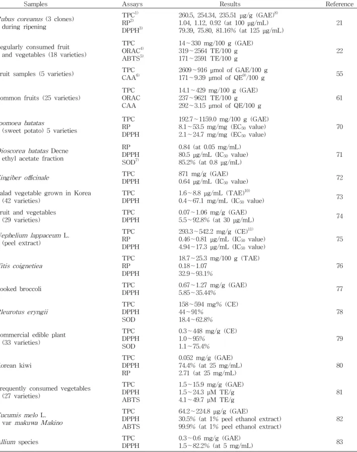
채소․과일 시료에 대한 적용 사례
앞서 언급한 assay를 이용하여 채소와 과일을 대상으로 실시한 항산화능 측정 적용사례를 Table 1에 제시하였다.
TPC assay와 DPPH assay는 제시되어진 연구에 높은 빈도 로 사용되었으며, 이는 항산화능 측정에 가장 보편적으로 이용되는 방법이 TPC assay와 DPPH assay임을 시사한다.
폴리페놀의 벤젠 고리에 치환되어 있는 여러 개의 hydroxyl group은 free radical과의 환원 반응에 참여함으로써 항산화 활성이 나타나므로 정확한 TPC 측정은 시료의 항산화능을 예측할 수 있는 좋은 지표가 된다. 특히 폴리페놀 화합물은 주로 채소와 과일에 함유되어 있으므로 채소와 과일을 시료 로 하는 항산화능 연구에 있어서 TPC의 정확한 측정이 매우 중요하다고 하겠다. 그러나 표준 검량 곡선에 사용되는 표준 물질이 다양하고 여러 가지 단위로 표현되어지고 있으므로 같은 시료에 대해서 연구한 결과에 있어서 비교가 어렵다.
DPPH assay 및 RP assay, ABTS assay의 원리는 산화
환원 반응으로 시료의 환원력에 의한 free radical 소거능을
흡광도로서 확인하는 실험 방법이다. DPPH assay와 ABTS
assay는 빠르고 간단하게 이용할 수 있는 측정법으로 시료의
환원력으로써 항산화 활성을 측정한다. 특히 ABTS assay
는 폭 넓은 pH 범위에서 측정이 가능하며(64), DPPH는 유기
용매에 녹는 반면, ABTS는 친수성 용매(hydrophilic sol-
vents)뿐만 아니라 소수성 용매(hydrophobic solvents)에도
용해되므로 다양한 식품 추출물의 항산화 활성 측정 시에
유리할 것이다(65). 따라서 RP assay는 간단하고 빠른 측정
Table 1. Application of antioxidant capacity assay in fruit and vegetable
Samples Assays Results Reference
Rubus coreanus (3 clones) during ripening
TPC
1)RP
2)DPPH
3)260.5, 254.34, 235.51 μg/g (GAE)
8)1.04, 1.12, 0.92 (at 100 μg/mL)
79.39, 75.80, 81.16% (at 125 μg/mL) 21 Regularly consumed fruit
and vegetables (18 varieties)
TPC ORAC
4)ABTS
5)14∼330 mg/100 g (GAE) 319∼2564 TE/100 g
171∼2591 TE/100 g 22
Fruit samples (5 varieties) TPC
CAA
6)2609∼916 μmol of GAE/100 g
171∼9.39 μmol of QE
9)/100 g 55
Common fruits (25 varieties) TPC ORAC CAA
14.1∼429 mg/100 g (GAE) 237∼9621 TE/100 g
292∼3.15 μmol of QE/100 g 61
Ipomoea batatas
(sweet potato) 5 varieties
TPC RP DPPH
192.7∼1159.0 mg/100 g (GAE) 8.1∼53.5 mg/mg (EC
50value)
2.1∼24.7 mg/mg (EC
50value) 70
Dioscorea batatas Decne ethyl acetate fraction
RP DPPH SOD
7)0.84 (at 0.05 mg/mL) 80.5 μg/mL (IC
50value)
85.2% (at 0.8 μg/mL) 71
Zingiber officinale TPC DPPH 871 mg/g (GAE)
0.64 μg/mL (IC
50value) 72
Salad vegetable grown in Korea
(42 varieties) TPC
DPPH 1.6∼8.8 μg/mL (TAE)
10)0.4∼67.1 mg/mL (IC
50value) 73
Fruit and vegetables
(29 varieties) TPC
DPPH 0.07∼1.06 mg/g (GAE)
5.5∼92.8% (at 30 μg/mL) 74
Nephelium lappaceum L.
(peel extract)
TPC RP DPPH
293.3∼542.2 mg/g (CE)
11)0.46∼0.81 μg/mL (IC
50value)
4.94∼17.3 μg/mL (IC
50value) 75
Vitis coignetiea TPC RP DPPH
18.7∼25.3 mg/100 g (TAE) 0.18∼1.07
32.9∼93.1% 76
Cooked broccoli TPC
DPPH 0.67∼1.27 mg/g (GAE)
5.85∼35.44% 77
Pleurotus eryngii TPC DPPH SOD
158∼594 mg% (CE) 44∼91%
18.4∼62.8% 78
Commercial edible plant (33 varieties)
TPC DPPH SOD
0.3∼448 mg/g (CE) 1.0∼95%
1.1∼75.4% 79
Korean kiwi TPC
DPPH RP
0.052 mg/g (GAE) 74.4% (at 25 mg/mL)
2.71 (at 25 mg/mL) 80
Frequently consumed vegetables (27 varieties)
TPC DPPH ABTS
1.5∼15.9 mg/g (GAE) 1.5∼24.3 μM TE/g
4.1∼49.7 μM TE/g 81
Cucumis melo L.
var makuwa Makino
TPC DPPH ABTS
64.2∼224.8 μg/g (GAE)
30.5% (at 1% peel ethanol extract)
99.9% (at 1% peel ethanol extract) 82
Allium species TPC
DPPH 0.3∼0.6 mg/g (GAE)
1.5∼82.2% (at 5 mg/mL) 83
1)
TPC stands for total phenolic contents.
2)RP stands for reducing power.
3)DPPH stands for DPPH radical scavenging activity.
4)
ORAC stands for oxygen radical absorbance capacity.
5)ABTS stands for ABTS cation radical scavenging activity.
6)
CAA stands for cellular antioxidant activity.
7)SOD stands for SOD-like activity.
8)
GAE stands for gallic acid equivalent.
9)QE stands for quercetin equivalent.
10)
TAE stands for tannic acid equivalent.
11)CE stands for catechin equivalent.
법임에도 불구하고 DPPH assay나 ABTS assay의 free radical 소거능으로 환원력에 의한 항산화 활성 측정을 대신 할 수 있으므로 적용되어진 연구는 미흡했다.
SOD-like activity assay의 경우, 조추출물(crude ex- tracts)의 항산화 활성 분석에 있어서 혼합물의 간섭이 유발 될 수 있다는 단점으로 채소와 과일을 대상으로 한 항산화능 측정에는 다소 드물게 사용되는 경향이 있었다. SOD-like activity assay는 채소와 과일로부터 분리 정제한 화합물의 항산화 활성 측정 시에는 유용한 방법이라 사료된다. ORAC assay는 재현성이 뛰어난 측정법으로 실험 방법이 표준화되 어 있고 단위가 TE로 통일되어 있다는 장점이 있다. 또한, 여러 가지 다른 radical을 이용하여 항산화 활성을 측정할 수 있으며, 특히 채소나 과일 등 천연 물질로부터 추출한 시료에 있어서 시간과 농도에 따른 항산화 활성을 확인할 수 있다.
산화적 스트레스와 그로 인해 유발되는 여러 만성 질병, 또한 이러한 질병의 예방과 치료를 위한 항산화제의 작용 메카니즘을 이해하기 위해서 살아 있는 세포에서 산화적 스 트레스를 평가하는 분석적 방법이 매우 중요하다(66). 그러 나 실험동물이나 인체를 대상으로 실시하는 실험은 비용이 비싸며, 시간이 많이 소요된다는 단점이 있어 식품이나 식이 보충제의 항산화 효과를 초기에 스크리닝(screening)하기에 는 부적합하다(67). Comet assay는 H
2O
2로써 인위적으로 산 화적 스트레스를 유발하여 적은 수의 세포에서 간단하고 빠 르게 DNA 손상을 민감하게 감지할 수 있는 실험방법이다.
그러나 Comet assay는 기능성식품이나 항산화제를 대상으 로 실시하는 intervention 연구에 주로 사용되고 있었으며, 채소나 과일 추출물을 백혈구에 직접 처리하여 항산화능을 측정한 실험은 드문 실정이었다. 한편, Comet assay를 포함 하여 앞서 언급한 실험 방법들은 폭넓게 이용되고 있음에도 불구하고 실험을 수행하는 pH나 온도 등이 생리적 환경에 적합하지 않으며 생물학적 이용 가능성이나 흡수, 항산화 물질의 대사는 전혀 고려하지 않고 있다(67). 그러므로 각종 세포주(cell line)를 이용하여 식품과 식이 보충제, phyto- chemical 등의 생물학적 활성을 검토하고, 지질 과산화 생성 물이나 DNA 부가 생성물을 포함한 세포의 산화적 손상을 측정할 수 있는 여러 가지 방법이 이용되고 있다(55,68). 세 포 배양을 통한 실험은 비용 면에서 저렴하고 상대적으로 빠르게 결과를 도출할 수 있으며 세포내의 흡수나 분포, 대 사 등에 대한 평가도 가능하다. 이러한 측면에서, CAA assay 는 채소, 과일을 포함한 식물 추출물의 항산화 활성 측정 시 생리학적으로 적절한 실험 방법이라고 사료된다(69).
요 약
건강한 삶에 대한 현대인의 관심이 나날이 고조되고 있으 며, 이에 따라 노화와 질병의 예방에 효과가 있는 항산화제
의 연구가 활발히 이루어지고 있다. 특히 천연물이나 식품을 소재로 한 식이성 항산화제에 대한 연구는 꾸준히 증가하고 있는 추세이며, 천연물의 소재나 연구 분야의 폭이 매우 넓 다. 따라서 다양한 식품 소재의 항산화능을 조사할 수 있는 측정법의 중요성이 크게 부각되고 있다. 현재 약용 식물이나 상용 채소, 과일을 시료로 하여 여러 가지 radical이나 target molecule에 대한 항산화능을 측정할 수 있는 다수의 생체 외(
in vitro) 측정법이 사용되고 있다. 이에 본 총설에서는 널리 사용되고 있는 항산화능 측정법의 특징을 분석하고 적 용 사례를 검토하였다. 식품의 구성 성분은 단일물질이 아닌 복합체로 구성되어 있으므로 하나의 측정법으로 항산화능 을 평가할 수가 없다. 그러므로 채소와 과일을 포함한 여러 가지 식물성 식품 추출물의 항산화 활성을 정확히 측정하기 위해서는 각 실험에 사용되는 radical과 기전(mechanism), 실험 조건(온도, pH, 실험기기, 시료추출방법, 소요 시간, 비 용) 등을 고려하여 적절히 수행되어야 할 것이다. 그러나 이 들 측정법의 다양성과 사용 단위의 차이로 인해 각 시료의 항산화능을 객관적으로 비교하는데 어려움이 있는 실정이 다. 따라서 향후 항산화능 측정법의 표준화와 표현 단위의 통일이 절실히 요구된다. 또한
in vitro에서 항산화능을 나타 내는 채소, 과일 등의 생체 내 활성은 다양한 biomechanism 과 polymerphism으로 인해 그 효과를 예측하기가 매우 어렵 다. 따라서
in vitro에서의 항산화능 screening 결과를 바탕 으로
in vivo에서의 그 효과를 검증하는 연구가 부가적으로 시행되어야 할 것이라 사료된다.
감사의 글
본 연구는 2011년도 경남대학교 학술논문게재연구비 지 원으로 이루어졌으며, 이에 감사드립니다.
문 헌