인체위암 세포에서 PI3K/AKT 신호 전달계 차단에 의한 동충하초 유래 Cordycepin의 Apoptosis 유발 효과 증진
이혜현1․정진우1․최영현1,2
1동의대학교 항노화연구소
2동의대학교 한의과대학 생화학교실
Inhibition of PI3K/AKT Signaling Pathway Enhances Cordycepin-Induced Apoptosis in Human Gastric Cancer Cells
Hye Hyeon Lee1, Jin-Woo Jeong1, and Yung Hyun Choi1,2
1Anti-Aging Research Center, Dongeui University
2Department of Biochemistry, Dongeui University College of Korean Medicine
ABSTRACT The phosphatidylinositol 3-kinase (PI3K)/Akt signaling pathway plays a crucial role in cancer occurrence by promoting cell proliferation and inhibiting apoptosis. In the present study, we evaluated the effect of a PI3K inhibitor, LY294002, on the chemosensitivity of gastric cancer cells to cordycepin, a predominant functional component of the fungus Cordyceps militaris, in AGS human gastric cancer cells and investigated possible underlying cellular mechanisms.
Our results revealed that cordycepin inhibited viability of AGS cells in a concentration-dependent manner and induced apoptosis, as determined by apoptotic cell morphologies and fluorescence-activated cell sorting analysis associated with attenuated activation of the PI3K/Akt signaling pathway. Treatment with cordycepin in combination with a subtoxic concentration of LY294002 enhanced cordycepin-induced cytotoxicity and apoptotic potentials in AGS cells. Sensitiza- tion of LY294002 to cordycepin-induced apoptosis was accompanied by activation of caspases (caspases-3, -8, and -9) and was concomitant with poly(ADP-ribose) polymerase cleavage. Moreover, LY294002 up-regulated pro-apoptotic Bax and enhanced truncation of Bid in cordycepin-treated AGS cells, which was connected with increased loss of mitochondrial membrane potential and release of cytochrome c from mitochondria to the cytosol. Taken together, these results indicate that inhibition of the PI3K/Akt signaling pathway could augment cordycepin-induced apoptosis in human gastric cancer cells by up-regulating caspase activity through mitochondrial dysfunction.
Key words: cordycepin, PI3K/Akt, AGS cells, apoptosis, caspase
Received 15 February 2016; Accepted 2 March 2016
Corresponding author: Yung Hyun Choi, Department of Biochemis- try, Dongeui University College of Korean Medicine, Busan 47227, Korea
E-mail: [email protected], Phone: +82-51-850-7413
서 론
동충하초는 전통 동양의학에서 임상적으로 허약 증상, 만 성기관지염, 거담과 천식, 폐결핵, 빈혈 및 병후 허약 등에 처방되어 왔다(1). Cordycepin(3'-deoxyadenosine)은 동 충하초 Cordyceps 속에 속하는 균에서 분리되었으며, 구조 적으로 deoxyadenosine과 매우 유사한 adenosine nu- cleoside의 ribose 3번 탄소에 산소가 없는 adenosine an- alogue이다(2). Cordycepin의 효능은 1970년대 핵산 합성 억제, 특히 polyadenylation 억제 효과가 알려지면서 다양 한 생리활성에 대한 연구가 활발히 진행되어 왔다(3). Cor- dycepin의 강력한 항암 활성은 이미 많은 논문에서 보고된
바 있는데, 이는 암세포의 능동적 죽음에 속하는 apoptosis 유도와 매우 밀접한 관계를 가진다(4-13). Apoptosis가 유 도되는 동안 나타나는 가장 일반적인 현상이 cysteine- dependent aspartate-directed protease 군에 속하는 caspase의 활성 증가이다(14,15). 일반적으로 caspases는 initiator caspases(caspase-8, -9 및 -10)와 effector caspases(caspase-3, -6 및 -7)로 구분되는데, initiator caspases의 활성 증가에 의하여 effector caspases가 활성 형으로 전환되는 caspase cascade 경로를 거치게 된다. 특 히 initiator caspases 중에서 caspase-8과 -9는 apopto- sis의 주요한 두 가지 경로인 extrinsic 및 intrinsic path- way의 개시를 알리는 데 관여하는 대표적인 caspase이다 (16,17). 활성화된 effector caspases는 DNA repair에 관 여하는 poly(ADP-ribose) polymerase(PARP)를 포함한 다양한 기질 단백질의 절단을 초래하며 궁극적으로 세포의 죽음을 종결짓는다(18).
Caspase의 활성 유도와 연관된 cordycepin의 apoptosis
유도는 이미 다양한 종류의 암세포에서 확인된 바 있는데, 그 과정에 calcium-calpain-caspase 경로가 관여하기도 하며(7), 활성산소종의 형성이 핵심적인 역할을 하기도 한다 (9,12). 또한 cordycepin에 의한 암세포의 apoptosis 유도 에는 autophagy가 관여하기도 하며(8,10,11), 특히 유방암 세포에서는 estrogen receptor가 비의존적으로 apoptosis 를 유도하였다(8). 한편 특정 약물에 의한 암세포의 apop- tosis 유도는 세포주기의 교란과 동반되기도 하는데, cor- dycepin의 경우 암세포의 종류에 따라 S기 arrest(19) 또는 G1기 arrest(13)를 유발하기도 하여 암세포에 따른 차이를 보여주고 있다(4,20). 아울러 cordycepin은 항암제 저항성 의 극복 가능성을 보여주고 있으며(21,22), cordycepin의 apoptosis 유도에는 일반적으로 extrinsic 및 intrinsic path- way의 두 경로를 모두 거치는 것으로 알려져 있으나, 암세 포의 종류에 따른 약간의 차이점이 있음을 알 수 있다(13, 23,24).
한편 세포 내 다양한 신호 전달계 중에서 phosphoinosi- tide 3-kinase(PI3K)는 대표적인 downstream effector인 Akt와 함께 세포의 생존과 증식에 핵심적인 역할을 한다 (25,26). 특히 위암 세포를 포함한 다양한 암세포의 악성 종양화 과정은 PI3K/Akt 신호계의 비정상적인 활성 증가와 연계되어 있으며(27-30), 암 화학요법에 대한 항암제 저항 성 취득과도 깊은 연관성을 지닌다(31,32). 특히 anti-apop- totic 단백질에 속하는 survivin과 Bcl-2 같은 유전자들의 발현이 Akt에 의하여 조절을 받고 있으며, caspase 활성 억제에도 관여한다(28,33,34). 따라서 PI3K/Akt 신호계의 차단은 암세포의 증식 억제와 apoptosis 유도를 위한 효율 적인 전략으로 인식되고 있다(30,35). 비록 cordycepin의 항암 활성과 연관된 PI3K/Akt 신호계에 대한 역할은 구체적 으로 알려진 바 없으나, 최근 본 연구실에서 cordycepin에 의한 인체 전립선암세포의 이동과 침윤성의 억제는 PI3K/
Akt 신호계의 활성 억제와 연관성이 있었으며(36), 백혈병 세포의 telomerase 활성 억제에도 PI3K/Akt 신호계의 불 활성화와 직접 관련성이 있음을 보고한 바 있다(37). 또한 Pan 등(38)은 cordycepin에 의한 간질 세포종의 apoptosis 유도가 PI3K/Akt 신호계에 의하여 조절 받을 수 있음을 제 시한 바 있다. 이들의 결과에서 PI3K/Akt 신호계의 차단은 cordycepin에 의한 암세포의 전이 억제, telomere 길이의 신장 억제 및 apoptosis 유도에 모두 유의적인 감수성 증대 효능을 나타내었다. 본 연구에서는 cordycepin의 항암 활성 과 연관된 PI3K/Akt 신호계의 역할에 대한 추가적인 자료를 얻기 위하여 인체위암 세포를 대상으로 cordycepin에 의한 세포증식 억제에 따른 apoptosis 유도에 PI3K/Akt 신호계 가 연관성이 있는지를 조사하였으며, PI3K/Akt 신호계 억 제제를 이용한 cordycepin에 의한 apoptosis 유도 효능 증 대 가능성을 조사하였다.
재료 및 방법
세포배양 및 생존율 측정
본 연구에 사용된 AGS 인체위암 세포는 American Type Culture Collection(Manassas, VA, USA)에서 구입하였으 며, 10% fetal bovine serum, penicillin 및 streptomycin 등이 혼합된 RPMI 1640 배지(Gibco-BRL, Grand Island, NY, USA)를 이용하여 37oC, 5% CO2 조건하에서 배양하였 다. Cordycepin(MW, 251.2; product no. C3394)과 LY 294002는 Sigma-Aldrich Co.(St. Louis, MO, USA) 및 Calbiochem(San Diego, CA, USA)에서 구입하였으며 di- methyl sulfoxide(DMSO, Sigma-Aldrich Co.)에 녹인 후 적정 농도로 배지에 희석하여 처리하였다. 최종 DMSO의 농도는 세포독성을 나타내지 않는 0.5% 이내로 사용하였다.
Cordycepin 단독 및 LY294002와의 복합처리에 따른 AGS 세포의 생존에 미치는 영향을 비교하기 위하여 적정 조건에 서 배양 후, 2,000 rpm으로 5분간 원심분리 하여 상층액을 제거하고 세포를 모은 다음 0.5% trypan blue 용액(Gibco BRL)을 2분간 처리한 후 hemocytometer를 이용하여 도립 현미경(inverted microscope, Carl Zeiss, Oberkochen, Germany)으로 살아있는 세포를 계수하였다.
핵의 형태 변화를 통한 apoptosis 유도 확인
전형적인 apoptosis가 유발되었을 경우 핵의 형태적 변 화를 관찰하기 위하여 준비된 세포들을 phosphate-buf- fered saline(PBS)으로 2~3회 수세 후 0.1 M phosphate buffer(pH 7.2)에 희석된 2.0% paraformaldehyde 고정액 (Sigma-Aldrich Co.)으로 1시간 세포들을 고정하였다. 고 정 후 세포들을 PBS로 수회 수세한 다음 0.05 M phosphate buffer(pH 7.2)에 2.5 μg/mL의 농도로 희석된 4,6-dia- midino-2-phenylindole(DAPI, Sigma-Aldrich Co.) 용액 으로 상온 암하에서 15분간 염색하였다. 이를 다시 PBS로 수회 수세한 후 형광현미경(fluorescence microscope, Carl Zeiss) 하에서 핵의 형태를 비교하였다.
Apoptosis 유발의 정량적인 비교
Cordycepin과 LY294002의 단독 및 복합 처리에 의한 apoptosis 유발 정도를 정량적으로 비교하기 위하여 준비된 세포들을 모은 다음 2,000 rpm으로 5분간 원심분리 하여 상층액을 제거하였다. 이들 세포를 PBS를 이용하여 2~3회 정도 세척하고 Annexin V binding buffer(Becton Dickin- son, San Jose, CA, USA)에 부유시킨 다음 Annexin V- fluorescein isothiocyanate(FITC) 및 propidium iodide (PI)를 처리하여 암실에서 20분 동안 반응을 시켰다. 반응이 끝난 후 세포를 35-mm mesh를 이용하여 단일 세포로 분리 한 다음 flow cytometer(FACSCalibur, Becton Dickinson) 에 적용해 apoptosis가 유발된 세포(V+/PI–)를 형광반응에 따라 분석하였다. 아울러 세포주기 분포도에서 apoptosis가
유발된 세포의 집단에 해당하는 sub-G1기의 세포 빈도를 정량하기 위하여 Cycletest Plus DNA Reagent kit(Becton Dickinson)을 이용하여 고정 및 염색을 하여 4°C, 암실에서 30분 동안 반응시켰다. 반응시킨 세포를 flow cytometer에 적용해 형광반응에 따른 cellular DNA content 및 histo- gram을 분석하였다.
단백질의 분리 및 western blot analysis
특정 단백질의 발현 변화 분석을 위해 25 mM Tris-Cl (pH 7.5), 250 mM NaCl, 5 mM ethylenediaminetetra acetic acid, 1% nonidet P-40, 0.1 mM sodium orthova- nadate, 2 μg/mL leupeptin 및 100 μg/mL phenylmethyl- sulfonyl fluoride가 함유된 lysis buffer를 이용하여 단백질 을 분리하였다. 아울러 세포질 및 미토콘드리아 단백질을 분리하기 위하여 mitochondrial fractionation kit(Active- motif, Carlsbad, CA, USA)을 사용하였다. 단백질 농도는 Bio-Rad 단백질 정량 시약(Bio-Rad, Hercules, CA, USA) 을 이용하여 정량한 다음 동량의 Laemmli sample buffer (Bio-Rad)를 섞어서 sample을 만들었다. 분리된 단백질들 의 농도를 측정한 후, western blot analysis를 위해 동량의 단백질들을 sodium dodecyl sulfate(SDS)-polyacryl- amide gel electrophoresis를 이용하여 분리하고 PVDF membrane(Schleicher & Schuell, Keene, NH, USA)으로 전이시켰다. 각각의 membrane에 적정 항체 및 enhanced chemiluminescence(ECL, Amersham Corp., Arlington Heights, IL, USA) 용액을 이용하여 단백질들의 발현 변화 를 조사하였다. 본 실험에 사용된 1차 항체들은 Santa Cruz Biotechnology Inc.(Santa Cruz, CA, USA) 및 Calbio- chem(Cambridge, MA, USA)에서 구입하였으며, 2차 항체 들은 Amersham Corp.에서 구입하였다.
In vitro caspase activity 측정
Apoptosis 유발 연계 caspase의 활성 정도가 cordyce- pin 단독 및 LY294002와의 복합 처리에 따라 어떠한 변화 를 보이는지를 알아보기 위하여 준비된 세포를 모은 뒤 상기 와 같은 방법으로 단백질을 추출하고 정량하였다. Caspase activity assay kit은 R&D Systems(Minneapolis, MN, USA)에서 구입하였으며, 150 μg의 단백질이 함유된 50 μL 의 sample에 기질 100 μM이 함유된 reaction buffer[40 mM HEPES(pH 7.4), 20% glycerol(v/v), 1 mM EDTA, 0.2% NP-40 and 10 mM DL-DTT] 50 μL를 혼합하여 각 sample 당 총량이 100 μL가 되게 하였다. 여기에 각 caspase 기질 5 μL를 첨가하여 37°C, 암실에서 3시간 동안 반응시킨 후 ELISA reader를 이용하여 405 nm의 흡광도에 서 반응의 정도를 측정하였다. 본 실험에 사용한 기질은 caspase-3의 경우에는 Asp-Glu-Val-Asp(DEVD)-p-ni- troaniline(pNA)이었고, caspase-8의 경우에는 Ile-Glu- Thr-Asp(IETD)-pNA였으며, caspase-9은 Leu-Glu-His-
Asp(LEHD)-pNA였다.
Mitochondrial membrane potential(MMP, Δψm)의 분석 Cordycepin 단독 및 LY294002와의 복합 처리에 따른 MMP 값 변화 정도를 측정하기 위하여 준비된 세포들에 10 μM의 5,5',6,6'-tetrachloro-1,1',3,3'-tetraethyl-imida- carbocyanine iodide(JC-1, Sigma-Aldrich Co.) 용액을 처리하여 20분 동안 상온에서 반응시켰다. 반응이 끝난 후 상층액을 제거하고 PBS를 첨가하여 세포를 부유시킨 다음 flow cytometer에 적용해 MMP의 변화를 측정하였다.
통계처리
모든 실험결과는 평균±표준편차로 표시하였고 Sigma Plot(Systat Software Inc., San Jose, CA, USA) Student t-test를 이용하여 통계적 유의성을 얻었다. 모든 통계처리 의 유의성은 P<0.05 수준에서 검증하였다.
결과 및 고찰
Cordycepin에 의한 AGS 세포의 생존율 저하 및 apopto- sis 유도
생명체의 정상적인 생명유지와 발생과정에 필수적인 ap- optosis는 병리학적인 요인에 의한 세포의 죽음인 necrosis 에 비하여 다양한 세포 내외 인자들이 관여하는 신호전달과 정을 통한 능동적인 세포 자멸 현상이다. 전형적인 apopto- sis가 일어난 세포에서 볼 수 있는 가장 일반적인 변화는 형태학적으로 세포의 크기가 축소되며, 핵의 응집과 DNA의 단편화이다(16). Cordycepin 단독 처리가 AGS 세포의 생 존에 미치는 영향을 분석하기 위하여 다양한 농도(0~25 μg /mL)로 24시간 처리한 후, trypan blue 용액으로 염색하고 hemocytometer를 이용하여 살아있는 세포의 수를 계수하 였다. Fig. 1A에 나타낸 바와 같이 AGS 세포에 cordycepin 의 처리 농도 증가에 따라 trypan blue 용액에 염색이 되지 않은 세포의 수가 유의적으로 감소하였다.
Cordycepin 처리에 따른 AGS 세포의 생존율 억제가 apoptosis 유발과 직접적인 연관이 있는지를 확인하기 위하 여 정상 및 cordycepin이 함유된 배지에서 배양된 세포를 대상으로 핵산에 특이적으로 결합하는 형광물질인 DAPI 염 색을 하였다. Fig. 1B에 나타낸 결과에서 알 수 있듯이 정상 배지에서 자란 AGS 세포에서는 핵의 형태가 뚜렷하게 염색 되었으나 cordycepin이 처리된 세포의 경우는 처리 농도의 증가에 따라 apoptosis가 일어난 세포에서 전형적으로 관찰 되는 염색질 응축에 의한 apoptotic body의 형성이 점차 증가하여 DNA의 단편화가 초래되었음을 알 수 있었다. 아 울러 cordycepin 처리에 따른 apoptosis 유발 정도를 An- nexin V 염색으로 통하여 확인한 결과, cordycepin 처리 농도의 증가에 따라 점차 증가하여 20 μg/mL 및 25 μg/mL 처리군에서 정상배지에서 배양된 세포에 비하여 약 9.1배
A
B
C
Fig. 1. Inhibition of cell viability and induction of apoptosis by cordycepin in AGS cells. Cells were treated with the indicated concentrations of cordycepin for 24 h. (A) Living cell number were determined by hemocytometer counts of trypan blue ex- cluding cells. Results are expressed as percentage of the vehicle treated control±SD of three separate experiments (*P<0.05 vs.
untreated control). (B) The cells were fixed and stained with DNA specific fluorescent dye DAPI solution. The stained nuclei were observed under a fluorescent microscope (Original magni- fication, 400×). (C) To quantify the degree of apoptosis in- duced by cordycepin, cells were stained with FITC-conjugated Annexin V and PI for DNA flow cytometry analysis. Apoptotic cells are determined by counting the % of Annexin V+/PI– cells and the percent of Annexin V+/PI– cells. Results shown are ex- pressed as the mean of two independent experiments.
Fig. 2. Inactivation of PI3K/Akt signaling pathway by cordyce- pin in AGS cells. (A) After treatment with the indicated concen- trations of cordycepin for 24 h, the cells were lysed and then equal amounts of cell lysates were separated on SDS-polyacryla- mide gels and transferred to PVDF membranes. The membranes were probed with the indicated antibodies and the proteins were visualized using an ECL detection system. Actin was used as an internal control.
및 18.9배로 증가하였다(Fig. 1C). 이러한 결과는 cordy- cepin 처리에 의한 AGS 세포의 생존율 억제가 apoptosis 유도와 연계되었음을 보여주는 것이다. 아울러 본 연구의 결과에서 나타난 AGS 위암 세포에서 cordycepin의 apop- tosis 유도 활성의 정도는 전립선암세포나 대장암세포 등에 비해 저농도에서 apoptosis 유도능을 보였으며, 백혈병 세 포들과 비교했을 경우 유사한 정도의 감수성임을 알 수 있었 다(9,11,13).
AGS 세포에서 cordycepin의 apoptosis 유발에 미치는 PI3K/Akt 신호계의 영향
이상에서 관찰된 cordycepin에 의한 AGS 세포의 생존율 억제 및 apoptosis 유도가 PI3K/Akt 신호계와 연관성이 있 는지를 조사하기 위하여 PI3K 및 PI3K의 하위 분자인 Akt
의 인산화에 미치는 cordycepin의 영향을 조사하였다. Fig.
2에 나타낸 바와 같이 PI3K 및 Akt 전체 단백질 발현의 변화 는 나타나지 않았으나 cordycepin 처리 농도의 증가에 따라 PI3K뿐만 아니라 Akt의 인산화형이 점차 감소하여 백혈병 세포나 간질 세포종 및 전립선암세포 등에서 관찰된 경우 (36-38)와 같이 cordycepin의 항암 활성은 세포 생존에 주 요한 역할을 하는 PI3K/Akt 신호계 활성 억제와 관련이 있 음을 알 수 있었다. 특히 PI3K/Akt 신호 경로 억제는 다양한 항암제의 처리 농도를 낮춤으로써 독성을 경감시키는 방법 으로 유용한 수단으로 인식되고 있기에(30,35), 세포독성을 나타내지 않는 범위의 PI3K 억제제인 LY294002 농도(10 μM)를 선정하여 PI3K/Akt 신호계의 불활성화가 cordyce- pin에 의한 AGS 세포의 apoptosis에 미치는 영향을 조사하 였다. Fig. 3의 결과에서 알 수 있듯이 LY294002는 cordy- cepin 단독 처리에 의한 염색질의 응축과 apoptosis 유발군 에 해당하는 세포의 빈도를 향상시켰으며, cordycepin에 의 한 세포 생존율도 저하시켰다. 즉 PI3K/Akt 신호계의 차단 은 cordycepin에 의한 세포독성을 더욱 증진시키는 효과를 보여주었음을 알 수 있었다.
AGS 세포에서 cordycepin에 의한 caspase 활성에 미치 는 PI3K/Akt 신호계의 역할
Cysteine-dependent aspartate-directed proteases에 속하는 caspase는 세포가 정상적으로 생존 및 증식할 경우, 핵과 mitochondria의 외막에 불활성 상태인 proenzyme 형 태로 존재하지만 다양한 세포 내외부 apoptosis 자극으로 활성화되면 선택적 기질 단백질들의 분해를 유발함으로써 apoptosis를 촉진하는 역할을 한다(39,40). Caspase는 ex- trinsic apoptosis pathway와 intrinsic apoptosis path- way의 initiator caspase인 caspase-8 및 -9와 effector
A
B
Fig. 3. PI3K inhibitor, LY294002, enhanced cordycepin-induced AGS cell apoptosis. AGS cells were treated with LY294002 (10 μM) and cordycepin (25 μg/mL), alone or in combination, for 24 h. (A) Apoptosis was then analyzed by staining with DAPI and flow cytometry analysis. Results shown are expressed as the mean of two independent experiments. (B) Living cell number were determined by hemocytometer counts of trypan blue excluding cells. Results are expressed as percentage of the vehicle treated control±SD of three separate experiments (*P<
0.05 vs. untreated control; #P<0.05 vs. cordycepin-treated group).
A
B
Fig. 4. PI3K inhibitor, LY294002, increased cordycepin-induced activation of caspases and degradation of PARP in AGS cells.
(A) AGS cells were treated with LY294002 (10 μM) and cordy- cepin (25 μg/mL), alone or in combination, for 24 h. The cel- lular proteins were separated by SDS-polyacrylamide gel elec- trophoresis and transferred to PVDF membranes. The mem- branes were probed with the indicated antibodies, and the pro- teins were visualized using an ECL detection system. Actin was used as an internal control. (B) The cell lysates obtained from cells grown under the same conditions as (A) were assayed for in vitro caspase-3, -8, and -9 activity using DEVD-pNA, IETD- pNA, and LEHD-pNA, respectively, as substrates. The relative fluorescent products were measured. Data are means±SD from representative experiments performed at least three times (*P<
0.05 vs. untreated control; #P<0.05 vs. cordycepin-treated group).
caspase인 caspase-3 및 -7 등으로 나누어지는데, 먼저 initiator caspase가 활성화되면 하위단계에 있는 effector caspase를 활성화함으로써 세포의 apoptosis를 유발한다 (14,15). PARP는 활성화된 effector caspase에 의하여 분 해되는 대표적인 기질 단백질로 정상적인 DNA 수복이나 유전자 안정성 유지에 핵심인자로 작용하지만, 116 kDa의 분자량을 가진 PARP 단백질이 85 및 24 kDa의 단편으로 잘리게 되면 PARP의 기능은 상실하게 되며, PARP의 단편 화는 caspase 활성 의존적 apoptosis 유발의 생화학적 표식 자로 사용이 되고 있다(18). 따라서 본 연구에서는 대표적인 apoptosis 유발 두 경로의 initiator caspase(caspase-8 및 -9)와 effector caspase인 caspase-3의 발현에 미치는 LY294002 및 cordycepin 각각의 영향과 복합 처리에 따른 영향을 조사하였다. 본 연구의 결과는 몇몇 선행연구에서 관찰된 바와 같이(11,12,23) cordycepin이 단독 처리된 배 지에서 배양된 AGS 세포에서 caspase-8 및 -9뿐만 아니라 caspase-3의 불활성형 단백질의 발현이 모두 감소하였으 며, 이러한 감소 효과는 LY294002와 cordycepin 복합 처 리군에서 더 강하게 관찰되었다(Fig. 4A). 그리고 비록 caspase-8의 활성형 단백질의 발현은 검출되지 않았지만 caspase-9 및 -3의 활성형 발현도 다소 증가하였으며, 증
가의 정도는 cordycepin 단독 처리군에서보다 LY294002 및 cordycepin 복합 처리군에서 매우 높게 나타났다. 따라 서 caspase의 활성 여부를 정량적으로 확인하기 위하여 in vitro caspase 활성을 분석한 결과, 조사된 3가지 caspase 의 활성이 각각의 단독 처리군에 비하여 복합 처리군에서 유의적으로 높게 증가하였다(Fig. 4B). 아울러 LY294002 와 cordycepin 복합처리군에서 PARP 단백질의 단편화도 매우 증가하여(Fig. 5A), PI3K/Akt 신호계의 차단은 cor- dycepin에 의한 caspase 활성을 증가시켜 apoptosis 유발 효과를 높였음을 알 수 있었다. 이러한 PI3K/Akt 신호계 차단에 의한 cordycepin의 apoptosis 유도에서 caspase 활 성 증가에 의한 감수성 증대 효과는 간질종세포에서 관찰된 것과 유사하였음을 알 수 있었다(37).
A
B
C
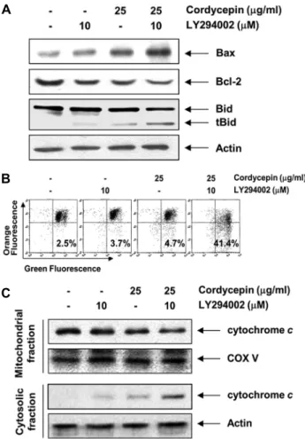
Fig. 5. PI3K inhibitor, LY294002, enhanced cordycepin-induced mitochondrial dysfunction in AGS cells. (A) Total cell lysates were prepared from cells treated with LY294002 (10 μM) and cordycepin (25 μg/mL), alone or in combination, for 24 h and western blot analysis was performed using antibodies against Bax, Bcl-2 and Bid, and ECL detection. Actin was used as an internal control. (B) The cells were stained with 10 μM JC-1 for 20 min at 37°C. The mean JC-1 fluorescence intensity was then detected using a flow cytometer. Data represent the means of two independent experiments. (C) Cytosolic and mitochon- drial extracts were prepared from cells grown under the same conditions resolved by SDS-polyacrylamide gels, transferred to PVDF membranes, and probed with the anti-cytochrome c anti- bodies and an ECL detection system. Actin and cytochrome c oxidase subunit 4 (COX Ⅳ) were used as internal controls for the cytosolic and mitochondrial fractions, respectively.
AGS 세포에서 cordycepin에 의한 미토콘드리아 기능에 미치는 PI3K/Akt 신호계의 역할
미토콘드리아의 기능 손상과 연관된 apoptosis 조절에 중요한 역할을 하는 Bcl-2 family에 속하는 단백질들에는 apoptosis를 억제하는 anti-apoptotic 단백질(Bcl-2 및 Bcl- xL 등)과 apoptosis를 유발하는 pro-apoptotic 단백질 (Bax, Bak 및 Bad 등)로 구성되어 있으며, 그들은 dimer의 형태로 결합하여 균형을 이루고 있다. 그러나 apoptosis 유 도 신호에 따라 이들 사이에 균형이 깨지게 되면 미토콘드리 아 내부로부터 세포질로 cytochrome c와 같은 apoptosis 를 유발하는 단백질이 방출되어 caspase의 활성 및 DNA의 단편화와 연관된 endonuclease와 같은 효소의 활성이 증가 한다(16,17). 따라서 본 연구에서는 PI3K/Akt 신호계 억제 에 의한 cordycepin의 apoptosis 유발 증대와 연관된 기전 해석을 위하여 Bcl-2 family에 속하는 Bax 및 Bcl-2의 발 현 변화를 조사하였다. Fig. 5A의 결과에서 알 수 있듯이 LY294002 및 cordycepin 단독 처리로 Bax의 발현이 크게 증가하지 않았으나, LY294002와 cordycepin이 복합 처리 된 조건에서 배양된 AGS 세포의 Bax 발현은 매우 증가하였 다. 이와는 반대로 Bcl-2의 발현은 LY294002 및 cordy- cepin 단독 처리군에 비하여 복합 처리군에서의 상대적인 발현 억제가 관찰되었다.
한편 extrinsic apoptosis pathway에서 initiator cas- pase인 caspase-8이 활성화되면 caspase cascade를 통 한 effector caspase를 활성화하거나 Bid의 truncation에 의한 단편화(tBid)의 형성으로 미토콘드리아의 기능 이상을 유발함으로써 intrinsic pathway를 통한 apoptosis의 증폭 유발에도 관여한다(14,17). 본 연구의 결과에 의하면 cor- dycepin 단독 처리군에 비하여 LY294002 단독 처리군에서 는 tBid의 발현이 거의 관찰되지 않았으며, LY294002와 cordycepin 복합 처리군에서는 tBid의 발현이 매우 증가하 였다(Fig. 5A). 이러한 변화가 미토콘드리아의 기능 이상과 연관성이 있는지를 조사하기 위하여 MMP 값의 변화를 JC-1 염색을 통하여 분석한 결과, LY294002와 cordyce- pin 단독 처리군에서는 미토콘드리아 기능 손상의 지표인 MMP 값의 손실이 다소 증가(각각 1.5 및 1.9배)하였으나, 복합 처리군에서는 약 17배 정도 증가하였다(Fig. 5B). 이러 한 미토콘드리아의 기능 손상은 cytochrome c의 세포질 유리와 밀접한 연관성이 있으므로 LY294002와 cordyce- pin 단독 및 복합 처리된 배지에서 배양된 AGS 세포들의 세포질 및 미토콘드리아 단백질을 분리하여 cytochrome c 의 발현 변화를 조사하였다. Fig. 5C에 나타난 결과에서 알 수 있듯이 미토콘드리아에서 세포질로의 cytochrome c 이 동이 각각의 단독 처리군에서는 미비하였으나 복합 처리군 에서는 세포질에서의 발현이 매우 증가하였으며, 상대적으 로 미토콘드리아에서의 발현은 감소하였음을 확인하였다.
따라서 본 연구의 결과는 LY294002와 cordycepin 복합
처리에 따른 Bcl-2에 대한 Bax의 상대적인 발현 증가와 Bid의 활성 증대로 인하여 미토콘드리아 막 교란이 증대하 였으며, 이에 따라 apoptosis 유도에 핵심적인 역할을 하는 cytochrome c의 세포질로의 이동에 따라 AGS 세포가 ap- optosis를 유도하였음을 알 수 있었다. 특히 Bcl-2 family 의 anti-apoptotic 단백질의 발현이 Akt의 활성에 의하여 조절 받는 점을 고려할 경우(28,33,34), PI3K/Akt 신호계의 차단은 cordycepin에 의한 extrinsic pathway를 통한 in- trinsic pathway의 증폭을 통하여 apoptosis 유발 효과를 더욱 증진했음을 알 수 있었다.
요 약
PI3K/Akt 신호계는 세포 생존의 조절에 필수적인 경로로 대부분 암세포에서 활성이 증대되어 있다. 본 연구에서는 동충하초의 주요 생리활성 물질인 cordycepin에 의한 AGS 인체 위암 세포의 apoptosis 유도에 미치는 PI3K/Akt 신호 계의 역할을 조사하였다. 본 연구의 결과에 의하면 cordy- cepin의 처리 농도의 증가에 따라 AGS 세포의 생존율은 억제되었으며, 이는 apoptosis 유도와 밀접한 관계가 있음 을 핵의 형태적 변화와 flow cytometry 분석을 통하여 확인 하였다. 이러한 cordycepin의 apoptosis 유도 효과는 PI3K /Akt 신호계의 활성 저하와 연관성이 있었으며, 세포독성을 나타내지 않는 범위의 PI3K/Akt 신호계 저해제인 LY294002 를 cordycepin과 동시 처리하였을 경우, cordycepin에 의 한 apoptosis 유발을 더욱 증대시켰다. 그리고 cordycepin 에 대한 LY294002의 apoptosis 유발 증대는 caspases (caspase-3, -8 및 -9)의 활성 증가와 poly(ADP-ribose) polymerase 단백질의 분해 증가를 촉진했다. 또한 cordy- cepin이 처리된 AGS 세포에서 LY294002는 apoptosis 유 도에 관여하는 Bax의 발현을 증가시켰고 apoptosis 억제에 관여하는 Bcl-2의 발현은 감소시켰으며, 이는 미토콘드리 아 기능 손상과 미토콘드리아에서 세포질로의 cytochrome c 유리를 증대시켰다. 따라서 PI3K/Akt 신호계의 활성 차단 은 cordycepin의 항암 활성을 더욱 상승시켰으며, 이는 미 토콘드리아 기능 손상과 caspase의 활성 증대를 통하여 이 루어짐을 알 수 있었다.
감사의 글
이 논문은 2015년도 정부(미래창조과학부)의 재원으로 한 국연구재단의 지원을 받아 수행된 기초연구사업임(No.
2015R1A2A2A01004633).
REFERENCES
1. Xiao JH, Zhong JJ. 2007. Secondary metabolites from Cor- dyceps species and their antitumor activity studies. Recent Pat Biotechnol 1: 123-137.
2. Cunningham KG, Manson W, Spring FS, Hutchinson SA.
1950. Cordycepin, a metabolic product isolated from cul- tures of Cordyceps militaris (Linn.) Link. Nature 166: 949.
3. Nair CN, Owens MJ. 1974. Preliminary observations per- taining to polyadenylation of rhinovirus RNA. J Virol 13:
535-537.
4. Thomadaki H, Tsiapalis CM, Scorilas A. 2005. Polyadenylate polymerase modulations in human epithelioid cervix and breast cancer cell lines, treated with etoposide or cordyce- pin, follow cell cycle rather than apoptosis induction. Biol Chem 386: 471-480.
5. Thomadaki H, Scorilas A, Tsiapalis CM, Havredaki M.
2008. The role of cordycepin in cancer treatment via in- duction or inhibition of apoptosis: implication of poly- adenylation in a cell type specific manner. Cancer Chemother
Pharmacol 61: 251-265.
6. Chen LS, Stellrecht CM, Gandhi V. 2008. RNA-directed agent, cordycepin, induces cell death in multiple myeloma cells. Br J Haematol 140: 682-691.
7. Chen Y, Chen YC, Lin YT, Huang SH, Wang SM. 2010.
Cordycepin induces apoptosis of CGTH W-2 thyroid carci- noma cells through the calcium-calpain-caspase 7-PARP pathway. J Agric Food Chem 58: 11645-11652.
8. Choi S, Lim MH, Kim KM, Jeon BH, Song WO, Kim TW.
2011. Cordycepin-induced apoptosis and autophagy in breast cancer cells are independent of the estrogen receptor. Toxicol Appl Pharmacol 257: 165-173.
9. Jeong JW, Jin CY, Park C, Hong SH, Kim GY, Jeong YK, Lee JD, Yoo YH, Choi YH. 2011. Induction of apoptosis by cordycepin via reactive oxygen species generation in hu- man leukemia cells. Toxicol In Vitro 25: 817-824.
10. Li Y, Li R, Zhu S, Zhou R, Wang L, DU J, Wang Y, Zhou B, Mai L. 2015. Cordycepin induces apoptosis and autoph- agy in human neuroblastoma SK-N-SH and BE(2)-M17 cells.
Oncol Lett 9: 2541-2547.
11. Lee HH, Kim SO, Kim GY, Moon SK, Kim WJ, Jeong YK, Yoo YH, Choi YH. 2014. Involvement of autophagy in cor- dycepin-induced apoptosis in human prostate carcinoma LNCaP cells. Environ Toxicol Pharmacol 38: 239-250.
12. Lee HH, Park C, Jeong JW, Kim MJ, Seo MJ, Kang BW, Park JU, Kim GY, Choi BT, Choi YH, Jeong YK. 2013.
Apoptosis induction of human prostate carcinoma cells by cordycepin through reactive oxygen species-mediated mi- tochondrial death pathway. Int J Oncol 42: 1036-1044.
13. Lee SY, Debnath T, Kim SK, Lim BO. 203. Anti-cancer effect and apoptosis induction of cordycepin through DR3 pathway in the human colonic cancer cell HT-29. Food Chem Toxicol 60: 439-447.
14. Fiandalo MV, Kyprianou N. 2012. Caspase control: protag- onists of cancer cell apoptosis. Exp Oncol 34: 165-175.
15. Hensley P, Mishra M, Kyprianou N. 2013. Targeting cas- pases in cancer therapeutics. Biol Chem 394: 831-843.
16. Lavrik IN. 2000. Systems biology of apoptosis signaling networks. Curr Opin Biotechnol 21: 551-555.
17. Fulda S, Debatin KM. 2006. Extrinsic versus intrinsic apop- tosis pathways in anticancer chemotherapy. Oncogene 25:
4798-4811.
18. Duriez PJ, Shah GM. 1997. Cleavage of poly(ADP-ribose) polymerase: a sensitive parameter to study cell death. Bio- chem Cell Biol 75: 337-349.
19. Wang XA, Xiang SS, Li HF, Wu XS, Li ML, Shu YJ, Zhang F, Cao Y, Ye YY, Bao RF, Weng H, Wu WG, Mu JS, Hu YP, Jiang L, Tan ZJ, Lu W, Wang P, Liu YB. 2014.
Cordycepin induces S phase arrest and apoptosis in human gallbladder cancer cells. Molecules 19: 11350-11365.
20. Liao Y, Ling J, Zhang G, Liu F, Tao S, Han Z, Chen S, Chen Z, Le H. 2015. Cordycepin induces cell cycle arrest and apoptosis by inducing DNA damage and up-regulation of p53 in Leukemia cells. Cell Cycle 14: 761-771.
21. Lee HH, Jeong JW, Lee JH, Kim GY, Cheong J, Jeong YK, Yoo YH, Choi YH. 2013. Cordycepin increases sensi- tivity of Hep3B human hepatocellular carcinoma cells to TRAIL-mediated apoptosis by inactivating the JNK signal- ing pathway. Oncol Rep 30: 1257-1264.
22. Chen YH, Hao LJ, Hung CP, Chen JW, Leu SF, Huang BM.
2014. Apoptotic effect of cisplatin and cordycepin on OC3 human oral cancer cells. Chin J Integr Med 20: 624-632.
23. Tian X, Li Y, Shen Y, Li Q, Wang Q, Feng L. 2015. Apop- tosis and inhibition of proliferation of cancer cells induced
by cordycepin. Oncol Lett 10: 595-599.
24. Baik JS, Kwon HY, Kim KS, Jeong YK, Cho YS, Lee YC.
2012. Cordycepin induces apoptosis in human neuroblastoma SK-N-BE(2)-C and melanoma SK-MEL-2 cells. Indian J Biochem Biophys 49: 86-91.
25. Chan CH, Jo U, Kohrman A, Rezaeian AH, Chou PC, Logo- thetis C, Lin HK. 2014. Posttranslational regulation of Akt in human cancer. Cell Biosci 4: 59.
26. Markman B, Tao JJ, Scaltriti M. 2013. PI3K pathway in- hibitors: better not left alone. Curr Pharm Des 19: 895-906.
27. Singh SS, Yap WN, Arfuso F, Kar S, Wang C, Cai W, Dharmarajan AM, Sethi G, Kumar AP. 2015. Targeting the PI3K/Akt signaling pathway in gastric carcinoma: A reality for personalized medicine?. World J Gastroenterol 21:
12261-12273.
28. Shi M, Zhang H, Li M, Xue J, Fu Y, Yan L, Zhao X. 2011.
Normal endometrial stromal cells regulate survival and apo- ptosis signaling through PI3K/AKt/Survivin pathway in en- dometrial adenocarcinoma cells in vitro. Gynecol Oncol 123:
387-392.
29. Michl P, Downward J. 2005. Mechanisms of disease: PI3K/
AKT signaling in gastrointestinal cancers. Z Gastroenterol 43: 1133-1139.
30. Almhanna K, Strosberg J, Malafa M. 2011. Targeting AKT protein kinase in gastric cancer. Anticancer Res 31: 4387- 4392.
31. Heavey S, O'Byrne KJ, Gately K. 2014. Strategies for co- targeting the PI3K/AKT/mTOR pathway in NSCLC. Cancer Treat Rev 40: 445-456.
32. Steelman LS, Stadelman KM, Chappell WH, Horn S, Bä- secke J, Cervello M, Nicoletti F, Libra M, Stivala F, Martelli AM, McCubrey JA. 2008. Akt as a therapeutic target in
cancer. Expert Opin Ther Targets 12: 1139-1165.
33. Ye Q, Cai W, Zheng Y, Evers BM, She QB. 2014. ERK and AKT signaling cooperate to translationally regulate sur- vivin expression for metastatic progression of colorectal cancer. Oncogene 33: 1828-1839.
34. Tang C, Lu YH, Xie JH, Wang F, Zou JN, Yang JS, Xing YY, Xi T. 2009. Downregulation of survivin and activation of caspase-3 through the PI3K/Akt pathway in ursolic acid- induced HepG2 cell apoptosis. Anticancer Drugs 20: 249- 258.
35. Yoeli-Lerner M, Toker A. 2006. Akt/PKB signaling in can- cer: a function in cell motility and invasion. Cell Cycle 5:
603-605.
36. Jeong JW, Jin CY, Park C, Han MH, Kim GY, Moon SK, Kim CG, Jeong YK, Kim WJ, Lee JD, Choi YH. 2012.
Inhibition of migration and invasion of LNCaP human pros- tate carcinoma cells by cordycepin through inactivation of Akt. Int J Oncol 40: 1697-1704.
37. Jang KJ, Kwon GS, Jeong JW, Kim CH, Yoon HM, Kim GY, Shim JH, Moon SK, Kim WJ, Choi YH. 2015. Cordy- ceptin induces apoptosis through repressing hTERT expres- sion and inducing extranuclear export of hTERT. J Biosci Bioeng 119: 351-357.
38. Pan BS, Wang YK, Lai MS, Mu YF, Huang BM. 2015.
Cordycepin induced MA-10 mouse Leydig tumor cell apop- tosis by regulating p38 MAPKs and PI3K/AKT signaling pathways. Sci Rep 5: 13372.
39. Stennicke HR, Salvesen GS. 1999. Catalytic properties of the caspases. Cell Death Differ 6: 1054-1059.
40. Lavrik IN, Golks A, Krammer PH. 2005. Caspases: pharma- cological manipulation of cell death. J Clin Invest 115:
2665-2672.