주사전자현미경을 이용한 거머리 , Limnotrachelobdella sinensis 감염 붕어 , Carassius auratus 의 조직병리학적 관찰
박정준ㆍ변주영*ㆍ김정호**ㆍ최혜승ㆍ박명애ㆍ김석렬†
국립수산과학원 병리연구과, *(주)한국수력원자력, **강릉원주대학교 해양과학기술학부
Histopathological observation of the crucian carp Carassius auratus with infected leech Limnotrachelobdella sinensis by scanning electron microscope
Jung Jun Park, Ju Young Byoun*, Jung Ho Kim**, Hye Sung Choi, Myoung Ae Park and Seok Ryel Kim†3) Pathology Division, National Fisheries Research and Development Institute, Busan 619-902, Korea
*Safety and Environment Department, Korea Hydro and Nuclear Power Co., Ltd, Seoul 135-791, Korea
**Faculty of Marine Bioscience and Technology, Gangneung-Wonju Nat. Univ. Gangneung, Gangwon-do 210-702, Korea
All of the crucian carp, Carassius auratus and 50% of common carp. Cyprinus carpio examined in this study were infected with the leeches, Limnotrachelobdella sinensis. Especially, the infection of C. carpio with L. sinensis was the first report in Korea. The gill of C. auratus showed increased hydrophic degeneration of epithelial cell in the filament, blood congestion, hyperplasia of epithelial cell in the filament and lamellae. In the SEM observation, gill filament was transformed to the cylinder form by the lamellae fusion. The lamellae surface showed degeneration, fragmentation of microridges. The extracellular cartilaginous matrix of the filaments was exposed by the collapse of epidermal layer. In the 18S rRNA analysis of L. sinensis, the relationships among these groups are not clear and not concord with their morphological classification.
Key words :Limnotrachelobdella sinensis, Carassius auratus, Gill, SEM
2010년 3~4월 충청북도 청원군 소재의 미호천과 병천천에 서식하는 자연산 붕어, Carassius auratus가 대량 폐사하였고, 폐사한 붕어 개체의 아가미뚜껑 내 부에 길이 3~4 cm가량의 기생성 거머리가 부착되어 있어 이들 특성에 대하여 보고하였다 (Park et al., 2010). 감염된 붕어는 유영이 느리고 아가미뚜껑이 양쪽 모두 벌려져 있었으며, 아가미의 심한 빈혈과 새엽의 말단부위 괴사로 아가미 흑화 등의 임상적 증
†Corresponding Author : Seok Ryel Kim
Tel : +82-51-720-2483, Fax : +82-51-720-2498 E-mail : [email protected]
상을 나타내었다 (Park and Kim, 2002; Park et al., 2010).
이 거머리는 국내에서 1986년 처음 보고되었으며 외부 형태를 바탕으로 Trachelobdella sinensis로 동정하 여 보고하였으나 (Rhee, 1986; Park and Kim, 2002), 이후 재실험을 거쳐 Limnotrachelobdella sinensis로 재 분류되었다 (Nagasawa et al., 2009). 현재까지, Limnotrachelobdella 속에는 5종이 알려져 있는데; L.
sinensis, L. turkestanica, L. okae, L. fujianensis와 L.
taimeni, 이들 중 L. sinensis, L. turkestanica와 L. taimeni는 담수어에 기생하고, L. okae와 L. fujianensis는 해산어에
기생함이 보고되어 있다 (Lukin, 1976; Yang, 1987).
이들 어류 거머리들은 근래까지 어류 병원체로 인식되지 않았으나, 숙주로부터 흡혈활동을 하고, 후 흡반을 숙주에 고착시킴으로써 어체에 심한 상처를 유발하며, 흡혈활동과 고착 특성으로 유발된 상처를 통해 2차 병원체의 유입을 야기할 수 있는 어류 병원 체로 증명되었다 (Burreson, 2006). 그렇지만, 거머리 에 대한 숙주의 병리학적 증상에 대한 연구는 Park et al. (2010) 의 연구 이외에는 거의 없는 실정이다.
이에 본 연구에서는 병천천에서 채집된 잉어과 어 류의 거머리 감염률과 이들 흡혈활동의 대상이 되는 숙주 아가미의 조직병리학적인 증상을 주사전자현미 경과 광학현미경으로 관찰하고 이를 보고하고자 한다.
재료 및 방법
시료 채집
2011년 3월 충청북도 청원군 병천천 (36°38′52″N, 127°20′41″E) 에서 자망을 사용하여 채집하였다 (Fig.
1).
Fig. 1. Sampling area.
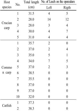
채집된 시료는 잉어 Cyprinus carpio 10마리 (평균 체장: 35.8 cm ± 1.3 cm), 붕어 Carassius auratus 5마리
(평균체장: 29.7 cm ± 1.7 cm) 와 메기 2마리 (평균체 장: 37.8 cm ± 1.0 cm) 였으며, 현장에서 거머리 유무를 확인한 후 (Table 1) 70% 에탄올, 10% 중성포르말린 그리고 1.0% glutaraldehyde로 고정하였다.
Host
species No. Total length (cm)
No. of Leech on the operculum Left Rigth
Crucian carp
1 31.5 4 5
2 28.0 14 12
3 28.0 3 4
4 30.0 4 7
5 31.0 4 4
Commo n carp
1 35.7 2 0
2 37.0 2 4
3 35.0 5 1
4 34.0 7 5
5 37.0 2 3
6 38.5 0 0
7 35.5 0 0
8 37.0 0 0
9 37.0 0 0
10 32.0 0 0
Catfish 1 37.3 0 0
2 38.3 0 0
Table 1. Average number of leech on the gill from different fish species
조직학적 분석
실험실로 옮겨온 시료들은 모두 24시간 이내에 고정액을 교환하였다. 광학현미경 관찰을 위한 시료 들은 관찰 부위를 1 cm2의 크기로 다듬고, 동일한 고정액으로 8시간 동안 재고정하였다. 이후 시료들 은 24시간 동안 흐르는 물에 수세한 후 일반적인 파라 핀 포매법을 이용하여 포매하였다. 포매된 시료들은 3㎛의 두께의 절편을 제작하여 Herris’s hematoxylin-0.5%
eosin (H-E) 과 AB-PAS (pH 2.5) 염색을 시행하였다.
주사전자현미경 (SEM) 관찰을 위한 시료들은 0.5 cm2의 크기로 다듬고, 2.5% glutaraldehyde로 재고정 후 0.1M phosphate buffer로 수세한 후 1% osmium tetroxide (OsO4) 로 후고정하였다. 이후 에탄올을 이 용하여 단계적으로 탈수 시키고, amyl acetate로 치환 하였다. 이들 시료들은 CO2 가스를 이용하여 건조 및 금이온 증착을 통하여 SEM (JSM-7500F, Hitachi, Japan) 으로 관찰하였다.
유전학적 분석
거머리의 유전학적 동정을 위하여 거머리 일부를 절제하여 DNA 분리에 사용하였다. DNA 분리는 High pure PCR template preparation kit (Roche, Germany) 를 이용하여 제조사에서 제공된 매뉴얼에 따라 DNA 를 분리하였다. 분리된 DNA를 template로 하여 universal 18S rRNA primer (F: 5'-AGATTAAGCCATGCATGCGT-3', R:
5'GCAGGTTCACCTACGGAAA-3') 를 이용하여 PCR을 실시한 후 product의 염기서열을 분석하여, NCBI web site (www.ncbi.nlm.nih.gov) 에서 유전학적 분류를 시도하였다.
결 과
충북 청원군 병천천에서 채집된 시료 중 붕어 5마 리에서 모두 거머리가 관찰되었고, 잉어는 10마리 중 5마리에서 거머리의 기생이 확인되었으나, 메기 에서는 관찰되지 않았다. 또한, 붕어 및 잉어 모두 30cm 전후의 2년생어 이상에서 기생하고 있음이 확 인되었다 (Table 1). 채집된 붕어의 아가미뚜껑에서 는 어류거머리의 일종인 L. sinensis가 평균 12마리가 기생하고 있었고, 거머리가 가장 많이 부착되어 있는 어체는 양쪽아가미 뚜껑에 26마리가 기생하였다. 감
염 잉어에서는 평균 6마리의 거머리가 기생하였고, 거머리가 가장 많이 부착되어 있는 어체는 12마리가 기생하였다 (Figs. 2A and B). 감염된 붕어, C. auratus 는 체색이 검게 변하고 체표손상이 보이지 않았지만
Fig. 2. The leech, Limnotrachelobdella sinensis on the gill of the common carp Cyprinus carpio (A) and crucian carp, Carassius auratus (B). A and B:
Attached leech on the inner surface of the operculum of the host. C: Affected gills of crucian carp showing severe necrosis (asterisk) and anemia (arrowhead).
아가미뚜껑 양쪽 모두 벌어져 있었다. 거머리가 기생 하고 있는 붕어의 경우 아가미가 창백하게 변하여 육안적으로도 빈혈 증세를 확인할 수 있었고, 새엽의 말단부위는 괴사로 인하여 검은색을 띠고 있었다 (Fig. 2C).
거머리의 흡혈로 인하여 아가미는 심각한 손상이 나타났다. 새엽 상피세포의 증가로 인하여 새엽의 기저부는 인접한 새엽과 붙어 있었으며, 새판에 존재 하는 모세혈관의 확장 및 부분적인 새판의 파괴가 관찰되었다 (Figs. 3A and B). 거머리의 흡혈활동으로 인하여 새판 일부분과 새엽이 소실되고, 새판 상피세 포들은 수종변성 (hydrophic degeneration) 과 함께 울 혈이 관찰되었다. 또한 새엽의 모세혈관에는 다수의 혈구들이 존재하고 있었다 (Figs. 3C and D). 새판 및 새엽 상피세포의 증식으로 인하여 새판의 융합이 발생하게 되면 새판의 구분이 불분명해졌으며, 새판 에 존재하는 연골기질이 소실되어 연골모세포만 남 아있는 조직상이 관찰되었다. 모세혈관에 존재하는 다량의 혈구들은 융합된 새엽들 사이에서도 관찰되 었다 (Figs. 3E and F). 새엽의 말단부위에는 모든 새판 이 융합되었으며, SEM으로 관찰한 결과 새판 하나가 원통모양으로 변형되었다 (Figs. 4A and B). 새판 상피 세포의 증식이 계속 진행되면 새판융합과 함께 인접 하는 새엽도 융합되었다. 새판 상피세포들은 모두 괴사되었고, 모세혈관은 파괴되었다. 새판의 연골기 질은 괴사되어 응축된 연골모세포만 존재하였다 (Figs. 4C and D). SEM으로 붕어 아가미 표면을 관찰 한 결과 새엽의 자유면에 존재하는 미세융기 (microridge) 가 부분적으로 파괴되었으며, 거머리의 흡혈로 인하 여 손상된 부분도 관찰되었다 (Fig. 5A). 또한 새엽 상피층이 파괴된 부분에는 연골기질이 외부로 노출 되었으며, 주변에는 출혈로 인하여 다수의 혈구들이 산재하고 있었다 (Fig. 5B).
Fig. 3. Histopathological gill lesions of the crucian carp, Carassius auratus. A, C and E: Scanning electron microscopy.
B, D, F: Light microscopy. H-E stain. A and B: Hyperplasia of the epithelial cell of gill filaments (asterisk) and absence of blood cells in the capillaries (arrow head). C and D: Gill lamellae (Gl) fusion (asterisk), gill lamellae distortion and hypertrophy of the epithelial cell of the lamellae (arrowhead).
E and F: Gill lamellar fusion (arrowhead) and hyperemia in gill filaments. Cl, capillary lumen.
Fig. 4. Histopathological gill lesions of the crucian carp, Carassius auratus. A and C: Scanning electron microscopy.
B and D: Light microscopy. H-E stain, AB-PAS (pH 2.5) reaction. A and B: Gill lamellae fusion and increased blood cell in the capillary lumen. C and D: Gill filament (Gf) fusion (black asterisk), diappearance of the extracellular cartilaginous matrix (white asterisk). Gl, gill lamellae.
Fig. 5. Histopathological gill lesions of the crucian carp, Carassius auratus. A: Short irregular microridges (Mr) and partially disrupting. B: Exposure extracellular cartilaginous matrix (Ecm) by the destruction of the filament epithelial cell. Bc blood cell.
L. sinensis의 18S rRNA 분석에 의한 유적학전 분류 를 시도하였으나 현재까지 어류거머리들의 유전정 보가 다양하게 등재되어 있지 않은 실정으로 Piscicolidae (어류거머리 속) 의 다른 종으로 동정되어, 형태학적 분류와 일치하지 않았다.
고 찰
병천천의 붕어 및 잉어에 기생하는 거머리 L.
sinensis는 1월부터 관찰되기 시작하여 5월 중순까지 관찰되었고, 이후부터는 어체에서 관찰되지 않았다.
L. sinensis는 중국과 일본에서는 보고되어 있으며, 주로 겨울과 봄에 L. sinensis의 피해사례가 보고된 바 있다 (Yang, 1987; Ogawa et al., 2007). L. sinensis의 생활사는 아직 명확하게 보고된 바 없지만, 일반적으 로 어류에 기생하는 거머리는 자웅동체로 1년생이 며, 이들은 숙주에 기생하면서 교미를 하는데, 이때 다른 개체의 정자를 제공받아 체내 수정을 한다. 그리 고 숙주를 떠나 수초의 키틴질 cocoon에 점착성 알을 낳는다 (Lukin, 1976). 부화한 거머리는 첫 번째 숙주 를 찾기 전 1주 또는 더 이상의 기간을 수중에서 부유 생활을 한다 (Burreson, 2006). 본 연구의 붕어에서 관찰된 L. sinensis는 1월부터 5월 초까지 기생단계로 관찰되었지만, 5월 말부터 12월까지는 붕어에서 관
찰되지 않아, 이 시기는 부화, 부유생활 및 다른 숙주 에서 성장하는 단계로 생각된다. Ogawa 등 (2007) 도 이들이 1년생으로서 12월부터 이듬해 4월까지 어 류에 기생하고, 그 이후의 기간에는 감염성이 없는 상태로 존재하는 것 같다고 보고하였는데, 이는 국내 에서 발생하는 경향과 매우 유사하다.
L. sinensis는 최초로 1896년 중국 Chinese carp에서 분리되어 Blanchard에 의해 T. sisnesis로 명명되어 보 고되었다. 이후 L. sinensis는 극동아시아 (중국, 러시 아, 일본 및 한국) 에서 보고되었는데, 중국에서는 Anhui, Jiangsu, sunan과 Heilongjiang 지역의 잉어, C. carpio와 붕어, C. auratus에서 보고되었고 (Yang, 1987), 러시아 에서는 Amur강과 Khanka호의 Amur carp C. carpio haematopterus와 Prussian carp C. auratus gibelio에서 보고되었다. 일본에서는 1910년 Tokyo만에서 T.
sisnensis로 분류하여 처음 보고하였으나 (Oka, 1910), 이후 형태 및 서식 특성 등 재분류하여 T. okae (현재 L. okae) 로 재명명되었고 (Moore, 1924), 2000년 Yodo 강의 crucian carps C. autatus langsdorfii와 C. cuvieri에 서도 관찰되었다 (Ogawa et al., 2007). 본 연구를 통해 서 국내 잉어에서도 처음으로 L. sinensis가 기생함이 확인되었고, 현재까지 결과에 의하면 L. sinensis는 잉 어과 어류에 종 특이성이 있는 기생성 거머리로 생각 된다. 또한, 잉어과 어류 중에서도 L. sinensis에 의한 감염성 및 피해정도는 붕어가 잉어보다 폐사량이 많 고, 아가미 손상도 심하게 나타났다.
어류의 아가미는 수질 환경뿐만 아니라 다양한 병원체에 의해서 아주 민감하게 반응하는 중요한 기 관이다 (Bhagwant and Elahee, 2002). 어류 아가미의 조직병리학적인 증상들은 크게 퇴행성 병변 (regressive changes) 과 진행성 병변 (progressive changes) 으로 나눌 수 있는데, 퇴행성 병변은 아가미 손상의 원인이 제거되더라도 쉽게 회복되지 않는 증상으로서 상피
세포의 수종변성 및 탈락, 공포화, 새판, pillar cell, 점액세포의 괴사 등이다. 진행성 병변은 손상의 원인 이 제거되면 회복이 가능한 증상들로서 상피세포의 비대, 세포 표면의 작은 돌기형성, 점액세포의 증가, 새엽의 곤봉화 현상 등이다. 이러한 증상 중에서 상피세 포의 수종변성은 염증에 반응하기 위한 방어기작이며, 상피세포의 비대 및 세포표면의 작은 돌기형성은 물리, 화학적인 스트레스에 의한 일차적인 반응이다. 또한 점액세포의 증가 및 새엽상피세포의 증식으로 인한 새 판 융합은 만성적인 세균과 기생충 혹은 화학물질 노출 에 의해서 발생될 수 있다 (Takashima and Hihiya, 1995).
미세융기 구조는 새엽의 pavement cell 표면에 존 재하는 지문모양의 구조로서 점액세포에서 분비된 점액물질들이 아가미 표면에서 제거되지 않고 유지 시켜 주는 기능을 한다. 따라서 아가미를 통하는 물의 흐름을 원활하게 하며, 점액물질이 유지됨에 따라 병원체의 침입으로부터 보호할 수 있게 도와주는 역 할을 한다 (Olson, 1995; Eiras-Stofella et al., 2001;
Evans et al., 2005). 본 연구의 붕어들은 부분적으로 아가미 표면의 미세융기 구조들이 파괴되었다. 따라 서 이러한 구조의 손상은 점액세포의 유지를 방해하 여 병원체가 쉽게 침입할 수 있는 계기가 된다.
L. sinensis가 숙주 아가미뚜껑 표피에 부착 기생하 면서 흡혈활동을 하게 되면 거머리의 두께만큼 아가 미뚜껑이 열리게 되며, 이로 인하여 아가미는 수중에 계속 노출되게 된다. 또한 아가미의 손상으로 인하여 부족한 산소를 보충하기 위해 붕어들은 수면에 머무 르며 입오름 현상을 보이는데 이때 열려 있는 아가미 뚜껑 때문에 아가미와 공기가 직접적으로 맞닿으면 아가미 조직의 손상은 더욱 심화되는 것으로 생각된 다. 하지만 L. sinensis에 감염된 붕어는 흡혈활동에 의한 물리적 자극으로 아가미에 1차적 손상이 되고, 손상된 아가미 조직에 다른 병원체에 2차 감염되어,
폐사에 이를 것으로 생각되어진다.
L. sinensis의 18S rRNA 분석에서 유전학적 분류가 형태학적 분류와 일치하지 않고 불분명하였는데, Ogawa 등 (2007) 도 어류거머리들의 유전학적 분류 에서 이들 종간의 유전적 계통 관계는 분명하지 않아, 더 많은 유전학적 정보가 요구된다고 보고하여 Piscicolidae의 다양한 유전정보 연구 및 공유의 필요 성을 제시하였다.
요 약
충북 청원군 병천천에서 채집된 시료 중 붕어 5마 리에서 모두 거머리가 관찰되었고, 잉어는 10마리 중 5마리에서 거머리의 기생이 확인되었다. 잉어에 서의 거머리 기생은 국내에서 처음으로 확인되었다. 붕어 아가미는 거머리의 흡혈로 인하여 새엽 상피세 포의 증가, 새판에 존재하는 모세혈관의 확장,새판 일부분과 새엽이 소실, 새판 상피세포들은 수종변성 (hydrophic degeneration), 울혈, 새판 및 새엽 상피세포 의 증식 등이 관찰되었다. SEM으로 관찰한 결과 새 판이 인접하는 새판과 점진적으로 융합되어, 결국은 새엽 하나가 원통모양으로 변형되었다. 또한, 붕어 아 가미 새엽의 자유면에 존재하는 미세융기의 부분적인 파괴가 관찰되었다. 새엽 상피층이 파괴된 부분에는 연골기질이 외부로 노출되었고, 그 주변에는 다수의 혈구들이 산재하고 있었다. L. sinensis의 18S rRNA 분석은 형태학적 분류와 일치하지 않았다.
감사의 글
본 연구는 국립수산과학원 (수산동물질병 역학 및 진단연구 : RP-2011-AQ-082) 에 의하여 수행되었습 니다.
참고문헌
Bhagwant, S. and Elahee, K.B.: Pathologic gill lesions in two edible laggon fish species,l Mulloidichthys flavolineatus and Mugil cephalus, from the bay of Poudre d’Or, Mauritius. Western Indian Ocean J. Mar. Sci., 1:35-42, 2002.
Burreson, E.R.: Phylum Annelida: Hirudinea as vectors and disease agents. In: Fish diseases and disorders.
Vol. 1. Protozoan and metazoan infections. 2nd edition. Woo, P.T.K., CAB International, Wallingford, pp. 566-591. 2006.
Eiras-Stofella, D.R. and Fank-de-Carvalho, S.M.:
Morphology of gills of the seawater fish Cathorops spixii (Agassiz) (Ariidae) by scanning and transmission electron microscopy. Revta. Bras.
Zool., 19:1215-1220, 2002.
Evans, D.H., Piermarini, P.M. and Choe, K.P.: The multifunctional fish gill: dominant site of gas exchange, osmoregulation, acid-base regulation, and excretion of nitrogenous waste. Physiol. Rev., 85:91-177, 2005.
Lukin, E.I.: Leeches of the fresh and salt water basins.
In Leeches. Fauna of USSR, Leninigrad, Nauka, Vol. 1, pp. 305-315. 1976.
Moore, J.P.: Notes on some Asiatic leeches (Hirudinea) principally from China, Kashimir, and British India.
Proc. Acad. Nat. Sci. Philad., 76:343-388, 1924.
Nagasawa, K., Park, S.-W., Kim, Y.-G. and Kim, H.J.:
Limnotrachelobdella sinensis, a leech associated with mortality in a wild population of Japaneses curcian carp Carassius cuvieri in Korea. J. Grad.
Sch. Biosp. Sci., 48:49-53, 2009.
Ogawa, K., Rusinek, O. and Tanaka, M.: New record of the leech Limnotrachelobdella sinensis infecting freshwater fish from Japanese water. Fish Pathol., 42:85-89, 2007.
Oka, A.: Synopsis der Japaneschen Hirudineen, mit Diagnosen der neuen Species. Annot. Zool. Japon., 7:165-182, 1910.
Olson, K.R.: Scanning electron microscopy of the fish gill.
In Fish Morphology: Horizon of New Research, Munshi, J.S.D., Dutta, H.M., Oxford and IBH Publishing Co. Pvt. Ltd, New Delhi, India, pp. 31-45.
1995.
Park, M.A., Kim, S.R., Kim, M.S., Kim, J.H. and Park, J.J.: Histopathological observation of the crucian carp, Carassius auratus by the leech, Limnotrachelobdella sinensis. J. Fish Pathol., 23:399-407, 2010.
Park, S.-W. and Kim, Y.-G.: The case report on the leech, Trachelobdella sp. infestation in wild crucian carp (Carassius cuvieri) of Chungnam province in Korea. J. Fish Pathol., 15:117-119, 2002.
Rhee, J.K.: Trachelobdella sinensis Blanchard, 1986 found from Cyprinus carpio nudus in Korea. Korean J. Parasitol., 24:216-217, 1986.
Takashima, F. and Hibiya, T.: V. Gills. In An atlas of fish histology: normal and pathological features, Kodaxnsha Ltd., Tokyo, pp. 66-71. 1995.
Yang, T.: On the genus Limnotrachelobdella Epshtein, 1968 and a new species from south China sea.
Acta Hydrobiol. Sin., 11:268-273, 1987.
Manuscript Received : August 18, 2011 Revised : September 19, 2011 Accepted : September 19, 2011