유향 추출물이 혈관내피세포 부착단백질 발현에 미치는 영향
이숭인1․권강범1․한종현2․류도곤1,3*
1 : 원광대학교 한의과대학 한방생리학교실, 2 : 약리학교실, 3 : 한국전통의학연구소
Effects of Olibanum Extracts
on Vascular Cell Adhesion Molecules Expression
Soong In Lee1, Kang Beom Kwon1, Jong Hyun Han2, Do Gon Ryu1,3*
1 : Department of Physiology, 2 : Department of Pharmacology, College of Oriental Medicine, Wonkwang University, 3 : Reserch Center of traditional Korean medicine
In order to validate the use of Olibanum as an anti-inflammatory drug in the traditional Korean medicine, I have investigated the effect of water-soluble extract of Olibanum (EO) on the expression of pro-inflammatory vascular cell adhesion molecule-1 (VCAM-1) in human umbilical vein endothelial cells (HUVECs) stimulated with tumor necrosis factor-α. The extract inhibited dose-dependently VCAM-1 expression without its cytotoxic effect on HUVECs, as measured by a flow cytometer using fluorescence-enhanced anti-VCAM-1 antibody, and significantly decreased mRNA levels of VCAM-1, as determined using reverse transcription polymerase chain reaction. These results suggest that Olibanum may have therapeutic potential in the control of endothelial disorders caused by inflammation.
Key words : olibanum, human umbilical vein endothelial cells, vascular cell adhesion molecule-1
* 교신저자 : 류도곤, 전북 익산시 신용동 344-2 원광대학교 한의과대학
․E-mail : tkryu@wku.ac.kr, ․Tel : 063-850-6846
․접수 : 2011/03/04 ․수정 : 2011/03/23 ․채택 : 2011/05/30
서 론
乳香(Olibanum)은 橄欖科(감람나무과 ; Burseraceae)에 속한 矮小灌木인 乳香나무 및 同屬 近緣植物의 膠狀樹脂로 性味는 辛.
苦, 溫 無毒하고 心.肝.脾 三經에 작용하며 活血止痛. 伸筋. 消腫.
生肌 등의 효능이 있어 痛經, 經閉, 胃脘痛,風濕痺痛, 腸癰, 瘡癰 久潰不斂 등의 병증을 치료하는 것으로 알려져 있다1). 실험적으 로 乳香은 면역조절효과2,3), 세포고사 유도효과4,5)등 다양한 효능 이 있는 것6-9)으로 보고되고 있다.
혈관염은 혈관에 염증이 발생하여 혈관내경이 좁아지고 조 직의 허혈 및 괴사를 유도하는 질환으로 크기, 위치 및 형태에 관계없이 모든 혈관을 침범하므로 다양한 임상소견을 초래한다
10-16). 대식세포, 혈관 평활근세포, T-림프구, B-림프구 등의 여러
세포들과 여기에서 분비되는 TNF-α (tumor necrosis factor-alpha), interleukin 등의 여러 cytokine과 성장인자 및 화 학주성인자들이 복합적으로 혈관에서의 염증발생에 관여하는 것 으로 보이며 특히, TNF-α는 혈관 내피세포를 자극하여 IL-8
(interleukin-8) 및 MCP-1 (monocyte chemoattractant protein-1) 과 같은 chemokine을 생성시키고 세포 표면에 VCAM-1 및 ICAM-1과 같은 세포부착분자의 발현을 유도하여 초기 혈관 염 증반응을 매개한다. 이러한 ICAM-1 및 VCAM-1은 혈관 내피세 포에 면역세포들이 부착할 수 있도록 유도한다.
특히 VCAM-1은 혈관 염증질환 발생시에 과량으로 발현 되어 과다한 염증반응을 유발하게 된다. VCAM-1의 발현은 유 전자 수준에서 조절이 가능하며, 일부 혈관염 치료제는 이러한 특성을 이용하여 발현을 억제시킴으로써 효과적인 약효를 나 타낸다17-24).
본 연구에서는 인체 내피세포(HUVEC)에서 세포 부착 분자 의 생성에 대한 乳香 추출물(EO, extract of Olibanum)의 유의성 있는 결과를 얻었기에 이를 보고하고자 한다.
재료 및 방법
1. 시약 및 재료
배양배지 (RPMI 1640), antibiotic/antimycotic 및 Trypsin-EDTA는 Gibco/BRL (Grand Island, NY, USA)로부터 구입하였다. SRB (Sulforrhodamine B)는 Molecular Probe 사
TNF-α (tumor necrosis factor-alpha) cytokine은 R&D (Minneapolis, MN, USA) 사로부터 구입하였고 ICAM-1 (intracellular adhesion molecule-1) 및 VCAM-1(vascular cell adhesion molecule-1) 항체는 Santa Cruz 사 (Santa Cruz, CA, USA)로부터 구입하여 사용하였다. Sodium bicarbonate 및 기본 시약들은 Sigma 사 (St. Louis, Missouri, USA)로부터 구입하여 사용하였다. 모든 세포배양 용기 및 튜브는 Falcon 사 (Becton Dikinson, San Jose, CA, USA)로부터 구입하여 사용하였다.
ICAM-1 및 VCAM-1 ELISA Kit는 R&D 사로부터 구입하여 사 용하였다.
2. 세포배양
인체 배꼽정맥 내피세포 (human umbilical vein endothelial cells ; HUVEC)를 Clonetics 사 (Cambrex Bio Science Rockland, USA)로부터 구입하여 10% FBS, antibiotic/antimycotic (100 U/
㎖ penicillin, 25 ㎍/㎖ amphotericin D, 100 ㎍/㎖
streptomycin) 및 sodium bicarbonate가 첨가된 RPMI 1640 배지 에서 95% 공기, 5% CO2, 습기가 충분한 37℃의 대기로 배양하였 다. 세포는 10 cm 배양접시에 2×10⁶ 개가 되도록 2-3일에 한 번 씩 분주하여 배양하였다.
3. 유향 물 추출물 제조
본 연구에 사용된 乳香은 원광대학교 익산 한방병원에서 구 입하여 사용하였다. 유향 100 g과 3차 증류수 0.9 L를 둥근바닥 플라스크에 넣고 냉각기를 부착한 다음 3시간 동안 전탕한 후 3,000 rpm에서 20분간 원심분리 한 상층액을 취해 회전 진공 농 축기로 감압 농축하였다. 농축된 시료는 동결 건조기에서 건조하 여 18.8 g을 얻은 후 DMSO에 녹여 사용하였다.
4. ICAM-1 및 VCAM-1의 ELISA 분석
HUVEC 세포 표면 부착 분자 (ICAM-1, VCAM-1)의 발현을 ELISA (enzyme linked immunosorbant assay) kit를 이용하여 분석하였다. 모든 분석과정은 지침서의 순서에 따라 수행하였다.
5. Western Blotting
포집된 세포를 PBS (phosphate-buffered saline)으로 세척한 다음 세포 파쇄용액 (50 mM Tris, pH 8.0, 110 mM NaCl, 5 mM EDTA, 1% Triton X-100, PMSF 100 ㎍/㎖)으로 용혈시킨 다음 원심분리하여 단백질을 얻었다. Bradford 분석방법을 이용하여 정량한 다음 60 ㎍을 취하여 동량의 sample buffer (125 mM Tris pH 6.8, 4% SDS 20% glycerol, 10% 2-mercaptoethanol) 혼합한 다음 100℃에서 5분 동안 가열하여 단백질 변성을 유도하였다.
변성된 단백질을 1.2% acrylamide gel에서 전기영동을 수행한 다 음 nitrocellulose membrane (Amersharm Pharmacia 사)으로 전 위시키고 5% skim milk/TBS-T로 상온에서 1시간 동안 반응시켜 비특이적인 항체반응을 억제시켰다. ICAM-1과 VCAM-1 및 β
시간 30분 동안 반응시키고 세척한 다음anti-mouse or goat IgG conjugated Horseradish peroxidase 이차 항체(secondary antibody)를 1% skim milk/TBS-T에서 1:5,000으로 희석하여 membrane과 상온에서 1시간 동안 반응시킨다. TBS-T로 세척한 다음 ECL reagent (ECL, Amersham, Buckinghamshire, England)로 발색시킨 다음 X-ray film에 감광시키는 방법으로 분 석하였다.
6. RT-PCR
전체 RNA를 Trizol reagent (Gibco/BRL)를 이용하여 분리 하고 Gene Quant (Amersham Pharmacia Biotech, Freiburg, Germany)로 정량하였다. 5 ㎍ RNA를 취하여 70℃에서 불활성 화 시킨 다음 1X 1st strand buffer, 0.5 mM dNTP, 15 ㎍/μL Oligo(dT), 10 U/μL reverse transcriptase 및 2 U/μL RNase inhibitor와 혼합하여 42℃에서 40분 동안 reverse transcription (RT)을 수행하여 cDNA를 합성하고 다시 70℃에서 10분 동안 불 활성화 시켰다. ICAM-1 및 VCAM-1 유전자 발현을 확인하기 위 하여 RT로부터 만든 cDNA를 1X PCR buffer, 0.2 mM dNTP, PCR primer 및 0.03 U/μL Taq Polymerase를 혼합하여 polymerase chain reaction (PCR, GeneAmp RNA PCR kit, erkin-Elmer Corp., Weiterstadt, Germany)을 시켜 분석하였다.
PCR 수행시 온도 및 시간은 94℃/1 min (1cycle); 94℃/ 1 min, 57℃/40 sec, 70℃/ 40 sec (27 cycles); 72℃/7 min이었고 각각 primer는 ICAM-1 : sense 5‘-TGA AGG CCA CCC CAG AGG ACA AC-3’, antisense 5’ATT ATG ACT GCG GCT GCT GCT ACC-3’, VCAM-1 : sense 5’-CCC TTG ACC GGC TGG AGA TT-3’, antisense 5’-CTG GGG GCA ACA TTG ACA TAA AGT G-3’ 및 GAPDH sense 5‘-GAG TCA ACG GAT TTG GTC GT-3’, antisense 5’-GTT GTC ATG GAT GAC CTT GG-3’이었 다. PCR 후 얻어진 생성물을 0.5 ㎍/㎖ EtBr (ethidium bromide) 이 포함된 1.5% agarose gel 상에서 전기영동하여 각각 발현된 유전자의 band를 확인하였다.
7. 세포 염색
HUVEC 세포를 well당 1×10⁵ 개가 되도록 계측하여 4-well 에 seeding한 다음 10% FBS가 포함된 RPMI 1640 배지를 이용하 여 배양하였다. 자극 처리 후 70% Ethanol로 세포를 고정한 다음 PBS로 세척하고 SRB를 처리하여 상온에서 10분 동안 세포를 염 색하였다. 염색된 세포를 다시 PBS로 세척한 다음 형광현미경을 이용하여 조사하였다 (PALM, Microlaser Technologies, Bernried, Germany).
8. 통계분석
실험 결과는 mean±S.D.로 표시하였으며 유의성 검정은 student's t-test에 의하였으며 p 값이 0.05 이하인 것만 유의성이 있는 것으로 하였다.
결 과
1. 乳香의 세포독성 효과
HUVEC 세포에서 乳香 물 추출물 (EO)의 세포독성을 조사 하였다. EO을 최소 50 ㎍/㎖에서 최대 200 ㎍/㎖까지 처리한 다 음 24시간 후에 이들 세포를 CV 로 염색하여 세포독성 및 세포 수를 확인하였다. Fig. 1에 보인 바와 같이 HUVEC 세포는 EO의 처리농도 및 처리시간에 의해 어떠한 영향도 없었다. 즉, 세포독 성으로 보이는 세포형태 및 세포 수에 있어서 유의한 변화를 관 찰 할 수 없었다.
Fig. 1. Effects of EO on viability and cell number. HUVEC cells were cultured for 24 h in the absence or presence of EO (50-200 ㎍/㎖). After 24-h treatment, the cells were stained with CV, and then examined under an inverted microscope. There is no significant change in either cell morphology or cell number. Similar results were observed in three independent experiments.
2. 乳香이 ICAM-1 및 VCAM-1 발현에 미치는 효과
EO를 농도별로 HUVEC 세포에 24시간 전처리한 다음 TNF-α로 자극하고 24시간 후에 이들 세포를 ICAM-1 및 VCAM-1 항체로 각각 표지하여 세포부착분자들의 발현을 FACS 방법으로 조사하였다. Fig. 2에 보인 결과와 같이 EO는 ICAM-1 발현에 큰 변화를 주지 못했지만 VCAM-1 발현을 유의하게 감 소시켰다.
VCAM-1 발현 억제효과는 EO의 농도에 의존하고(Fig. 2B), EO의 전처리 시간에 의존함을 알 수 있었다(Fig. 3). EO의 VCAM-1 최대 억제효과는 농도 200 ㎍/㎖과 24시간 전처리 실 험에서 관찰되었다.
3. 乳香이 ICAM-1 및 VCAM-1 단백질 생성에 미치는 효과 EO를 농도별로 HUVEC 세포에 24시간 전처리한 다음 12 시간 TNF-α로 자극하고, 이들 세포로부터 단백질을 분리하여 ICAM-1 및 VCAM-1 단백질 생성 정도를 Western blot 방법으로 조사하였다.
Fig. 4에 보인 결과와 같이 EO는 TNF-α 자극에 의한 VCAM-1 단백질 생성을 억제시켰으나, ICAM-1 단백질 생성에 영향을 주지 못했다.
Fig. 2. Effects of EO on ICAM-1 and VCAM-1 expression in TNF-α -stimulated HUVEC cells.(A) The cells were pre-treated for 24 h with either PBS or EO at the concentrations ranging from 50 ㎍/㎖ to 200 ㎍/㎖, and then stimulated for 24 with 50 ng/㎖ of TNF-α. After staining the cells with either FITC-ICAM-1 antibody, the expression of ICAM-1 was determined by FACS. (B) The cells were pre-treated for 24 h with either PBS or EO at the concentrations ranging from 50 ㎍/㎖ to 200 ㎍/㎖, and then stimulated for 24 with 50 ng/㎖ of TNF-α. After staining the cells with either FITC-VCAM-1 antibody, the expression of VCAM-1 was determined by FACS. Values are the mean±S.E. of duplicate determinations from three separated experiments.
Fig. 3. Effects of EO pre-treatment on VCAM-1 expression in TNF-α -stimulated HUVEC cells.The cells were pre-treated for indicated times with either PBS or EFT at 200 ㎍/㎖, and then stimulated for 24 with 50 ng/㎖ of TNF-α. After staining the cells with FITC-VCAM-1 antibody, VCAM-1 expression was determined by FACS. Values are the mean±S.E. of duplicate determinations from three separated experiments.
Fig. 4. Effects of EO on the expressions of ICAM-1 and VCAM-1 proteins in TNF-α-stimulated HUVEC cells. HUVEC cells were pre-treated for 24h with either PBS or EFT at 200 ㎍/㎖, and then stimulated for 12 with 50 ng/㎖ of TNF-α. After staining the cells with either ICAM-1 or VCAM-1 antibody, their expressions were determined by Western blot analysis.
Similar results were observed in three independent experiments.
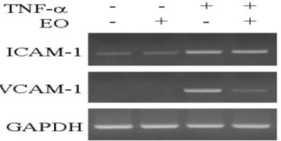
다음 6시간 TNF-α로 자극하고, 이들 세포로부터 mRNA를 분리 하여 EO가 ICAM-1 및 VCAM-1 mRNA 발현에 미치는 효과를 RT-PCR 방법으로 조사하였다. Fig. 5에 보인 결과와 같이 EO는 ICAM-1 mRNA 발현에 영향을 주지 못했으나, VCAM-1 mRNA 발현을 효과적으로 억제시켰다.
Fig. 5. Effects of EO on ICAM-1 and VCAM-1 mRNA expressions in TNF-α -stimulated HUVEC cells. The cells were pre-treated for 24 h with either PBS or EO at 200 ㎍/㎖, and then stimulated for 6 with 50 ng/㎖ of TNF-α. Each mRNA expression was determined by RT-PCR analysis. Similar results were observed in three separated experiments.
고 찰
乳香(Olibanum)은 活血止痛. 伸筋. 消腫. 生肌 등의 효능이 있어 痛經, 經閉, 胃脘痛, 風濕痺痛, 腸癰, 瘡癰久潰不斂 등의 병 증을 치료하는 것으로 알려져 있으며1)실험적으로 면역조절효과
2,3), 세포고사 유도효과4,5)등 다양한 효능이 있는 것6-9)으로 보고 되고 있다. 본 논문에서 혈관염 발생에 중요한 역할을 하는 부착 단백 질인 VCAM-1과 ICAM-1의 발현에 대한 乳香 추출물의 효 과를 조사하였다.
혈관염은 혈관에 염증이 발생하여 혈관내경이 좁아지고 조 직의 허혈 및 괴사를 초래하는 질환이다. 크기, 위치 및 형태에 관계없이 모든 혈관을 침범하므로 다양한 임상소견을 초래한다
11-16). 면역기전이 발병에 중요한 역할을 하고 있는 것으로 추정된
다. 면역복합체의 형성과 침착 및 그에 따른 보체와 중성구의 활 성화, 중성구, 라이소자임 효소에 대한 항체, 항내피세포 항체, 세포면역과 육아종형성, 감염, 종양 및 독소에 의한 혈관 손상 및 수반된 혈관 기능의 장애 등이 혈관염의 발병기전에 관여하는 것으로 생각되고 있다17,18,25-27).
대식세포, 혈관 평활근세포, T-림프구, B-림프구 등의 여러 세포들과 여기에서 분비되는 TNF-α, interleukin 등의 여러 cytokine, 여러 성장인자, 화학주성인자들 이 복합적으로 혈관에 서의 염증발생에 관여하는 것으로 보인다. 특히, TNF-α는 혈관 내피세포를 자극하여 IL-8 및 MCP-1과 같은 chemokine 생성을 유도하고 세포 표면에 VCAM-1 및 ICAM-1과 같은 세포부착분 자의 발현 유도하여 초기 혈관 염증반응을 매개한다17-23).
최근 연구결과에 의하면 TNF-α에 의해 활성화된 혈관 내피 세포에서 분비되는 chemokine의 생성을 차단시키거나 또는 세 포부착분자들의 발현을 차단시킴으로써 초기 혈관 염증반응을
에 따라 점차 용량을 줄여 나가게 된다. 스테로이드의 사용 기간이 나 부작용을 줄이기 위하여, 혈관 내피세포에서 분비되는 chemokine의 생성을 차단시키거나 또는 세포부착분자들의 발현을 차단시킬 수 있는 여러 가지 제제들이 병합 투여되기도 한다23,24). ICAM-1 및 VCAM-1은 혈관 내피세포에 면역세포들이 부착 할 수 있도록 한다. 특히 VCAM-1은 혈관 염증질환 발생시에 과 량으로 발현되어 과다한 염증반응을 유발한다. VCAM-1의 발현 은 유전자 수준에서 조절되며, 일부 혈관염 치료제는 이 유전자 의 발현을 효과적으로 억제시킴으로써 약효를 나타낸다. 본 실험 에서 乳香 추출물은 TNF-α에 의하여 증가한 VCAM-1의 발현을 유의하게 억제하였다. 그러나 ICAM-1의 발현은 억제하지 못하 였다. 乳香에 의한 VCAM-1의 발현 억제는 단백질뿐만 아니라 mRNA 수준에서도 나타났다. 본 실험의 결과로 乳香을 혈관염 의 치료제로서 개발하는데 실험적 기초를 마련하였다 할 수 있 으며 앞으로 혈관내피세포에서 DNA 수준에서의 乳香 추출물의 효과를 밝히고자 한다.
결 론
乳香 물 추출물이 혈관 내피세포의 세포부착분자 발현에 미 치는 효과를 조사하여 다음과 같은 결과를 얻었다.
200 ㎍/㎖의 乳香 추출물을 24시간 혈관 내피세포 (HUVEC)에 처리하였으나 어떤 독성도 관찰되지 않았으며 혈관 내피세포의 ICAM-1 발현을 억제시키지 못했지만, VCAM-1 발 현을 농도 및 전처리 시간 의존적으로 억제시켰다. 또한 乳香 추 출물은 VCAM-1 단백질의 발현을 억제시켰으나 ICAM-1 발현억 제에는 효과가 없었다.
이상의 실험결과로부터 乳香은 혈관 내피세포의 염증성 VCAM-1 유전자 발현을 저해시켜 항염증 효과를 나타내는 것으 로 사료된다.
감사의 글
이 논문은 2011학년도 원광대학교 교비지원에 의해서 수행 되었습니다.
참고문헌
1. 신민교. 임상본초학. 서울, 남산당, pp 739-740, 1997.
2. Pungle, P., Banavalikar, M., Suthar, A., Biyani, M., Mengi, S. Immunomodulatory activity of boswellic acids of Boswellia serrata Roxb. Indian J Exp Biol 41(12):1460-1462, 2003.
3. Badria, F.A., Mikhaeil, B.R., Maatooq, G.T., Amer, M.M.
Immunomodulatory triterpenoids from the oleogum resin of Boswellia carterii Birdwood. Z Naturforsch
58(7-8):505-516, 2003.
4. Hostanska, K., Daum, G., Saller, R. Cytostatic and apoptosis-inducing activity of boswellic acids toward malignant cell lines in vitro. Anticancer Res 22(5):
2853-2862, 2002.
5. Liu, J.J., Nilsson, A., Oredsson, S., Badmaev, V., Zhao, W.Z., Duan, R.D. Boswellic acids trigger apoptosis via a pathway dependent on caspase-8 activation but independent on Fas/Fas ligand interaction in colon cancer HT-29 cells. Carcinogenesis 23(12):2087-2093, 2002.
6. Sharma, S., Thawani, V., Hingorani, L., Shrivastava, M., Bhate, V.R., Khiyani, R. Pharmacokinetic study of 11-Keto beta-Boswellic acid. Phytomedicine 11(2-3):255-260, 2004.
7. Lakshmi Niranjan Reddy V., Ravinder, K., Srinivasulu, M., venkateshwar Goud, T., Malla Reddy, S., Srujankumar, D., Prabhakar Rao, T., Suryanarayana Murty, U., Venkateswarlu, Y. Two new macrocyclic diaryl ether heptanoids from Boswellia ovalifoliolata. Chem Pharm Bull 51(9):1081-1084, 2003.
8. Kimmatkar, N., Thawani, V., Hingorani, L., Khiyani, R.
Efficacy and tolerability of Boswellia serrata extract in treatment of osteoarthritis of knee a randomized double blind placebo controlled trial. Phytomedicine 10(1):3-7, 2003.
9. Culioli, G., Mathe, C., Archier, P., Vieillescazes, C. A lupane triterpene from frankincense (Boswellia sp., Burseraceae). Phytochemistry 62(4):537-541, 2003.
10. Park, Y.S., Lee, J.H., Bondar, J., Harwalkar, J.A., Safayhi, H., Golubic, M. Cytotoxic action of acetyl-11-keto-beta-boswellic acid (AKBA) on meningioma cells. Planta Med 68(5):397-401, 2002.
11. Uriarte, S.M., Molestina, R.E., Miller, R.D., Bernabo, J., Farinati, A., Eiguchi, K., Ramirez, J.A., Summersgill, J.T.
Effects of fluoroquinolones on the migration of human phagocytes through Chlamydia pneumoniae-infected and tumor necrosis factor alpha-stimulated endothelial cells.
Antimicrob Agents Chemother 48(7):2538-2543, 2004.
12. Yang, Y.Y., Hu, C.J., Chang, S.M., Tai, T.Y., Leu, S.J.
Aspirin inhibits monocyte chemoattractant protein-1 and interleukin-8 expression in TNF-alpha stimulated human umbilical vein endothelial cells. Atherosclerosis 174(2):
207-213, 2004.
13. Li, W.G., Gavrila, D., Liu, X., Wang, L., Gunnlaugsson, S., Stoll, L.L., McCormick, M.L., Sigmund, C.D., Tang, C., Weintraub, N.L. Ghrelin inhibits proinflammatory responses and nuclear factor-kappaB activation in human endothelial cells. Circulation 11;109(18):2221-2226, 2004.
14. Wagner, A.H., Guldenzoph, B., Lienenluke, B., Hecker, M.
CD154/CD40-mediated expression of CD154 in endothelial
cells: consequences for endothelial cell-monocyte interaction. Arterioscler Thromb Vasc Biol 24(4):715-720, 2004.
15. Murugesan, G., Sandhya Rani M.R., Gerber, C.E., Mukhopadhyay, C., Ransohoff, R.M., Chisolm, G.M., Kottke-Marchant K. Lysophosphatidylcholine regulates human microvascular endothelial cell expression of chemokines. J Mol Cell Cardiol 35(11):1375-1384, 2003.
16. Walton, K.A., Cole, A.L., Yeh, M., Subbanagounder, G., Krutzik, S.R., Modlin, R.L., Lucas, R.M., Nakai, J., Smart, E.J., Vora, D.K., Berliner, J.A. Specific phospholipid oxidation products inhibit ligand activation of toll-like receptors 4 and 2. Arterioscler Thromb Vasc Biol 23(7):
1197-1203, 2003.
17. Poddar, R., Sivasubramanian, N., DiBello, P.M., Robinson, K., Jacobsen, D.W. Homocysteine induces expression and secretion of monocyte chemoattractant protein-1 and interleukin-8 in human aortic endothelial cells: implications for vascular disease. Circulation 103(22):2717-2723, 2001.
18. Li, Y., Chi, L., Stechschulte, D.J., Dileepan, K.N.
Histamine-induced production of interleukin-6 and interleukin-8 by human coronary artery endothelial cells is enhanced by endotoxin and tumor necrosis factor-alpha.
Microvasc Res 61(3):253-262, 2001.
19. Volin, M.V., Harlow, L.A., Woods, J.M., Campbell, P.L., Amin, M.A., Tokuhira, M., Koch, A.E. Treatment with sulfasalazine or sulfapyridine, but not 5-aminosalicyclic acid, inhibits basic fibroblast growth factor-induced endothelial cell chemotaxis. Arthritis Rheum 42(9):
1927-1935, 1999.
20. Kayal, S., Lilienbaum, A., Poyart, C., Memet, S., Israel, A., Berche, P. Listeriolysin O-dependent activation of endothelial cells during infection with Listeria monocytogenes: activation of NF-kappa B and upregulation of adhesion molecules and chemokines. Mol Microbiol 31(6):1709-1722, 1999.
21. Lakshminarayanan, V., Beno, D.W., Costa, R.H., Roebuck, K.A. Differential regulation of interleukin-8 and intercellular adhesion molecule-1 by H2O2 and tumor necrosis factor-alpha in endothelial and epithelial cells. J Biol Chem 272(52):32910-32918, 1997.
22. Goebeler, M., Yoshimura, T., Toksoy, A., Ritter, U., Brocker, E.B., Gillitzer, R. The chemokine repertoire of human dermal microvascular endothelial cells and its regulation by inflammatory cytokines. J Invest Dermatol 108(4):
445-451, 1997.
23. Lukacs, N.W., Strieter, R.M., Elner, V., Evanoff, H.L., Burdick, M.D., Kunkel, S.L. Production of chemokines,
2767-2773, 1995.
24. Brown, Z., Gerritsen, M.E., Carley, W.W., Strieter, R.M., Kunkel, S.L., Westwick, J. Chemokine gene expression and secretion by cytokine-activated human microvascular endothelial cells. Differential regulation of monocyte chemoattractant protein-1 and interleukin-8 in response to interferon-gamma. Am J Pathol 145(4):913-921, 1994.
25. Zhao, B., Stavchansky, S.A., Bowden, R.A., Bowman, P.D.
Effect of interleukin-1beta and tumor necrosis factor-alpha
26. Fiuza, C., Bustin, M., Talwar, S., Tropea, M., Gerstenberger, E,. Shelhamer, J.H., Suffredini, A.F.
Inflammation-promoting activity of HMGB1 on human microvascular endothelial cells. Blood 101(7):2652-2660, 2002.
27. Unger, R.E., Krump-Konvalinkova V., Peters, K., Kirkpatrick, C.J. In vitro expression of the endothelial phenotype: comparative study of primary isolated cells and cell lines, including the novel cell line HPMEC-ST1.6R.
Microvasc Res 64(3):384-397, 2002.