Vol.27 No.1 p57-62, June. 2010
폐포단백증 1예
우대형⋅박정은⋅류영하⋅김현정⋅신경철⋅정진홍⋅이관호
영남대학교 의과대학 내과학교실A Case of Pulmonary Alveolar Proteinosis
Dae-Hyung Woo, Jung-Eun Park, Yung-Ha Ryu, Hyun-Jung Kim, Kyeong-Cheol Shin, Jin Hong Chung, Kwan Ho Lee
Department of Internal Medicine,
College of Medicine, Yeungnam University, Daegu, Korea
1)
-Abstract-
Pulmonary alveolar proteinosis (PAP) is a rare disorder that’s characterized by accumulation of surfactant components in the alveolar space. Idiopathic PAP is recognized as an autoimmune disease that’s due to impaired alveolar macrophage function and this caused by autoantibodies against granulocyte-macrophage colony-stimulating factor (GM-CSF). We report here a case of pulmonary alveolar proteinosis that was deemed interstitial lung disease at the initial diagnosis. A 61-year-old man presented with intermittent blood tinged sputum and dyspnea on exertion. The man was a painter for 30 years and he had a 10 pack-years smoking history. Chest computerized tomography (CT) revealed multifocal ground-glass opacity with interstitial thickening at both lungs. His pulmonary function tests and methacholine test revealed non specific results. He was diagnosed with interstitial lung disease on the basis of the chest CT finding and occupational history. However, seven months later, his symptoms progressed. Follow-up chest CT was performed. Wedge resection via video- assisted thoracoscopic surgery (the anterior basal segment of the left lower lobe) was done.
Microscopic examination showed large groups of alveoli with excessive amounts of surfactant and a complex mixture of protein and lipid (fat) molecules. Finally, he was diagnosed as having pulmonary alveolar proteinosis.
책임저자:신경철, 대구광역시 남구 대명5동 317-1, 영남대학교 의과대학 내과학교실 Tel: (053) 620-3850, Fax: (053) 654-8386, E-mail: [email protected]
Key Words: Pulmonary alveolar proteinosis, ChestCT
The FVC and FEV1 are normal. The FEV1/FVC ratio is relatively normal
Fig. 1. PFT results of the subject.
This result does not indicate an asthma component
Fig. 2. Methacholine challenge results of the subject.
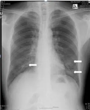
Ground glass opacities and fine reticular infiltration are noted at both lower lung fields.
Fig. 3. Initial chest X-ray of the subject.
서 론
폐포단백증은 폐포내에 PAS(Periodic acid- Schiff) 염색 양성인 불용성의 인지질이 풍부 한 단백질과 같은 물질이 폐포와 기관지에 침 착하여 가스 교환의 장애를 일으키는 것을 특 징으로 하는 질환으로 1958년 Rosen 등
1)에 의 해 27예가 처음 보고되었고, 국내에서는 1986 년 유 등
2)에 의해 1예가 보고 된 후 몇 차례 보고된 적이 있는 질환이다.
저자들은 30년 동안 페인트공으로 일해 왔 던 환자로, 약 2개월간의 운동시 호흡곤란을 주소로 내원하여 간질성 폐질환으로 진단하고, 치료하였던 환자가 증상의 호전이 없고 영상학 적으로 병변이 진행되어 비디오 흉강경하 폐생 검 검사로 폐포단백증으로 진단된1예를 경험하 였기에 문헌고찰과 함께 보고하는 바이다.
증 례
61세 남자가 내원 2달전부터 간헐적인 혈담
과 운동시에 심해지는 호흡곤란이 있어 본원
외래를 방문하였다. 내원 당시 의식은 명료하
였고, 객담과 기침은 없었으며, 호흡곤란도 없
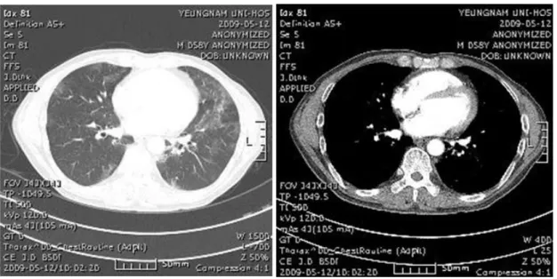
Fig. 5. Follow-up chest CT of the subject. The progression of multifocal ground glass opacities with interstitial thickening in both lungs is noted.
Fig. 4. Initial chest CT (09.05.12)of the subject. Multifocal ground glass opacities with interstitial thickening are predominately seen at both lower lungs.
었다. 직업력상 페인트공으로 30년간 일을 하였 으며, 10갑년의 흡연력이 있었다. 일반혈액검사 에서 백혈구 6,140 /mm
3, 혈색소 15 g/dL, 혈소 판 227,000 /mm
3이었으며, 총 IgE 3013 KU/L 및 간기능 검사를 포함한 혈액검사와 소변검
사에서 특이소견은 없었다. 시행한 폐기능검사
상 특이 소견은 없었고(Fig. 1), Methacholine
검사에서도 특이 소견 없었다(Fig. 2). 흉부엑
스선검사에서 양측 폐의 간유리성 음영과 미세
한 망상의 침윤이 관찰되었고(Fig. 3), 흉부전
Fig. 6. Gross findings of the wedge resection biopsy specimen of the left lower lobe of the subject. Protein deposition is noted in it.
Fig. 7. The pathology findings were large groups of alveoli with excessive amounts of surfactant and a complex mixture of protein and lipid(fat) molecules (A), positive PAS staining (B) and negative GMS staining (C), (D).
산화단층촬영(Fig. 4)에서는 양측 폐하에서 다 수의 폐실질이 두꺼워지는 양상으로 보여 간질 성 폐질환으로 진단되었다.
7개월 뒤 환자는 호흡곤란 증상 지속되어 외래를 방문 하였고, 외래에서 시행한 흉부전 산화단층촬영(Fig. 5)에서 병변이 진행하였으 며, 조직학적 확인을 위해 비디오 흉강경하 조 직검사를 시행하였다. 검체의 육안적 모습은 지방질이 침착되어 보였으며(Fig. 6), 조직학적 검사에서는 폐포내에 지방조직과 단백질이 침 착된 전형적인 폐포단백증의 소견을 보였고 (Fig. 7), PAS 염색에 양성, GMS(Grocott's Methenamine Silver) 염색에 음성 소견 있어
폐포단백증으로 진단되었다. 현재 외래로 정기적
C D
A B
진료를 받고 있으며 임상 증상의 호전을 보이 고, 호흡 곤란의 정도가 심하지 않아 전폐 세척 술과 GM-CSF(Granulocyte/macrophage colony stimulating factor) 사용은 하지 않고 있다.
고 찰
폐포단백증은 비정형적이고 불용성인 인지 질이 풍부한 단백질과 같은 물질이 폐포와 기 관지세지에 침착함으로서 산소교환의 장애가 일어나는 질환이다.
3)이 질환은 Rosen 등
1)에 의해 1958년 처음 기술되었는데 병리조직 소견 이 워낙 독특하였기 때문에 과거부터 존재하였 던 질환을 발견한 것이 아니라 새로이 생겨난 질환이라고 생각하였다.
1)그 동안 이 질병의 원인에 대해 수많은 논 란이 있어 왔으나, 1994년 GM-CSF가 결핍된 유전자 결손 쥐(knockout mice)에서 인간의 폐 포단백증과 비슷한 조직 양상을 보이는 것이 알려지면서부터 폐포단백증의 발병 기전 및 치 료에 있어 중요한 진전이 있어 왔다.
3, 4)폐포단 백증은 100만명 당 3.7명의 유병률을 보이는 드문 질환으로
5)국내에서는 1986년부터 30여 편의 증례보고가 있어 왔다.
6, 7)이학적 검사 소견은 특징적이지는 않으나 약 50%에서 흡기 시 수포음이, 25%에서 청색 증이, 그 외 적은 빈도로 곤봉형 수지가 나타 나기도 한다. 말초혈액검사, 생화학 검사, 소변 검사는 일반적으로 정상범위로 나타나며 LDH 수치는 종종 상승되는 결과를 보여 질병의 중 증도를 나타내는 지표가 되기도 한다.
3)폐기능 검사에서는 주로 제한성장애의 양상으로 나타 나고, 단순 흉부 방사선에서 양측폐를 비교적 비슷한 정도로 침범하고 폐문 주위에서 주변부
로 침윤해 들어가는 박쥐 또는 나비 모양의 소 견을 보이며, 고해상도 컴퓨터단층촬영시 때때 로 간유리 모양의 침윤과 소엽간중격의 비후로 인한 보도블록 모양의 양상을 보인다. 방사선 소견이 심함에도 불구하고 증상이 경미할 수도 있어 그 소견이 일치하지는 않는다.
4)진단은 기관지경하 또는 개흉 폐생검을 시 행하여 확진할 수 있는데, 전형적인 광학현미 경 소견은 폐포벽과 간질 조직이 잘 유지되며 폐포 내에 PAS 염색양성으로 호산성 과립상 물질이 관찰된다. 폐포 안에 변형된 포말상의 폐포 대식세포가 발견되고 면역조직화학염색에 서 표면활성 단백질 양성의 소견을 보인다. 때 때로 이차감염이 동반되는 경우 림프구 침윤에 의한 폐포벽 비후나 드물게 폐포벽 섬유화 소 견을 보이기도 한다.
4)최근에는 임상증상, 검 사 소견, 방사선소견 등에서 폐포단백증이 의 심되어지는 경우, 기관지 폐포세척술을 시행하 여 육안적으로 탁한 우유빛 또는 회색빛 세척 액이 다량으로 보이고, 세포학적 검사에서 과 립상의 비세포성 호산성 단백질이 포말상 대식 세포 및 전자현미경 검사로 층판상 소체를 나 타내는 표면활성 단백질을 증명함으로써 확진 한다.
8)최신 치료인 GM-CSF를 피하투여 또는 흡 입시키는 방법은 적어도 절반이상에서는 효과 가 있다고 보지만, 치료 전후의 GM-CSF 자가 항체의 정량적인 변화와 GM-CSF 투여량과 자가항체의 정량적 관계 등이 명료하게 밝혀져 야 할 필요성이 제기되고 있다. 장기간의 GM- CSF 사용이 골수에 미치는 영향, 폐 섬유화의 가능성 등 부작용에 대해 좀 더 연구되어져야 할 필요성이 있다.
8, 9)본 증례는 환자의 직업력과 흉부전산화단층
촬영으로 판단했을 때 간질성 폐질환으로 오인 되었던 환자로, 증상의 지속적인 악화와 영상 학적으로 병변이 진행하여, 흉강경하 조직검사 시행 후 폐포단백증으로 진단하였다. 흔한 질 병이 아니고, 문진과 흉부전산화단층촬영만으 로 진단을 하기 힘든 경우로 조직학적 검사가 확진에 필요함을 보여 주는 증례이다.
참 고 문 헌
1. Rosen SH, Castleman B, Liebow AA. Pulmonary alveolar proteinosis. N Engl J Med 1958 Jun5;
258(23): 1123-42.
2. 유덕종, 최정윤, 이원식, 정태훈, 곽정석, 손태중.
Pulmonary alveolar proteinosis 1예. 제63차 추 계 결핵학술대회 초록집 1986.
3. Stanley E, Lieschke GJ, Grail D, Metcalf D, Hodgson G, Gall JA, et al. Granulocyte/
macrophage colony stimulating factor-deficient mice show no major perturbation of hemato- poiesis but develop a characteristic pulmonary pathology. Proc Natl Acad Sci USA 1994 Jun7;91(12):5592-6.
4. Trapnell BC, Whitsett JA, Nakata K.
Pulmonary alveolar proteinosis. N Engl J Med 2003 Dec25;349(26):2527-39.
5. Seymour JF, Presneill JJ. Pulmonary alveolar proteinosis: progress in the first 44 years. Am J Respir Crit Care Med 2002 Jul;166(2):215-35.
6. Kim G, Lee SJ, Lee HP, Yoo CG, Han SK, Shim YS, et al. The clinical characteristics of pulmonary alveolar proteinosis. experience at Seoul National University Hospital, and review of the literature. J Korean Med Sci 1999;14:
159-64.
7. Seo JH, Bahk JH. Whole Lung Lavage and Extracorporeal Membrane Oxygenation in a Patient with Pulmonary Alveolar Proteinosis and Lung cancer. Korean J Anesthesiol 2005;
48:549-52.
8. Wang BM, Stern EJ, Schmidt RA, Pierson DJ. Diagnosing pulmonary alveolar proteinosis.
A review and an update. Chest 1997 Feb;111 (2):460-6.
9. Ioachimescu OC, Kavuru MS. Pulmonary alveolar proteinosis. Chron Respir Dis 2006;3 (3):149-59.