Article
http://dx.doi.org/10.4217/OPR.2020.42.1.015 March 2020
Scalarane-type Sesterterpenes from the Philippines Sponge Hyrtios sp.
Jae-Hyeong Choi
1,2, Hyi-Seung Lee
1,2*, and Wilfredo L. Campos
31
Marine Biotechnology Research Center, Korea Institute of Ocean Science & Technology Busan 49111, Korea
2
Department of Applied Ocean Science, University of Science and Technology Daejeon 34113, Korea
3
OceanBio Lab, University of the Philippines Visayas Iloilo 5023, Philippines
Abstract : The marine sponge Hyrtios sp. collected in the Philippines was extracted and partitioned. The resulting organic layer was purified by C
18reversed-phase column chromatography and HPLC to achieve the separation of nine scalarane-type sesterterpenes, including one new compound with eight known sca- larane analogs. The chemical structures of the isolated compounds 1−9 were elucidated by 1D and 2D NMR and MS data analysis. All nine compounds were evaluated for their antibacterial activities against three Gram-positive and three Gram-negative bacteria. The compound 3 exhibited potent antibacterial activities against Bacillus subtilis and Micrococcus luteus. The compounds 7 and 9 displayed considerable activities against Bacillus subtilis and the others had moderate results.
Key words : marine sponge, scalarane, sesterterpene, Hyrtios sp., antibacterial activities
1. Introduction
Marine organisms have received growing attention as potential sources for bioactive secondary metabolites exhibiting novel chemical structures. In particular, marine sponges are known to contain various classes of bioactive compounds, such as alkaloids, terpenoids, peptides, glycosides, and macrolides (Amina and Al Musayeib 2018). For example, spongistatin isolated from Hyrtios sp.
and halichondrin isolated from Halichondria okadai were previously reported as microtubule-destabilizing agents (Miller et al. 2010).
Scalarane-type sesterterpenoids are compounds pos- sessing a 6/6/6/6-tetracyclic or 6/6/6/6/5-pentacyclic fused ring system. Since scalarin, the first scalarane-type sesterterpene, was isolated from the sponge Cacospongia scalaris (Fattorusso et al. 1972), diverse subgroups of these skeletal structures have been discovered, including homoscalaranes (Fontana et al. 2000; Jaspars et al. 1997;
Roy et al. 2002) and isoscalaranes (Davis and Capon 1993; Song et al. 2008; De Rosa et al. 1994). Moreover, further research into the synthesis (Ungur and Kulciţki 2004) or semi-synthesis (Kamel et al. 2009) of the scaffolds of scalarane derivatives has been conducted.
Scalarane-type sesterterpenoids are commonly found in marine sponges belonging to the order Dictyoceratida (Liu et al. 2007; Gonzalez 2010), and are occasionally found in shell-less molluscs, such as nudibranchs (Gavagnin et al. 2004), likely due to their dietary source of sponges.
For this reason, scalarane-type sesterterpenoids have been regarded as a useful chemotaxonomical marker (Jaspars et al. 1997).
Scalarane-type sesterterpenoids exhibit an extensive spectrum of bioactivities, including anticancer, antibacterial, anti-inflammatory, and anti-fouling activities. In particular, the majority of scalaranic sesterterpenoids have a broad range of cytotoxicities against many cancer cell lines, and so play the key role as eco-physiological mediators in the chemical defensive mechanism of marine invertebrates (Gonzalez 2010).
*Corresponding author. E-mail : [email protected]
Herein, we report the isolation of a new compound with eight known scalarane-type sesterterpene derivatives (Fig.
1) that generally exhibit antibacterial activities against Gram-positive bacteria.
2. Materials and Methods
General experimental procedure
HPLC was performed using the YMC-Pack C
18column and a Shodex RI-101 detector. The optical rotations were obtained on a JASCO digital polarimeter.
1H and
13
C NMR spectra were recorded at 600 and 150 MHz on a Bruker spectrometer, respectively. UV spectra were measured using a Shimadzu UV-1650PC spectrophotometer.
IR spectra were recorded using a JASCO FT/IR-4100 spectrophotometer. High-resolution mass spectra (HRMS) were obtained with a Shimadzu LC/MS spectrometer at the Korea Basic Science Institute.
Biological material
The marine sponge of the genus Hyrtios (sample number 163PIL-229) was collected by hand using Scuba at a depth of 20 −30 m in April 2016, along the shore of Anda (N 9°43' E 124°31'), Bohol Island, the Philippines. The sponge was identified as Hyrtios sp. (order Dictyoceratida, family Thorectidae) by Dr. Young-A Kim. The specimen is stored in the Marine Biotechnology Research Center, Korea Institute of Ocean Science and Technology.
Extraction and isolation
The collected sponge Hyrtios sp. (1.0 kg, wet weight) was transported frozen and maintained at -20
oC until required for chemical analysis. The sponge was extracted twice with MeOH (2 L), and cut into pieces prior to extraction with DCM (2 L) and MeOH : DCM = 1 : 1 (2 L).
The extracts were then combined and the solvent was evaporated in vacuo to afford the crude extract (81.0 g), which was partitioned between n-BuOH and 3
rddistilled water to remove salts and water-soluble components. The n- BuOH layer (19.4 g) was further partitioned between n- hexane and 15% aqueous MeOH to give an aqueous MeOH layer (8.9 g). The 15% aqueous MeOH layer was separated into eight fractions by C
18reversed-phase flash column chromatography using a step gradient of MeOH/
H
2O (4:6, 5:5, 6:4, 7:3, 85:15, 1:0 v/v), then acetone and EtOAc. The 100% MeOH fraction was subjected to C
18reversed-phase flash column chromatography once again to give fractions mp 1 −24. Fraction mp 9 (75.0 mg) was purified by reversed-phase HPLC (YMC-Pack C
18, 250 × 4.6 mm, 55% MeCN in H
2O) to yield compounds 1 (4.7 mg), 2 (3.9 mg), and 5 (2.6 mg). Fraction mp 8 was purified using the same column (65% MeCN in H
2O) to give compounds 1 (6.4 mg), 2 (7.9 mg), and 8 (1.8 mg).
The combined fraction of mp 5 −7 due to their similar
1H
NMR spectra was purified under the same conditions
employed for mp 8 to afford compounds 6 (10.0 mg) and
8 (1.8 mg). Compound 7 (26.3 mg) was obtained from mp
Fig. 1. Structures of compounds 1 −9 isolated from Hyrtios sp.
11 using semi-preparative reversed-phase HPLC (YMC- Pack C
18, 250 × 10 mm, 65% MeCN in H
2O). Compounds 3 (8.0 mg) and 9 (5.5 mg) were purified from mp 14 by analytical reversed-phase HPLC (YMC-Pack C
18, 250 × 4.6 mm, 62% MeCN in H
2O). The combined fraction of mp 16 −17 was purified by analytical reversed-phase HPLC (YMC-Pack C
18, 250 × 4.6 mm, 65% MeCN in H
2O) to give compound 4 (7.0 mg).
Compound 1 : White amorphous solid; [α]
25D+10 (c 0.1, MeOH); IR ν
max3407, 2929, 1742, 1654, 1554, 1456, 1389, 1318, 1251, 1042 cm
-1; UV(MeOH) λ
max(log ε) 214 (3.68) nm; HRESIMS m/z 409.2716 [M + Na]
+(calcd for 409.2713, C
25H
38O
3Na
+);
1H NMR (CD
3OD and C
5D
5N, 600 MHz) and
13C NMR (CD
3OD and C
5D
5N, 150 MHz) see Table 1.
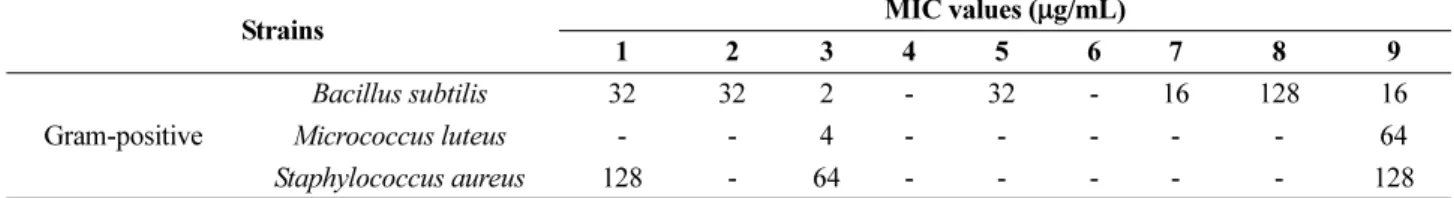
Minimum Inhibitory Concentration (MIC) assay The MIC assay is a useful method to evaluate the lowest concentration of antibacterial sources, as it is based on an antibacterial susceptibility test for bioactive natural products that cause inhibited bacterial growth or death.
The assay was carried out against six bacteria. The three Gram-positive bacteria, namely Bacillus subtilis (KCTC-1021), Micrococcus luteus (KCTC-1915), and Staphylococcus aureus (KCTC-1927), and three Gram- negative bacteria, namely Escherichia coli (KCTC-2441), Salmonella typhimurium (KCTC-2515), and Klebsiella pneumonia subsp. (KCTC-2690) were purchased from the Korean Collection for Type Cultures (KCTC). All six bacteria were streaked onto Mueller-Hinton agar (MHA) plates and cultured in a standing incubator for 24 h at 37
oC. A separated single colony was transferred from the MHA plates to Muller-Hinton broth, and incubated for 24 h at 37
oC and 170 rpm in a shaking incubator. In brief, the six bacteria were inoculated into 96-well plates and then treated with compounds 1 −9 diluted in DMSO (100 µL) in accordance with concentrations ranging from 128 to 0.25 µg/mL (i.e., 128, 64, 32, 16, 8, 4, 2, 1, 0.5, and 0.25 µg/mL). Subsequently, these mixtures were cultivated in a standing incubator for 24 h at 37
oC.
3. Results and Discussion
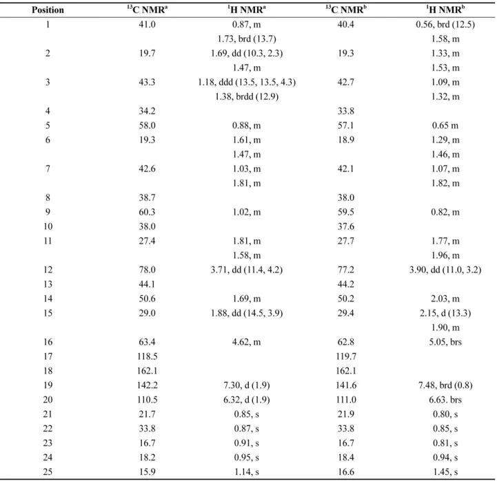
Compound 1 has a molecular formula of C
25H
38O
3, as deduced from its high-resolution electrospray ionization mass spectrometry (HRESIMS) data. The
1H NMR spectrum of 1 displayed resonance for two olefinic protons, seven methylene protons, five methine protons, and five
methyl groups (Table 1). The planar structure of 1 was determined by combined investigation of the COSY and HMBC correlations (Fig. 2). The COSY correlations of H
2-1/H
2-2/H
2-3, H
2-6/H
2-7, H-9/H
2-11/H-12, and H-14/
H
2-15/H
2-16 and the HMBC correlations from H
3-21 and H
3-22 to C-3, C-4 and C-5, from H
3-23 to C-1, C-5, C-9 and C-10, from H
3-24 to C-7, C-8, C-9 and C-14, and from H
3-25 to C-12, C-13, C-14 and C-18 confirmed the frame of a 6/6/6/6-membered ring system. The presence of a five-membered ring containing one oxygen atom was also elucidated by the HMBC correlations from H-19 to C-17, C-18 and C-20, from H-20 to C-17, C-18 and C-19, and from H-16 to C-17, C-18 and C-20. Furthermore, the HMBC correlations from H
3-21 and H
3-22 to C-3, C-4 and C-5 suggested the presence of a gem-dimethyl group on quaternary carbon C-4, thereby defining a 6/6/6/6/5- pentacyclic carbon skeleton. The structure of compound 1 shared the same carbon and heteroatom framework with 12-O-desacetylfuroscalarol, with the exception of differences in the stereochemistry at H-12. The unsaturated degree of compound 1 also indicated the presence of a pentacyclic ring system with a furan ring attached at C-17 and C-18.
Compound 1 has not been reported, but its 12-epimer has been already isolated from Hyrtios sp. and Hyrtios cf.
erectus (Cimino et al. 1978; Doi et al. 1993). The previously reported
1H and
13C NMR chemical shifts of the C-12 position in C
5D
5N (δ
C71.4 and δ
H4.62) were somewhat different from those of compound 1 (δ
C77.2 and δ
H3.90) in C
5D
5N. The stereochemistry of H-12 was therefore further investigated by the analysis of NOESY correlations (Fig. 3) and through comparison of the proton-proton coupling constants.
More specifically, the NOESY correlations of H-2/H- 11/H-22/H-23/H-25 and H-6/H-24/H-25 suggested that H-22, H-23, H-24, and H-25 are on the β-face, while the strong NOESY correlations of H-9/H-12/H-14 suggested that H-9 and H-14 are on the α-face and H-12 is an axial proton. In addition, the stereochemistry of H-12 (δ
H3.90) was confirmed as the down orientation by study of the coupling constant, which showed J value of 11.04 Hz due to the trans-diaxial coupling between the axial proton on H-12 (δ
H3.90) and the axial proton on H-11 (δ
H1.77);
such a coupling was not observed in the previously
reported 12-epimer (Lee et al. 2014). Furthermore, the
strong NOESY signals of H-14/H-15/H-16 indicated that
H-14, H-15, and H-16 present the same orientation as H-
12. A strong NOESY correlation was also observed
between H-12 and pseudoaxial proton H-16 due to the D
ring being a cyclohexene ring.
The other purified compounds, namely compounds 2 − 9, were previously reported to be sesterstatin 4 (2) (Pettit et al. 1998), heteronemin (3) (Kashman and Rudi 1977), 12-deacetyl-12-epi-deoxoscalarin (4) (Fontana et al. 1999), 16-hydroxyscalarolide (5) (Miyaoka et al. 2000), 16-O- deacetyl-16-epi-scalarolbutenolide (6) (Ryu et al. 1996), 12-O-deacetyl-19-deoxyscalarin (7) (Pettit et al. 1998), hyrtiosin A (8) (Yu et al. 2005), and hyrtiosal (9) (Iguchi et al. 1991) (Fig. 1). These known compounds were confirmed through a comparison of the obtained
1H NMR Table 1.
1H (600 MHz) NMR and
13C (150 MHz) NMR data for 1 (δ in ppm, J in Hz)
Position
13C NMR
a 1H NMR
a 13C NMR
b 1H NMR
b1 41.0 0.87, m 40.4 0.56, brd (12.5)
1.73, brd (13.7) 1.58, m
2 19.7 1.69, dd (10.3, 2.3) 19.3 1.33, m
1.47, m 1.53, m
3 43.3 1.18, ddd (13.5, 13.5, 4.3) 42.7 1.09, m
1.38, brdd (12.9) 1.32, m
4 34.2 33.8
5 58.0 0.88, m 57.1 0.65 m
6 19.3 1.61, m 18.9 1.29, m
1.47, m 1.46, m
7 42.6 1.03, m 42.1 1.07, m
1.81, m 1.82, m
8 38.7 38.0
9 60.3 1.02, m 59.5 0.82, m
10 38.0 37.6
11 27.4 1.81, m 27.7 1.77, m
1.58, m 1.96, m
12 78.0 3.71, dd (11.4, 4.2) 77.2 3.90, dd (11.0, 3.2)
13 44.1 44.2
14 50.6 1.69, m 50.2 2.03, m
15 29.0 1.88, dd (14.5, 3.9) 29.4 2.15, d (13.3)
1.90, m
16 63.4 4.62, m 62.8 5.05, brs
17 118.5 119.7
18 162.1 162.1
19 142.2 7.30, d (1.9) 141.6 7.48, brd (0.8)
20 110.5 6.32, d (1.9) 111.0 6.63. brs
21 21.7 0.85, s 21.9 0.80, s
22 33.8 0.87, s 33.8 0.85, s
23 16.7 0.91, s 16.7 0.81, s
24 18.2 0.95, s 18.4 0.94, s
25 15.9 1.14, s 16.6 1.45, s
a1H and 13C NMR spectra were measured in CD3OD
b1H and 13C NMR spectra were measured in C5D5N
Fig. 2. Key COSY and HMBC correlations of 1
and
13C NMR data with those reported in the literatures.
The minimum inhibitory concentration (MIC) assay method was then employed to test the antibacterial activities of nine compounds against three Gram-positive and three Gram-negative bacteria. The majority of compounds were highly active against Bacillus subtilis compared with the other bacteria examined. More specifically, compound 3 displayed the strongest activity against Bacillus subtilis and Micrococcus luteus with MIC values of 2 and 4 µg/mL, respectively. In addition, compounds 7 and 9 exhibited MIC values of 16 µg/mL against Bacillus subtilis. None of the nine compounds exhibited activity against the three Gram-negative bacteria.
4. Conclusions
Chemical analysis of the 1D and 2D NMR spectra recorded for nine scalarane-type sesterterpene derivatives isolated from the Philippines marine sponge Hyrtios sp.
led to the determination of their chemical structures. The carbon framework was found to belong to the 6/6/6/6/5- pentacyclic ring system-scalarane-type sesterterpenoids class. In addition, compound 1 was identified as a novel 12-epimer exhibiting an α-axial orientation at H-12 position. Known compounds were also isolated, and these were confirmed as sesterstatin 4 (2), heteronemin (3), 12- deacetyl-12-epi-deoxoscalarin (4), 16-hydroxyscalarolide (5), 16-O-deacetyl-16-epi-scalarolbutenolide (6), 12-O-
deacetyl-19-deoxyscalarin (7), hyrtiosin A (8), and hyrtiosal (9). The compounds 1 −9 were then investigated for their antibacterial activities against three Gram-positive bacteria (Bacillus subtilis, Micrococcus luteus, and Staphylococcus aureus) and three Gram-negative bacteria (Escherichia coli, Salmonella typhimurium, and Klebsiella pneumonia subsp.) using the minimum inhibitory concentration assay.
It was found that compound 3 showed potent activities against Bacillus subtilis and Micrococcus luteus, and the compounds 7 and 9 had moderate to good bioactivities against Bacillus subtilis. The other scalarane analogs displayed weak activities against the three Gram-positive bacteria, and none of the compounds exhibited activity against the three Gram-negative bacteria.
Acknowledgments
We would like to thank Dr. Young-A Kim (Hannam University) for identifying the sponge and the Korea Basic Science Institute in Ochang, Korea, for providing mass spectrometric data. This research was partially supported by the KIOST (PE99824), and the Ministry of Oceans and Fisheries, Republic of Korea (PM61620).
References
Amina M, Al Musayeib NM (2018) Biological and medicinal importance of sponge. Ray S (ed) In: Biological resource of waters. IntechOpen, London, pp 201−230
Fig. 3. Key NOESY correlations of 1
Table 2. MIC assay against Gram-positive bacteria
Strains MIC values ( µg/mL)
1 2 3 4 5 6 7 8 9
Gram-positive
Bacillus subtilis 32 32 2 - 32 - 16 128 16
Micrococcus luteus - - 4 - - - - - 64
Staphylococcus aureus 128 - 64 - - - - - 128
MIC values are the minimal inhibitory concentration inhibiting cell growth or inducing cell death