Ann Clin Microbiol Vol. 18, No. 2, June, 2015 http://dx.doi.org/10.5145/ACM.2015.18.2.52 pISSN 2288-0585⋅eISSN 2288-6850
Microbiological Characteristics according to Transudative and Exudative Effusion in Pleural Fluid Culture
Hyeun Gyeo Lee
1, Gyu Yel Hwang
1, Soon Deok Park
1, Young Uh
1, Juwon Kim
1, Kap Jun Yoon
1, Won-Yeon Lee
2Departments of
1Laboratory Medicine and
2Internal Medicine, Yonsei University Wonju College of Medicine, Wonju, Korea
A total of 1,132 pleural fluid culture results obtained from October 2012 to July 2014 were analyzed to eluci- date the microbiological characteristics according to transudative and exudative pleural fluid. The pleural fluid cultures were performed using aerobic and anae- robic blood culture bottles. The blood and pleural fluid for total protein, lactate dehydrogenase, and glucose measurement were submitted to laboratory at the same
time with pleural fluid cultures. The rates for culture positivity, anaerobes isolation, and polymicrobials be- tween transudative and exudative pleural fluid were 5.2% vs. 10.4%, 14.8% vs. 7.8%, and 14.8% vs. 10.9%.
(Ann Clin Microbiol 2015;18:52-55)
Key Words: Anaerobe, Culture, Exudate, Pleural fluid, Transudate
52
Received 16 October, 2014, Revised 11 February, 2015, Accepted 1 April, 2015
Correspondence: Young Uh, Department of Laboratory Medicine, Yonsei University Wonju College of Medicine, 162, Ilsan-dong, Wonju 220-701, Korea.
(Tel) 82-33-741-1592, (Fax) 82-33-731-0506, (E-mail) [email protected]
ⓒ The Korean Society of Clinical Microbiology.
This is an Open Access article distributed under the terms of the Creative Commons Attribution Non-Commercial License (http://creativecommons.org/licenses/by-nc/4.0) which permits unrestricted non-commercial use, distribution, and reproduction in any medium, provided the original work is properly cited.
흉막액(pleural fluid)은 누출액(transudate)과 삼출액(exudate) 으로 구분하면 발생 원인에 따른 진단과 치료 방침을 결정하는 데 도움을 줄 수 있다. 흉막액의 성상이 누출액일 경우에는 일 반적으로 추가 검사 없이 전신 질환에 대한 치료를 시행하지 만, 삼출액은 원인에 따른 치료가 필요하므로 세균, 결핵균, 진 균 등의 미생물배양과 C-반응단백, 아데노신탈아미노효소, 암 배아항원, 더 나아가 핵산증폭기반의 진단 검사를 추가하게 된 다[1].
흉막액의 누출액과 삼출액의 감별은 기본적으로 Light 기준 [2]을 적용하지만 검사 전 이뇨제를 사용하거나 흉수 적혈구수 가 10,000/mm3를 초과할 때는 누출액이 삼출액의 소견을 보일 수 있다[3]. 임상적으로 누출액이지만 흉막액과 혈청의 단백질 비가 0.5-0.65, 흉막액과 혈청의 젖산탈수소효소 비 0.6-1.0 사 이, 흉막액 젖산탈수소효소 수치가 혈청 젖산탈수소효소 참고 범위 상한치에서 2/3 이상인 경우에는 환자가 실제로 누출액의 선행 질환이 있는지를 확인해야 한다[4]. 그러므로 전신 질환에 의한 누출액이 의심되지만 Light 기준으로 누출액이 아닐 경우 에는 혈액과 흉수액의 단백질 차이를 계산하여 3.1 g/dL를 초 과하면 누출액으로 분류하고, 3.1 g/dL 이하일 때에는 혈액과 흉수액의 알부민 측정치의 차이가 1.2 g/dL를 초과하면 누출액 으로 분류하는 변경된 Light 기준을 제안하고 있다[4]. 또한 악
성 종양에 의한 흉수액은 드물게 울혈심부전과 같은 질환이 동 반되면 누출액의 양상을 보일 수 있다[5].
임상의는 흉수액을 세균 감염에 의한 삼출액으로 조기에 감별 진단하게 되면 신속하고 적절한 항균제 투여가 가능하게 되어 가슴관 삽입을 하지 않고 치료할 수 있다[6]. 그러나 흉막 감염의 초기에는 세균 침입 없이 삼출액이 생기게 되며, 이 시기에는 흉막액의 백혈구수도 낮고, 젖산탈수소효소 농도도 혈청 수치의 50% 이하이며 pH와 당 수치도 참고범위를 보이게 된다[7].
이에 본 연구에서는 누출액과 삼출액에서의 배양 결과를 비 교하여, 누출액에서 배양 양성인 검체에서의 특성을 분석하였 다.
2012년 10월부터 2014년 6월까지 흉막액 배양 검사가 의뢰 된 검체 중에서 혈청과 체액에서 총단백, 젖산탈수소효소, 당 검사를 동시에 의뢰한 검체를 대상으로 하였다. 흉막액 배양 검체는 입원 환자인 경우에는 응급실에서 의뢰한 검사를 포함 하여 입원 기간 중 처음 시행한 1,132검체에서의 결과를 분석 하였다. 흉막액의 세포수 검사와 화학검사는 BD Vacutainer K2 EDTA 5.4 mg 시험관(Becton Dickinson, Franklin Lakes, NJ, USA)에 채혈한 후 세포수를 검사한 다음 원심분리하여 얻 은 상청액으로 총단백, 젖산탈수소효소, 알부민과 당 검사를 시 행하였다[8]. 혈액 화학검사는 혈청을 이용하였다. 흉막액의 누
Hyeun Gyeo Lee, et al. : Early Diagnosis of Simple Pleural Effusion
53
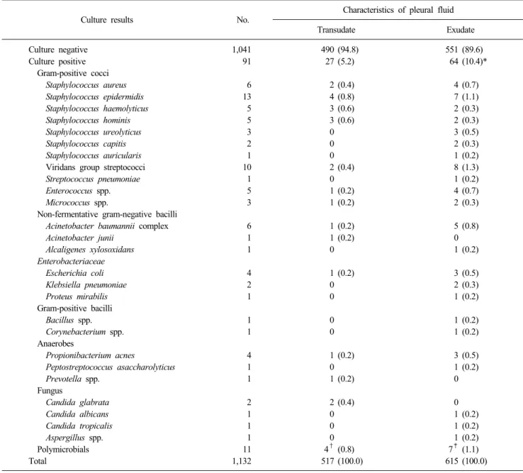
Table 1. Isolation rates of organisms according to transudate and exudate in pleural fluid specimens
Culture results No. Characteristics of pleural fluid
Transudate Exudate Culture negative
Culture positive Gram-positive cocci Staphylococcus aureus Staphylococcus epidermidis Staphylococcus haemolyticus Staphylococcus hominis Staphylococcus ureolyticus Staphylococcus capitis Staphylococcus auricularis Viridans group streptococci Streptococcus pneumoniae Enterococcus spp.
Micrococcus spp.
Non-fermentative gram-negative bacilli Acinetobacter baumannii complex Acinetobacter junii
Alcaligenes xylosoxidans Enterobacteriaceae Escherichia coli Klebsiella pneumoniae Proteus mirabilis Gram-positive bacilli Bacillus spp.
Corynebacterium spp.
Anaerobes
Propionibacterium acnes
Peptostreptococcus asaccharolyticus Prevotella spp.
Fungus
Candida glabrata Candida albicans Candida tropicalis Aspergillus spp.
Polymicrobials Total
1,041 91
6 13 5 5 3 2 1 10 1 5 3 6 1 1 4 2 1 1 1 4 1 1
2 1 1 1 11 1,132
490 27 2 4 3 3 0 0 0 2 0 1 1 1 1 0 1 0 0 0 0 1 0 1
2 0 0 0 4† 517
(94.8) (5.2)
(0.4) (0.8) (0.6) (0.6)
(0.4) (0.2) (0.2) (0.2) (0.2)
(0.2)
(0.2) (0.2)
(0.4)
(0.8) (100.0)
551 64
4 7 2 2 3 2 1 8 1 4 2 5 0 1 3 2 1 1 1 3 1 0
0 1 1 1 7‡ 615
(89.6) (10.4)*
(0.7) (1.1) (0.3) (0.3) (0.5) (0.3) (0.2) (1.3) (0.2) (0.7) (0.3) (0.8) (0.2) (0.5) (0.3) (0.2) (0.2) (0.2) (0.5) (0.2)
(0.2) (0.2) (0.2) (1.1) (100.0)
*P=0.001. †Streptococcus constellatus/K. pneumoniae/Clostridium perfringens (1), S. aureus/Streptococcus mitis/K. pneumoniae (1), Streptococcus anginosus/Prevotella buccae (1), and S. mitis/Streptococcus salivarius (1). ‡Enterococcus faecium/Morganella morganii/Cryptococcus laurentii (1), Streptococcus parasanguis/Neisseria sicca/Lactobacillus acidophilus (1), S. aureus/Acinetobacter baumannii (1), Staphylococcus warneri/S.
anginosus (1), S. aureus/A. baumannii (1), E. faecalis/Serratia marcescens (1), and S. aureus/Neisseria cinerea (1).
출액과 삼출액의 구분은 Light 기준[2]을 적용하였다. 흉막액의 당과 젖산탈수소효소 농도는 각각 40 mg/dL와 1,000 U/L로 나 누어 분석하였다. 흉막액 배양은 흉막액을 무균적으로 채취한 후 BACTEC FX (BD, Sparks, MD, USA) 또는 BacT/ALERT 3D (bioMérieux, Durham, NC, USA) 호기성과 혐기성 배양병 에 접종한 다음 검사실로 보내져 지속적혈액배양감시장비에서 5일간 배양하였다[9,10]. 계대배양 배지에서 균이 증식되면 Microscan (Siemens Healthcare Diagnostics, Sacramento, CA, USA)과 Vitek 2 (bioMerieux, Marcy l’Etoile, France) 장비를
이용하여 상품화된 kit로 동정하였다. 통계분석은 IBM SPSS Statistics version 20 (SPSS Inc., Chicago, IL, USA)을 이용하였 다. 변수의 특성에 따라 이분 변수인 경우에는 chi-square와 Fisher’s exact test를 적용하였고, 연속 변수는 T-test를 적용하 여 Levene test에서 등분산(P>0.05) 유무에 따른 유의성을 양 쪽 검증하였다.
흉막 누출액과 삼출액에서 배양 양성 비율은 각각 5.2%
(27/517)와 10.4% (64/615)였다(P=0.001, Table 1). 흉막액 배양 양성률은 단백질 비가 0.5 미만과 0.5 이상인 두 군에서 차이가
54
Ann Clin Microbiol 2015;18(2):52-55Table 2. Parameter characteristics of transudate (n=517) and exudate (n=615) according to culture results in pleural fluid specimens
Parameters
Mean and P values
Transudate Exudate
Culture negative (490)*
Culture positive
(27)* P value Culture negative
(551)*
Culture positive
(64)* P value
Age (yr) Interval†
Aerobic culture volume Anaerobic culture volume Detection time (hr) RBC (103/μL) WBC (103/μL) Neutrophil (%) Lymphocyte (%) Glucose (mg/dL) <40 ≥40
Total protein (g/dL) Albumin (g/dL) LDH (U/L) <1,000 ≥1,000 Ratio (PF/serum) Glucose Total protein LDH
Difference (Serum – PF) Total protein (g/dL) Albumin (g/dL)
70.9 6.75 8.49 8.34 7.8 0.54
21.9 50.1 152.2 (0)*
(490)*
1.95 1.05 104.7 (490)*
(0)*
1.18 0.34 0.33 3.66 1.82
68.8 6.26 8.65 8.23 34.9 9.9 1.35
43.4 34.7 113.6 (4)*
(23)*
1.92 0.85 96.3 (27)*
(0)*
1.18 0.32 0.32 3.87 1.76
0.464 0.896 0.856 0.911 0.772 0.106 0.008*
0.024 0.002
0.000 0.887 0.065 0.260
0.974 0.422 0.594 0.384 0.623
66.8 5.27 7.74 7.76 101.8 2.80
36.1 50.2 122.0 (36)*
(515)*
3.82 2.03 591.0 (491)*
(60)*
0.97 0.63 1.98 2.18 0.93
66.4 6.55 6.53 6.45 28.2 106.0 6.47
68.6 20.6 81.8 (25)*
(39)*
3.51 1.67 3,085.3 (42)*
(22)*
0.59 0.59 11.24 2.29 1.10
0.853 0.602 0.063 0.050 0.941 0.072 0.000 0.000 0.000
0.000 0.115 0.000 0.005
0.000 0.000 0.157 0.002 0.366 0.035
*Numbers in parenthesis mean numbers of specimen. †Time interval between admission day and request day of pleural fluid culture.
Abbreviations: RBC, red blood cell; WBC, white blood cell; LDH, lactate dehydrogenase; PF, pleural fluid.
없었으나 젖산탈수소효소 비는 0.6 미만과 0.6 이상인 두 군에 서 통계적으로 유의한 차이를 보였다(P<0.001). 흉수액 배양 양성과 음성에 따라 유의하게 차이가 있는 변수로 삼출액에서 는 흉수의 호중구 백분율, 림프구 백분율, 당, 알부민, 젖산탈수 소효소 수치, 당농도 비, 젖산탈수소효소 비 및 혈청과 흉막액 의 알부민 농도 차이였으며, 누출액에서는 흉수의 호중구 백분 율, 림프구 백분율과 당 농도만이 유의한 차이가 있었다(P
<0.05, Table 2).
본 연구는 후향적으로 흉수액의 배양 결과를 기준으로 화학 검사와 흉수액 세포수 결과를 동시에 의뢰한 경우만을 분석하 였기에 환자의 정확한 진단명을 확인할 수 없었고 분리 원인균 의 임상적 의의를 평가하지 못하였다. 또한 삼출액의 많은 원인 을 차지하는 결핵과 암종에 대해서도 분석할 수 없었다. 분석 대상 미생물의 수와 종류가 많지 않았으나 본 연구에서 다음과 같은 몇 가지 고려할 내용이 있을 것 같다. 흉수액 배양 양성률 은 삼출액이 누출액에 비해 높았으나 통상적으로 피부 오염균 으로 간주하는 coagulase 음성 포도알균과 Propionibacterium
acnes의 분리 빈도는 오히려 삼출액에서 높았고, Bacillus spp.와 Corynebacterium spp.도 1균주씩이지만 삼출액에서만 분리되었 다. 향후 흉수액 배양에서 분리되는 coagulase 음성 포도알균의 임상적 의의에 대한 연구가 필요하다고 생각되었다. 흉막액에 서 무산소세균의 분리 비율은 증가하는 추세로 12-34%로 보고 하고 있다[7]. 본 연구에서 흉막액 무산소세균의 분리 비율은 누출액 14.8% (4/27), 삼출액 7.8% (5/64)로 누출액에서의 비율 이 높았다. 흉막액 무산소세균 감염은 지역사회획득에서 발생 빈도가 높고, 흡인폐렴이 선행 원인인 경우가 많기 때문에 구강 과 호흡기계에 상재하는 무산소세균의 분포에 따라 원인균의 분리 비율이 달라진다[7]. 또한 흉막액의 무산소세균이 단독으 로 감염을 일으킬 때는 임상 증상이 서서히 진행하며, 발열도 미약하다[7]. 이러한 흉막액 무산소세균 감염의 특성이 누출액 에서의 분리 빈도가 높은 것과 연관성이 있는지에 대하여 향후 분석이 필요할 것으로 생각되었다. 흉막액 배양에서 두 가지 이 상의 균이 분리되는 비율이 누출액 14.8%, 삼출액 10.9%로 전 체로는 12.1%였는데 이는 Maskell 등[11]의 12%와 같았다. 결
Hyeun Gyeo Lee, et al. : Early Diagnosis of Simple Pleural Effusion
55
=국문초록=
흉막액 배양에서 누출액과 삼출액에 따른 세균학적 특성
연세대학교 원주의과대학 1진단검사의학교실, 2내과학교실 이현교1, 황규열1, 박순덕1, 어 영1, 김주원1, 윤갑준1, 리원연2
흉막의 누출액과 삼출액에 따른 흉막액 배양에서의 세균학적 특성을 규명하기 위해 2012년 10월부터 2014년 6월까지 혈액배양용 호기성과 무산소성 배지를 이용한 흉막액 배양 검사와 함께 혈청과 체액에서 총단백, 젖산탈수소효소와 당 검사를 의뢰한 1,132검체에서의 결과를 분석하였다. 흉수 누출액과 삼출액의 배양 양성률, 미호기세균 분리율과 다균증 빈도는 각각 5.2%와 10.4%, 14.8%와 7.8% 및 14.8%와 10.9%였다. [Ann Clin Microbiol 2015;18:52-55]
교신저자 : 어 영, 220-701, 강원도 원주시 일산동 162 원주기독병원 진단검사의학과
Tel: 033-741-1592, Fax: 033-731-0506 E-mail: [email protected]
론적으로 흉막액의 성상이 누출액일 경우에도 호기성과 무산소 배양을 시행하는 것이 흉막 감염의 정확한 진단에 필요하다.
REFERENCES
1. Karcher DS and McPherson RA. Cerebrospinal, synovial, serous body fluids, and alternative specimens. In: Henry JB, ed. Clinical Diagnosis and Management by Laboratory Methods. 22nd ed, Philadelphia; WB Saunders, 2011:480-506.
2. Light RW, Macgregor MI, Luchsinger PC, Ball WC Jr. Pleural effusions: the diagnostic separation of transudates and exudates.
Ann Intern Med 1972;77:507-13.
3. Porcel JM, Esquerda A, Martínez M, Rodríguez-Panadero F, Bielsa S. Influence of pleural fluid red blood cell count on the misidentification of transudates. Med Clin (Barc) 2008;131:770-2.
4. Light RW. The light criteria: the beginning and why they are useful 40 years later. Clin Chest Med 2013;34:21-6.
5. Ashchi M, Golish J, Eng P, O'Donovan P. Transudative malignant pleural effusions: prevalence and mechanisms. South Med J 1998;
91:23-6.
6. Kwon YS. Pleural infection and empyema. Tuberc Respir Dis (Seoul) 2014;76:160-2.
7. Davies HE, Davies RJ, Davies CW; BTS Pleural Disease Guideline Group. Management of pleural infection in adults:
British Thoracic Society Pleural Disease Guideline 2010. Thorax 2010;65 Suppl 2:ii41-53.
8. Lee D, Jang JY, Yoon KR, Kim H, Uh Y, Kim J, et al. Comparison of six clinical chemistry test results according to the treatment of EDTA anticoagulant in body fluid specimens. J Lab Med Qual Assur 2012;34:87-92.
9. Uh Y, Jang IH, Park SD, Kim KS, Seo DM, Yoon KJ, et al. Factors influencing the false positive signals of continuous monitoring blood culture system. Ann Clin Microbiol 2014;17:58-64.
10. Menzies SM, Rahman NM, Wrightson JM, Davies HE, Shorten R, Gillespie SH, et al. Blood culture bottle culture of pleural fluid in pleural infection. Thorax 2011;66:658-62.
11. Maskell NA, Batt S, Hedley EL, Davies CW, Gillespie SH, Davies RJ. The bacteriology of pleural infection by genetic and standard methods and its mortality significance. Am J Respir Crit Care Med 2006;174:817-23.