The Antimicrobial Activity of Bacterial-challenged Black Soldier Fly, Hermetia illucens
Kwanho Park1†, Eun-Young Yun2†, Seung-Won Park3 and Tae-Won Goo4*
1Department of Agricultural Biology, National Academy of Agricultural Science, RDA, Wanju-gun 565-851, Korea
2Graduate School of Integrated Bioindustry, Sejong University, Seoul 05006, Korea
3Department of Biotechnology, Catholic University of Daegu, Daegu 712-702, Korea
4Department of Biochemistry, School of Medicine, Dongguk University, Gyeongju 780-714, Korea
Received August 5, 2016 /Revised October 19, 2016 /Accepted November 1, 2016
In the larvae of the black soldier fly, Hermetia illucens, innate immunity mechanisms are activated in response to various pathogens and stimulants, resulting in the expression of antimicrobial peptides (AMPs). To induce the mass production of AMPs, H. illucens fifth instar larvae were immunized with five different kinds of bacteria. We isolated from the hemolymph of the H. illucens larvae after bacte- rial challenge, and their antimicrobial activities against Gram-positive bacteria (Staphylococcus aureus) and Gram-negative bacteria (Escherichia coli) were measured using the inhibition zone assay. Among these five different kinds of bacteria, the hemolymph of Bacillus subtilis-challenged H. illucens larvae showed the strongest antimicrobial activity against both Gram-positive bacteria and Gram-negative bacteria. The antimicrobial activity of the hemolymph of 1×10⁹ cfu/ml B. subtilis-challenged H. illucens peaks at 24 hr at 48 hr post-infection and gradually declines with time. Moreover, the immunized he- molymph also showed strong antimicrobial activity against various poultry pathogens such as S. enter- itidis, S. typhimurium, and S. pullorum. These results suggest that the expression of AMP genes in B.
subtilis-challenged H. illucens is up-regulated by innate immune responses, and that B. subtilis-chal- lenged H. illucens overexpressing AMPs may be useful as a feed additive in livestock diets to reduce the need for antibiotics.
Key words : Antimicrobial peptide, alternative antibiotics, innate immunity, Hermetia illucens
†Authors contributed equally.
*Corresponding author
*Tel : +82-54-703-7801, Fax : +82-54-770-2447
*E-mail : [email protected]
This is an Open-Access article distributed under the terms of the Creative Commons Attribution Non-Commercial License (http://creativecommons.org/licenses/by-nc/3.0) which permits unrestricted non-commercial use, distribution, and reproduction in any medium, provided the original work is properly cited.
Journal of Life Science 2016 Vol. 26. No. 12. 1409~1414 DOI : http://dx.doi.org/10.5352/JLS.2016.26.12.1409
서 론
동물용 항생제는 성장촉진용과 치료용으로 사용되어 왔으 며, 오늘날 집약적․기업적 축산으로 대규모화되는데 주도적 인 역할을 하였다. 그러나 가축사료 내 항생제 오남용으로 인 한 내성균 출현, 축산물 내 항생제 잔류, 가축의 질병저항성 약화, 분뇨로 유출된 항생제 잔류물로 인한 생태계 오염 등의 문제점이 대두되었다[2, 3, 17]. 이러한 동물용 항생제의 다양 한 문제점을 해소하기 위하여 유럽연합(European Union)에 서는 2006년 성장촉진용 항생제 사용을 전면 금지하였으며, 우리나라도 항생제 오남용 방지, 소비자의 축산물 위생․안전 성 요구, 국내 축산 경쟁력 제고를 위한 항생제 사용 규제 강화 및 무항생제 축산물 인증 정책 추진에 따른 대책을 마련하기 위하여 2014년부터 연차별로 항생제 허용품목을 단계적으로 감축하여 2011년 7월 1일에는 성장촉진용 항생제 사용이 전면
금지되었다. 그러나 성장촉진용 항생제 전면 금지로 인한 가 축 질병 발생 및 폐사로 인해 치료용 항생제 사용량이 많아져 인수공통항생제 사용 비중의 급격한 증가가 예상됨에 따라 내성균주를 퇴치할 수 있는 새로운 작용기작을 가지는 천연항 생제 개발이 시급이 요구되는 실정이다[13, 25].
동애등에를 포함한 곤충은 불량환경에서 오랜 진화의 역사 를 통해 병원균 침입에 대해 강력한 항균 펩타이드를 발현하 는 선천성 면역기전(innate immune system)을 보유하고 있 어, 화학항생제를 대체 할 수 있는 천연항생제 개발 소재로써 매우 우수하다[1, 6]. 곤충의 선천성 면역기전은 세포성 면역과 체액성 면역반응을 포함한다. 세포성 면역은 세균, 곰팡이, 원 생동물의 식균작용, 결절형성 및 캡슐형성을 포함하며, 체액 성 면역반응은 병원체 침입에 의해 다양한 종류의 단백질 및 펩타이드가 지방체 및 혈구세포에서 합성된 후 혈림프로 분비 됨으로써 병원체의 강력한 방어인자로 작용한다[4-6, 11]. 곤 충의 체액성 면역반응의 일환으로 분비되는 항균성 펩타이드 들은 아미노산 서열 및 구조에 따라 cecropin, defensin, pro- line-rich peptide, glycine-rich peptide 및 lysozyme류 등으로 분류되며, 나비목, 벌목, 파리목 및 딱정벌레목 등 다양한 곤충 종으로부터 발견되었다[7, 9, 10, 15, 19, 27]. 아메리카동애등에 (Hermetia illucens)는 환경정화곤충으로서 세균, 곰팡이, 바이 러스 등이 다량 포함된 유기성폐기물(음식물쓰레기, 축분폐기 물, 농업부산물 등)에 생존하면서 이들 유기성폐기물을 신속
Table 1. Sequences of primer used in real-time PCR analysis
Name Sequences
HiCec1 Forward Reverse
5'-TTGGTCAACGAGTTCGTGATGC-3' 5'-TCCTTGTTGTGGTGGTCCACCT-3' HiDef1 Forward
Reverse
5'-AGGTGGTGGAGCAGCATTAC-3' 5'-ACGACGTCCCAAAGCAATAC-3' Act5C Forward
Reverse
5’-AAGGACTCGTACGTGGGTG-3’
5’-CATCTTCTCACGGTTGGC-3’
하게 분해하기 위하여 생체방어를 위해 강력한 항균물질을 발현하는 기작을 보유하고 있어, 화학항생제를 대체 할 수 있 는 천연항생제의 개발 소재로써 매우 우수할 뿐만 아니라, 타 곤충에 비하여 성장속도가 매우 빠르며 별도의 사료비가 들지 않아 천연항생제의 저가 대량생산 공장으로 매우 적합하다.
그러나 동애등에를 포함한 곤충은 평상시에는 선천성 면역기 작을 작동시키지 않아, 항균 펩타이드를 거의 발현하지 않으 며, 생체의 체강 내로 병원균이 침입 시에만 항균 펩타이드를 다량으로 분비하여 병원균 침입에 대응한다[8, 20].
따라서 본 연구에서는 동애등에에 인위적으로 선천성 면역 기전을 유도하고 이에 의해 발현되는 항균 펩타이드를 새로운 천연항생제 개발을 위한 소재로 개발하기 위하여 다양한 병원 균에 대하여 항균활성과 특성을 분석하였다.
재료 및 방법
실험 곤충 및 균주
본 연구에 사용된 동애등에(H. illucens) 유충은 농촌진흥청 국립농업과학원 농업생물부에서 분양 받아 사육실(25~27℃, 상대습도 60)에서 사육된 동애등에를 사용하였다. 유충의 면 역유도를 위하여 사용된 균주는 Enterococcus faecalis (KACC 11859), Serratia marcescens (KACC 11961), Bacillus subtilis (KACC 17047), Bifidobacterium animalis (KACC 16637), Bifido- bacterium breve (KACC 16639) 등 총 5종의 균주를 사용하였 다. 항균활성 분석을 위해서는 그람양성균인 Staphylococcus aureus (KCCM 40881)와, 그람음성균인 Escherichia coli (KCCM 11234), Salmonella pullorum, Salmonella typhimurium, Salmonella enteritidis를 사용하였으며, 살모넬라균은 국립축산과학원으 로부터 분양받았다.
동애등에 유충의 면역유도
동애등에의 면역 유도를 위하여 직경 0.35 nm, 길이 40 mm 의 한방침(행림서원, 한국)에 균주를 묻힌 다음에 5령 유충의 10 체절 복부측면에서 머리 방향으로 찔렀다. 면역인 유도된 동애등에는 급이를 하지 않고 실온에서 2, 4, 8, 16, 24, 48 및 72시간 정치 후 각 시간별 혈림프를 이용한 항균활성 분석과 전사체 발현 분석 실험에 사용하였다.
동애등에 유충 분말로부터 항균물질 추출
면역이 유도된 동애등에 유충으로부터 항균활성물질을 추 출하기 위하여 마이크로파로 45분간 건조한 후 분쇄기를 이용 하여 분말로 제조한 다음 추출에 사용하였다. 동애등에 건조 분말(340 g)로부터 항균활성물질을 추출하기 위하여, 추출 욤 매(증류수 : 아세트산 = 80 : 20) 3.4 l를 첨가하고 30분간 가열 한 후 원심분리(4℃, 4,500rpm, 30 min)하여 상등액 회수하였 다. 회수한 상등액은 진공농축원심분리기를 이용하여 9시간
농축한 다음 멸균증류수에 녹여서 항균활성 분석에 사용하였다.
면역유도 동애등에 유충의 항균 활성 분석
면역유도 동애등에의 항균활성 검정은 방사선 확산 분석법 (RDA: radial diffusion assay)과 병원균에 대한 최저성장저해 농도(MIC: minimum inhibitory concentration)를 측정하여 검정하였다. RDA 분석에 사용된 균주는 그람양성균인 Sta- phylococcus aureus와, 그람음성균인 Escherichia coli, Salmonella pullorum, Salmonella typhimurium, Salmonella enteritidis를 사용 하였다. RDA 분석을 위하여 멸균된 RDA용 underlay gel (9 mM sodium phosphate, 1 mM sodium citrate, pH7.4, 1% low electroendosmosis agarose, 0.03% TSB)에 배양된 각각의 균주 (4×10⁶ cfu/ml)을 혼합하여 100 mm 사각플레이트에 부어 굳 혔다. Underlay gel에 지름 3.5 mm의 구멍을 내어 시료를 10 μl씩 구멍에 넣었고, 항균 펩타이드가 확산되도록 37℃에서 3시간 배양한 후, 그 위에 RDA용 overlay gel (6% TSB, 1%
low electroendosmosis agarose)을 부어 굳힌 다음 37℃에서 18시간 동안 배양하였다. 이후 clear zone의 크기를 측정하여 항균 활성을 비교하였다.
MIC 분석을 위해서는 Staphylococcus aureus와 Escherichia coli를 사용하였다. 각각의 균들은 액체 배지에서 37℃, 200 rpm 조건으로 18시간 진탕배양한 후, 다시 동일한 조건에서 4×10⁶ cfu/ml 농도가 되도록 2시간 30분간 2차 배양하였다.
96-well microplate의 각 well에 90 μl의 세균 배양액(1×10⁶ cfu/ml)을 분주 한 후, 단계적으로 희석된 동애등에 추출물의 용액을 각 well당 10 μl씩 첨가한 다음 37℃에서 18시간 동안 배양한 후, 분광 광도계(600 nm)에서 흡광도를 측정하여 MIC 를 결정하였다. RNA 및 MIC 분석을 위한 대조구로는 곤충 유래 천연항생제 중 유일하게 상업화된 melittin (Sigma- Aldrich, USA)과 정제봉독(청진바이오, 한국)을 사용하였다.
전사체 발현 분석
세균 주사 후 시간 경과별 항균펩타이드 유전자의 전사체 검정을 위하여 면역유도 동애등에로부터 각각 total RNA를 분리한 후, high capacity cDNA reverse transcription kit (Applied Biosystems, IL, USA)를 이용하여 cDNA를 합성하 였다. 합성된 cDNA는 Power SYBRⓇ Green Maser Mix
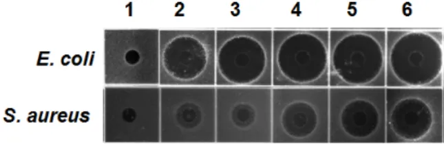
Fig. 1. Antimicrobial acitivity against E. coli and S. aureus of the hemolymph in various bacterial-challenged H. illu- cens larvae. Lane 1, non-immunized hemolymph; lane 2, hemolymph from E. faecalis-injected larvae; lane 3, he- molymph from S. marcescens -injected larvae; lane 4, he- molymph from B. animalis-injected larvae; lane 5, hemo- lymph from B. breve-injected larvae; lane 6, hemolymph from B. subtilis-injected larvae.
(Applied Biosystems, IL, USA)를 이용하여 AB step one (Applied Biosystems, IL, USA)를 통해 동애등에 유래 cecro- pin 1 (Hi Cec1) 및 defensin 1 (HiDef1)유전자를 증폭시켰다.
각각의 증폭 산물은 pGEM-T easy vector (Promega, USA)에 cloning한 후 염기서열 분석을 통하여 확인하였다. 이때 사용 한 프라이머(primer)는 Primer3 프로그램(http://simgene.com/
Primer3)을 사용하여 제작하였으며, 프라이머의 염기서열은 Table 1에 나타내었다. 초파리(Drosophila melanogaster) 유래의 actin 5C (DmAct5C)를 endogenous control로 사용하여 상대 정량 분석하였다[20].
결과 및 고찰
동에등에 생체 내 항균펩타이드 과량 발현을 위한 최적 균 주 선발
동애등에를 포함한 곤충은 체강 내로 병원균이 침입 시에만 항균펩타이드를 다량으로 분비하여 병원균 침입에 대응하는 반면에, 평상시에는 선천성 면역기전을 작동시키지 않아 항균 펩타이드를 거의 발현하지 않다. 따라서 동애등에 유충의 생 체 내 항군펩타이드를 인위적으로 과량 발현하기 위하여 E.
faecalis, S. marcescens, B. subtilis, B. animalis, B. breve 등 5종의 균주를 각각 1×10⁹ cfu/ml 농도로 적정한 다음 한방침에 묻혀 주사하였다. 각각의 균주를 주사한 동애등에 유충은 별도의 먹이를 급이하지 않고 24시간 동안 상온에 정치한 다음 혈림 프를 채취하여 각 균주에 따른 항균펩타이드 유도 효과에 의 한 항균활성을 비교 분석하였다. 항균활성 분석을 위하여 그 람양성균인 S. aureus와 그람음성균인 E. coli를 사용하였다.
그 결과, 곤충 병원균인 E. faecalis와 S. marcescens를 면역유도 제로 사용 시 그람음성균인 E. coli에 대해서는 비교적 높은 항균활성을 나타내었으나 그람양성균인 S. aureus에 대해서는 매우 약한 항균활성을 나타내었다. 이에 반해, 고초균인 B.
subtilis와 비퍼더스균인 B. breve 및 B. animalis를 면역유도제로 사용 시 곤충 병원균에 비해 그람양성 및 음성균 둘 다에 대해 높은 항균활성을 나타내었으며 특히 고초균인 B. subtilis은 5 종 균주 중 그람 양성 및 음성균에 대해서 가장 높은 항균활성 을 나타내었다(Fig. 1). 따라서 고초균에 의한 면역유도는 나머 지 4종 균주에 비하여 동애등에의 선천성 면역기전인 Toll 및 IMD pathway를 강력하게 유도함으로써 그람양성 및 음성균 에 대응하는 항균 펩타이드를 가장 다량으로 발현하는 것으로 추정되었다.
동에등에 생체 내 항균 펩타이드 과량 발현을 위한 최적 유 도 조건
상기 면역유도제로 사용한 5종의 균주 중 최대 항균 펩타이 드 발현을 나타낸 B. subtilis에 대하여 동애등에 생체 내 항균 펩타이드의 과량발현을 위한 최적 처리조건을 확립하기 위하
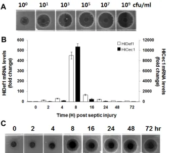
여 B. subtilis의 처리농도를 1×100~109 cfu/ml로 달리하여동애 등에 생체 내 항균 펩타이드가 최대로 발현되는 최적 처리농 도를 구명하고자 하였다. 또한, B. subtilis 주사 후 동애등에 생체 내 항균 펩티이드가 최대로 발현되는 시기를 구명하기 위해서는 선정된 최적 농도의 B. subtilis를 동애등에 유충의 복강에 주사하고 0~72시간 후에 동애등에 유충으로부터 각각 전사체 발현 분석과 E. coli에 대한 항균활성을 분석하고자 하 였다.
동애등에 생체 내 항균 펩타이드의 발현은 B. subtilis를 1×101 cfu/ml 농도로 주사한 동애등에의 혈림프에서 항균활 성을 나타내기 시작하여 1×10⁷ cfu/ml까지 농도 의존적으로 항균활성이 증가하다 이후 농도에서는 거의 일정 수준을 나타 내었다(Fig. 2A).
동애등에 생체 내 항균 펩타이드가 최대로 발현되는 시기를 결정하기 위한 전사체 발현분석은, B. subtilis (1×10⁹ cfu/ml) 를 주사하고 0, 2, 4, 8, 16, 24, 48 및 72시간 후에 동애등에 유충을 회수하여 total RNA를 분리한 다음 cDNA를 합성하였 다. 시간별 각각의 cDNA를 주형으로 real-time PCR를 수행하 여 동애등에 유래 항균 펩타이드 유전자인 defensin1 (HiDef1) 과 cecropin1 (HiCec1)의 전사체가 최대로 발현되는 시기를 결정하였다. 전사체 발현 분석 결과, HiDef1 및 HiCec1의 전사 체는 2시간 이후부터 증가하기 시작하여 8시간 후에 최대 발 현을 나타내다가 이후 급격하게 감소하였다. 즉, B. subtilis 주 사 후 8시간째 HiDef1 및 HiCec1의 전사체 발현은 0시간(면역 이 유도되지 않은 일반 동애등에)에 비하여 450.6배 및 10,732.9배 이상 증가하였다(Fig. 2B). 그러나 항균활성 분석 결과는 전사체 발현 분석과는 달리 B. subtilis 주사 후 24시간 째 가장 강한 항균활성을 나타내었으며 24시간 이후에도 매우 높은 수준으로 항균활성이 유지되었는데(Fig. 2C). 이것은 면 역유도제 주사에 의해 동애등에 생체 내 유도 발현된 HiDef1 과 HiCec1 이외의 또 다른 항균 펩타이드에 의한 영향으로 판단된다.
A
B
C
Fig. 2. (A) Determination of the optimal concentration of B. sub- tilis to induce the mass production of antimicrobial pep- tides into H. illucens. (B) Determination of the optimal concentration of B. subtilis to induce the mass production of antimicrobial peptides into H. illucens. (B) Determination of peak time for expression of anti- microbial peptides in the hemolymph of the H. illucens larvae after bacterial challenge. (C) Relative expression values of the HiCec1 and HiDef1 from RNA isolated from the hemolymph of control and bacteria-challenged H. illucens larvae at 2, 4, 8, 16, 24, 48 and 72 hr p.i. Data were statistically analyzed and compared with control using ANOVA. Values The bars show the mean±SE of relative mRNA expression levels.
A
B
C
Fig. 3. Characterization of antimicrobial peptides identified from immunized H. illucens. (A) Antimicrobial activity of the immunized hemolymph against S. enteritidis, S. enteritidis and S. enteritidis in a radial diffusion assay. Lane 1, non-immunized hemolymph; lane 2, melittin (1 μg); lane 3, hemolymph from B. subtilis-injected larvae. (B) Thermal stability of antimicrobial peptides. The immunized he- molymph was heat-treated at 100℃ for 10 (lane 1), 30 (lane 2), 60 (lane 3) and 120 min (lane 4), and then per- formed antimicrobial activity assay using heat-treated hemolymph. (C) pH stability of antimicrobial peptides.
A sample (2 μl) of hemolymph was mixed with 8 μl of pH 2 (lane 1), 5 (lane 2), 7 (lane 3), 9 (lane 4) and 11 buffer (lane 5), respectively. Antimicrobial activity of im- munized hemolymph (upper panel) and pH buffers (lower panel).
동에등에 유래 항균 펩타이드의 특성 분석
축산물은 고단백 영양식품이지만 적절한 취급이 이루어지 지 않거나 보관이 잘못될 경우 식중독균의 주요 매개체로써 작용하며, 식중독 발생의 77.1%가 살모넬라균이 원인으로 나 타났다. 주요 살모넬라 원인균은 Salmonella enteritidis로 1/3 이상을 차지하는 것으로 보고되고 있다. 살모넬라균은 주로 사람이나 동물의 장(腸)내에 기생하는 병원성 세균으로써, 실 제 국내 가금의 생체 및 환경에서도 S. enteritidis를 비롯한 다 양한 살모넬라가 분리되었다. 그리고 이러한 살모넬라균은 식 중독 발생뿐 만 아니라 가축의 생산성을 저하하는 요인으로 작용한다[14, 18].
동애등에 유래 항균 펩타이드가 살모넬라균에 대한 항균활 성을 나타내지는 확인하기 위하여 S. pullorum, S. typhimurium 및 S. enteritidis 3종의 살모넬라균에 대해 항균활성을 melittin 1ug과 비교 분석한 결과, 3종 모두에 대하여 melittin 1ug에 비해 훨씬 강력한 항균활성을 나타내었으며 특히, 국내 닭도 체로부터 분리한 24종의 살모넬라균 중 17종을 나타낸 S. en- teritidis (70.8%)에 대하여[15] 가장 높은 항균활성을 나타내었 다(Fig. 3A).
또 한편으로 면역이 유도된 동애등에 유충을 가축 성장촉진 용 항생제를 대체할 수 있는 천연항생제 개발 소재로써의 가 능성을 확인하기 위하여 면역유도 동애등에로부터 혈림프를 분리하여 열 안정성과 pH 안정을 확인하였다. 그 결과, 면역유 도 동애등에 유래 항균펩타이드는 100℃ 고온 장시간 처리에 서도 높은 안정성을 나타내었으며(Fig. 3B) [23], 고온 처리에 의해 분해되는 일부 산물은 열안정성이 낮은 attacin [20], leb- ocin [21], gloverin [12], transferrin [26] 등 고분자의 항균단백 질로 추정할 수 있었다[1, 6, 24]. 그리고 pH 안정성 분석 결과, 동애등에 유래 항균 펩타이드는 pH=2~11에서 높은 안정성을 나타내었으며(Fig. 3C), pH=7에서 항균활성이 pH=2 또는 pH=11에 비하여 항균활성이 낮은 것은 추출 혈림프 내의 일 부 존재하는 protease의 영향으로 판단되었다[23].
A
B
C
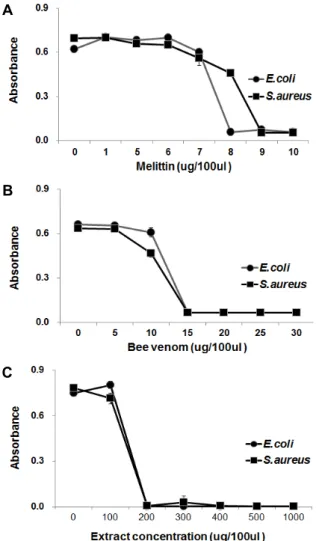
Fig. 4. Analysis of the minimal inhibitory concentration (MIC) of melittin (A), bee venom (B), and extract from immu- nized bacterial-challenged H. illucens larvae (C).
면역유도 동애등에 유래 항균펩타이드의 최소억제농도 분석 액체배지에서 자란 대수증식기의 그람음성균인 E. coli 및 그람양성균인 S. aureus를 1×10⁶ cell/ml가 되도록 희석하여, 90 μl씩 96-well plate에 분주한 후, serial dilution법으로 희석 한 시료를 농도별로 10 μl씩 주입한 다음, 37℃에서 24시간 동안 배양기에서 배양하여 흡광도(OD600)를 측정하였다. 그 결 과, melittin의 MIC 범위는 E. coli 및 S. aureus에 대해 각각 7~8 μg/100 μl 및 8~9 μg/100 μl로 측정되었으며(Fig. 4A), 정제봉독의 MIC 범위는 E. coli 및 S. aureus에 대해 둘 다 10~15 μg/100 μl으로 측정되었고(Fig. 4B), 동애등에 유래 항 균물질의 MIC 범위는 E. coli 및 S. aureus에 대해 둘 다 건조분 말 추출물 100~200 μg/100 μl로 측정되었다(Fig. 4C). 한편으 로, MIC assay에 의해 동애등에 건조분말 추출물 내 항균물질 을 정량적으로 분석한 결과, 동애등에 유충 생체 1 kg (건조분 말 340 g)으로부터 그람음성균인 E. coli에 대해서는 melittin 13.6 g, 정제봉독 25.5 g의 활성에 해당하는 항균활성물질이, 그람양성균인 B. subtilis에 대해서는 melittin 15.3 g, 정제봉독
25.5 g의 활성에 해당하는 항균활성물질이 존재하는 것으로 추정되었다.
따라서, 이상의 결과로부터 면역이 유도된 동애등에는 가축 성장촉진용 항생제를 대체할 수 있는 저가의 천연항생제 개발 소재로써의 매우 우수할 것으로 기대된다.
감사의 글
본 논문은 농촌진흥청 공동연구사업(PJ01084302)과 농림축 산식품부의 재원으로 농림수산식품기술기획평가원의 농생명 산업기술개발사업사업(315030-3)의 지원에 의해 이루어진 결 과물임을 밝힙니다.
References
1. Andreu, D. and Rivas, L. 1998. Animal antimicrobial pep- tides: an overview. Biopolymers 47, 415-433.
2. Baltzer, S. A. and Brown, M. H. 2011. Antimicrobial pep- tides: promising alternatives to conventional antibiotics. J.
Mol. Microbiol. Biotechnol. 20, 228-235.
3. Barton, M. D. 2000. Antibiotic use in animal feed and its impact on human health. Nutr. Res. Rev. 13, 279-299.
4. Boman, H. G. 1995. Peptide antibiotics and their role in in- nate immunity. Annu. Rev. Immunol. 13, 61-92.
5. Brogden K. A. 2005. Antimicrobial peptides: pore formers or metabolic inhibitors in bacteria. Nat. Rev. Microbiol. 3, 238-250.
6. Bulet, P., Hetru, C., Dimarcq, J. L. and Hoffmann, D. 1999.
Antimicrobial peptides in insects; structure and function.
Dev. Comp. Immunol. 23, 329-344.
7. Casteels, P., Ampe, C., Riviere, L., Damme, J. V., Elicone, C., Fleming, M., Jacobs, F. and Tempst, P. 1990. Isolation and characterization of abaecin, a major antibacterial re- sponse peptide in the honeybee (Apis mellifera). Eur. J.
Biochem. 187, 381-386.
8. Choi, Y. C., Park, K. H., Nam, S. H., Jang, B. G., Kim, J.
H., Kim, D. W. and Yu, D. J. 2013. The effect on growth performance of chicken meat in broiler chicks by dietary supplementation of black soldier fly larvae, Hermetia illucens (Diptera : Stratmyidae). J. Seric. Entomol. Sci. 51, 30-35.
9. Goo, T. W., Yun, E. Y., Kim, S. W., Choi, K. H., Kang, S.
W, Kwon, K., Yu, K. and Kwon, O. Y. 2008. Bombyx mori protein disulfide isomerase enhances the production of nue- cin, an antibacterial protein. BMB Rep. 41, 400-403.
10. Goo, T. W, Yun, E. Y, Kim, S. W., Choi, K. H, Kang, S.
W., Kwon, K., Choi, J. S. and Kwon, O. Y. 2008. Secretion of the antibacterial recombinant protein enbocin. Z.
Naturforsch C. 63, 284-288.
11. Hoffman, J. A., Kafatos, F. C., Janeway, C. A. and Ezekowitz, R. A. 1999. Phylogenetic perspectives in innate immunity. Science 284, 1313-1318.
12. Kaneko, Y., Furukawa, S., Tanaka, H. and Yamakawa, M.
2007. Expression of antimicrobial peptide genes encoding Enbocin and Gloverin isoforms in the silkworm, Bombyx
초록:세균에 의해 면역이 유도된 동애등에의 항균활성
박관호1†․윤은영2†․박승원3․구태원4*
(1농촌진흥청 국립농업과학원 농업생물부, 2세종대학교 일반대학원 바이오산업융합과, 3대구가톨릭대학교 생명공
학과, 4동국대학교 의과대학 생화학교실)
동애등에(Hermetia illucens)를 포함한 곤충은 불량환경에서 오랜 진화의 역사를 통해 병원균 침입에 대응하기 위하여 강력한 항균 펩타이드를 발현하는 선천성 면역기전을 보유하고 있다. 동애등에 생체 내 항균펩타이드를 인위적으로 유도하기 위하여, E. faecalis 포함한 5종의 세균을 한방침에 묻혀 주사하였다. 5종 세균에 의해 면역이 유도된 동애등에로부터 혈림프를 채취하여 각 세균에 따른 항균펩타이드 유도효과에 의한 항균활성을 그람양성 균인 S. aureus와 그람음성균인 E. coli에 대해 비교 분석하였다. 그 결과, 고초균인 B. subtilis은 5종 세균 중 그람 양성 및 음성균 둘 다에 대해서 가장 높은 항균활성을 나타내었다. B. subtilis (1×10⁹ cfu/ml)에 의해 면역이 유도 된 동애등에는 주사 후 24시간째 가장 강한 항균활성을 나타내었다. 또한 B. subtilis에 의해 면역이 유도된 동애등 에는 가금류의 주요 병원균인 S. enteritidis, S. typhimurium 및 S. pullorum에 대해서도 높은 항균활성을 나타내었 다. 이상의 결과로써, B. subtilis에 의해 면역이 유도된 동애등에는 가축 성장촉진용 항생제를 대체할 수 있는 사료 첨가제로써 매우 유용할 것으로 판단된다.
mori. Biosci. Biotechnol. Biochem. 71, 2233-2241.
13. Kang, H. K., Seo, O. S., Choi, H. C., Chae, H. S., Na, J. C., Bang, H. T., Kim, D. W., Park, S. B, Kim, M. J. and Jung, S. H. 2010. Effect of dietary supplementation of chlorella powder on production performances, blood components.
Kor J. Poult Sci. 27th Regular Conference 106-108.
14. Kim, A. R., Lee, Y. J., Kang, M. S., Kwag, S. I. and Cho, J. K. 2007. Dissemination and tracking of Salmonella spp.
in integrated broiler operation. J. Vet. Sci. 8, 155-161.
15. Kim, S. R., Lee, E. M., Yoon, H. J., Choi, Y. S., Yun, E. Y., Hwang, J. S., Jin, B. R., Lee, I. H. and Kim, I. 2007.
Antibactrial activity of peptides synthesized based on the Bombus ignitus abaecin, a novel prolinerich antimicrobial peptide. Int. J. Indust. Entomol. 14, 147-150.
16. Lee, H. W., Hong, C. H. and Jung, B. Y. 2007. Characteristics of Salmonella spp. isolated from poultry carcasses. Kor. J.
Vet. Serv. 30, 339-351.
17. Looft, T., Johnson, T. A., Allen, H. K., Bayles, D. O., Alt, D. P., Stedtfeld, R. D., Sul, W. J., Stedtfeld, T. M., Chai, B., Cole, J. R., Hashsham, S. A., Tiedje, J. M. and Stantona, T.
B. 2012. In-feed antibiotic effects on the swine intestinal microbiome. Proc. Natl. Acad. Sci. USA 109, 1691-1696.
18. Olsen, S. J., MacKinon, L. C., Goulding, J. S., Bean, N. H.
and Slutsker, L. 2000. Surveillance for foodborne disease outbreaks–United States, 1993~1997. MMWR Surveill Summ. 49, 1-51.
19. Otvos, L. Jr. 2000. Antibacterial peptides isolated from insects. J. Pept. Sci. 6, 497-511.
20. Park, S. I., Kim, J. W. and Yoe, S. M. 2015. Purification and characterization of a novel antibacterial peptide from black soldier fly (Hermetia illucens) larvae. Dev. Comp. Immunol.
52, 98-106.
21. Sugiyama, M., Kuniyoshi, H., Kotani, E., Taniai, K., Kadono- Okuda, K., Kato, Y., Yamamoto, M., Shimabukuro, M., Chowdhury, S. and Xu, J. 1995. Characterization of a Bombyx mori cDNA encoding a novel member of the atta- cin family of insect antibacterial proteins. Insect Biochem.
Mol. Biol. 25, 385-392.
22. Tanaka, H., Ishibashi, J., Fujita, K., Nakajima, Y., Sagisaka, A., Tomimoto, K., Suzuki, N., Yoshiyama, M., Kaneko, Y., Iwasaki, T., Sunagawa, T., Yamaji, K., Asaoka, A., Mita, K.
and Yamakawa, M. 2008. A genome-wide analysis of genes and gene families involved in innate immunity of Bombyx mori. Insect Biochem. Mol. Biol. 38, 1087-1110.
23. Walkenhorst, W. F., Klein, J. W., Vo, P. and Wimley, W.
C. 2013. pH Dependence of microbe sterilization by cationic antimicrobial peptides. Antimicrob. Agents Chemother. 7, 3312- 3320.
24. Yi, H. Y., Chowdhury, M., Huang, Y. D. and Yu, X. Q. 2014.
Insect antimicrobial peptides and their applications. Appl.
Microbiol. Biotechnol. 98, 5807-5822.
25. Yoo, J. H., Park, G. H., Sung, J. S., Song, H. N., Shin, S.
Y., Jung, W. H. and Jung, M. H. 2014. Feed additives in broiler diets to produce healthy chickens without in-feed antimicrobial compounds. CNU Journal of Agricultural Science 41, 441-453.
26. Yun, E. Y., Lee, J. K., Kwon, O. Y., Hwang, J. S., Kim, I., Kang, S. W., Lee, W. J., Ding, J. L., You, K. H. and Goo, T. W. 2009. Bombyx mori transferrin: genomic structure, ex- pression and antimicrobial activity of recombinant protein.
Dev. Comp. Immunol. 33, 1064-1069.
27. Zasloff, M. 2002. Antimicrobial peptides of muticellular organism. Nature 415, 329-344.