대한두경부종양학회지, 제37권 제1호, 2021. pp.1-10 Korean Journal of Head & Neck Oncology, Vol.37, No.1
https://doi.org/10.21593/kjhno/2021.37.1.1 ISSN 1229-5183(Print) / ISSN 2586-2553(Online)
두경부암의 양성자치료: 현재의 임상 적용 및 발전 방향
오동렬+
성균관대학교 의과대학 삼성서울병원 방사선종양학과
Proton Therapy for Head and Neck Cancer:
Current Clinical Applications and Future Direction
Dongryul Oh, MD, PhD+
Departments of Radiation Oncology, Samsung Medical Center, Sungkyunkwan University School of Medicine, Seoul, Korea
= Abstract =
Intensity-modulated radiation therapy (IMRT) using X-rays is a standard technique implemented for treating head and neck cancer (HN C). Compared to 3D conformal RT, IMRT can significantly reduce the radiation dose to surrounding normal tissues by using a highly conformal dose to the tumor. Proton therapy is a type of RT that uses positively charged particles named protons. Proton therapy has a unique energy deposit (i.e., Bragg peak) and greater biological effectiveness than that of therapy using X-rays. These inherent properties of proton therapy make the technique advantageous for HNC treatment. Recently, advanced techniques such as intensity-modulated proton therapy have further decreased the dose to normal organs with a higher conformal dose to the tumor. The usage of proton therapy for HNC is becoming widespread as the number of operational proton therapy centers has increased worldwide. This paper aims to present the current clinical evidence of proton therapy utility to HNC clinicians through a literature review. It also discusses the challenges associated with proton therapy and prospective development of the technique.
Key Words : Proton therapy⋅Intensity-modulated proton therapy⋅Radiation therapy⋅Head and neck cancer
Received
Accepted: May 9, 2021 : May 18, 2021
+Corresponding author: Dongryul Oh, MD, PhD
Department of Radiation Oncology, Samsung Medical Center, Sungkyunkwan University School of Medicine, #81 Irwon-ro, Gangnam-gu, Seoul, 06351, Korea
Tel: +82-2-3410-2612, Fax: +82-2-3410-2619 E-mail: [email protected]
Introduction
Radiation therapy (RT) is an integral part of a multi- disciplinary approach for the management of head and neck cancer (HNC). It is used as a definitive treatment with or without chemotherapy or adjuvant therapy following sur- gery or palliative treatment for HNC. Due to the rapid tech- nological development over the last two decades, in-
tensity-modulated RT (IMRT) using X-rays has been widely adopted as a standard technique for the treatment of HNC.
Compared to 3D conformal RT (3D-CRT), IMRT can re- duce the radiation dose to normal organs surrounding the tumor and deliver a highly conformal dose distribution.1) Many studies have shown that IMRT in HNC reduces acute and late RT-toxicity and improves treatment outcomes.2-5)
Proton therapy is a type of RT that uses the positively charged nuclei of a hydrogen atom called proton, and not X-rays. Proton therapy has the advantage of reducing the radiation dose to the surrounding normal tissue more effec- tively than therapy with X-rays due to a unique physical property of proton beams called the Bragg peak.6,7) In recent years, with an increase in the number of operational proton therapy centers worldwide, more than 220,000 patients have
Fig. 1. Physical properties of a proton beam. SOBP, Spread-out Bragg Peak.
A
B
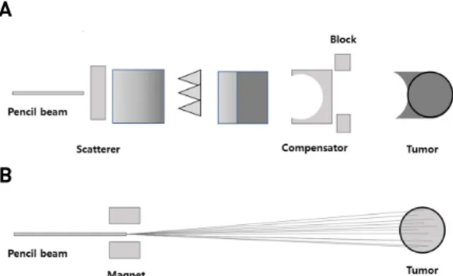
Fig. 2. Scattering and scanning techniques of proton therapy.
(A) Scattering. (B) Scanning.
undergone proton therapy.8) The growing usage of proton therapy is generating a significant level of clinical evidence regarding the effectiveness of this technique. In particular, proton therapy has shown relatively favorable treatment out- comes and toxicity profiles compared to thos e of the current standard IMRT technique, and it has been widely used in various clinical settings for the treatment of HNC.9-12) Furthermore, as proton therapy techniques have evolved, intensity-modulated proton therapy (IMPT), which provides a more conformal radiation dose distribution than standard proton therapy does, has become available.10) Currently, many prospective cohort or randomized controlled trials of proton therapy are ongoing worldwide. These studies address the issues of clinical efficacy, toxicity, and cost-effectiveness.13)
This review aims to introduce the basic characteristics of proton therapy to HNC clinicians and to present the cur- rent clinical evidence of proton therapy through a literature review. It also discusses current challenges of proton ther- apy and future directions for this technique.
Physical and biological properties of proton therapy:
the Bragg peak and RBE 1.1
Proton beams have a unique energy deposit compared to that of X-rays.6) X-rays deposit most of the energy near the surface of the body, and the absorbed dose is gradually decreased; however, it continues to deliver the energy until it leaves the patient’s body. In contrast, proton beams depos- it a small dose when entering the patient’s body, and the absorbed dose suddenly increases at a certain depth (i.e., the “Bragg peak”). Beyond this peak, the energy of the pro- ton beam is negligible (the no-exit dose) (Fig. 1). The initial
energy of the proton beam generated by a cyclotron de- termines the depth of the peak, which can precisely control the Bragg peak and localize it at the tumor site (target vol- ume). The Bragg peak is an inherent advantage of proton therapy. Consequently, this technique can spare the surround- ing normal tissue to a better degree during a target volume irradiation compared to that with therapy using X-rays.
Proton therapy is characterized by a greater biological effectiveness than that of X-ray therapy.14) Relative bio- logical effectiveness (RBE) is the ratio of the doses required by two different radiation types to cause the same level of a biological effect. When planning proton therapy, an RBE of 1.1 is generally assumed.15) This means that X-rays require a 10% higher dose than proton beams do to produce the same biological effect. This RBE of 1.1 is mainly based on experimental in vitro data. However, limited data exist on the RBE of various human cancers and normal tissues.
The use of a fixed RBE value of 1.1 for proton therapy imposes the limitation of not representing considerable tu- mor heterogeneity and different normal tissues.16,17)
What is IMPT?
As mentioned above, the Bragg peak is a narrow peak of the dose deposit; therefore, different methods are required to cover the whole tumor volume using this narrow peak.
Passively scattered proton therapy (PSPT), analogous to 3D-CRT using X-rays, uses a uniform spread-out Bragg peak (SOBP) to cover the tumor volume.18) For dos e con- formity, the lateral edge of the beam is blocked, and the distal edge of the beam is modified by a compensator as the tumor shape (Fig. 2). In contrast to PSPT, IMPT, similar
to IMRT with X-rays, uses a “pencil beam technique” or
“scanning technique.”10) It directly uses a pencil beam from a cyclotron without scattering the beam. It covers the tumor volume by using the electromagnetic control of a pencil beam. Similar to the 3D-printing technique, the irradiated dose can be deposited to each layer of the tumor volume voxel by voxel. The dose distribution can be modulated by the number of protons, beam energy, and off-axis position control of the electromagnetic deflection.
Clinical evidence of proton therapy in HNC
Skull base tumors
Skull base tumors, including chordoma and chondrosarcoma, have been treated with proton therapy for many decades.19) These tumors require high doses of radiation for effective tumor control, but their location is close to the brain, brain- stem, and optic apparatus, limiting the possibility of dose escalation. Many retrospective studies have shown excellent local control (LC) and survival outcomes with proton ther- apy in the treatment of skull base tumors. A large clinical s eries of 519 s kull bas e tumor cas es was included in a report from the Massachusetts General Hospital in 1999.20) The 5-year local relapse-free survival rate was 73% for chordo- ma and 98% for chondrosarcoma. The 5-year overall surviv- al (OS) rates were 80% and 91%, respectively. Similarly, the Paul Scherrer Institute reported the treatment outcomes of a scanning technique in proton therapy.21) Initially, they reported that the 3-year LC rates were 87.5% and 100% for chordoma (n=28) and chondrosarcoma (n=11), respectively.
The long-term follow-up results for chondrosarcoma showed that the actuarial 8-year LC and OS rates were 89.7% and 93.5%, respectively.22) Grade 3 or higher radiation-induced toxicity was reported in only 7.8% of patients. Recently, the risk of radiation-induced optic neuropathy was analyzed in 216 patients with skull base tumors, including chordoma and chondrosarcoma, who were treated with scanning pro- ton therapy.23) Radiation-induced optic neuropathy of any grade was observed in only 6.5% of patients with a median follow-up of 5.3 years when the median maximum dose was 59.5 Gy to the optic nerve and 58.8 Gy to the optic chiasm. Although proton therapy has shown better results
than those of X-ray therapy historically, recent IMRT or stereotactic body radiation therapy (SBRT) techniques also enable the delivery of high-dose irradiation to skull base tumors and sparing of adjacent critical normal organs.
Further studies are required to determine whether IMRT or SBRT results are comparable to those of proton therapy.
Paranasal sinus and nasal cavity cancers
Sinonasal cancers are a rare and heterogeneous group of tumors of various histological types, including squamous cell carcinoma, adenocarcinoma, adenoid cystic carcinoma, neuroendocrine carcinoma, and melanoma. Surgery with or without adjuvant RT is the mainstay of treatment for a re- sectable tumor, while definitive RT with or without chemo- therapy is used for unresectable tumors.24) Delivering a high dose of RT to the sinonasal site is challenging because of the surrounding normal tissues (both lenses and the retina, optic nerve, optic chiasm, brain, and brainstem). Proton ther- apy has a dosimetric advantage over X-ray therapy in spar- ing the surrounding normal organs. A meta-analysis by Patel et al. reported that proton therapy demonstrated higher 5-year OS (relative risk [RR] 1.51, 95% confidence interval [CI]
1.14-1.99; P=0.038) and disease-free survival (DFS) (RR 1.93, 95% CI 1.36 - 2.75; P=0.003) rates than did X-ray therapy.25) Additionally, a subgroup analysis showed that proton therapy showed significantly higher 5-year DFS (RR, 1.44, 95% CI 1.01-2.05; P=0.045) and locoregional control (LRC) (RR 1.26, 95% CI 1.05-1.51; P=0.011) rates com- pared to those of IMRT. Although this study demonstrated better treatment outcomes of proton therapy compared to that of X-ray therapy, the meta-analysis included only 43 cohorts from 41 non-comparative observational studies. The Proton Collaborative Group analyzed 69 sinonasal cancer patients who underwent curative proton therapy.26) This study included 27 patients who underwent re-irradiation.
For de novo proton therapy, the 3-year OS, freedom from disease progression (FFDP), and freedom from locoregional recurrence (FFLR) rates were 100%, 77.3%, and 92.9%, respectively. For re-irradiation, the 3-year OS, FFDP, and FFLR rates were 76.2%, 32.1%, and 33.8%, respectively.
There were no grade 3 or higher late toxicities. This registry data showed that proton therapy might be an effective and safe treatment for sinonasal cancers. One prospective cohort study reported physician-assessed toxicities (PATs) and pa-
tient-reported outcomes (PROs) of 64 patients with sino- nasal cancer treated with proton therapy.27) There was only one acute grade 3 neurologic PAT and no grade 3 or higher late PAT reported in the study. Significant worsening of PRO was notable in the acute and subacute periods, but no significant changes from baseline to the chronic period were noted. The 3-year LC, DFS, and OS rates were 88%, 76%, and 82%, respectively. Owing to the rarity of sino- nasal tumors, a randomized controlled trial for proton ther- apy compared with IMRT may be difficult. However, multi- ple retrospective and prospective registry studies are cur- rently being conducted.
Re-irradiation in HNC
Recurrent or secondary HNCs often require re-irradiation of tissues that had previously received a substantial radia- tion dose for salvage or adjuvant treatment. A significant cumulative dose to normal tissue can result in severe or fatal toxicity, including soft tissue or bone necrosis. Thus, radiation oncologists strive to minimize dose overlap with the previously irradiated target volume when planning a re-irradiation.28-30) Here, proton therapy has an inherent advantage.31) The MD Anders on Cancer Center (MDACC) reported the results of an initial study on 60 patients who were re-irradiated with proton therapy for HNC.32) The 1-year LR failure-free survival and OS rates were 68.4%
and 83.8%, respectively. Acute grade 3 toxicity occurred in 30% of patients, and the late grade 3 toxicity rate was 16.7% at the 1-year follow-up. Three patients died due to re-irradiation-related side effects. McDonald et al. also re- ported the clinical outcomes of re-irradiation with proton therapy in HNC cases.33) Sixty-one patients received a me- dian dose of 66 Gy for microscopic control and 70.2 Gy for gross tumors. The 2-year OS rate was 32.7%, and the 2-year local failure rate was 19.7%. Grade 3 or higher tox- icities occurred in 14.7% of patients in the acute period and 24.6% in the late period, and these included three deaths. A systematic review, including four studies of HNC re-irradiation, showed appropriate local/locoregional control and favorable toxicity profiles, including low feeding tube placement rates of 9-10%, of proton therapy versus histor- ical X-ray-based methods.34) The usage of proton re-irradi- ation for HNC is still limited, and long-term follow-up stud- ies on large populations will be needed. Therefore, the
MDACC has started a phase II randomized trial to compare the 2-year rate of grade 3 or higher toxicity between SBRT and IMRT/IMPT (NCT03164460).
Oropharyngeal cancer
Several single-institution retrospective studies have dem- onstrated favorable efficacy and low toxicity of proton ther- apy for oropharyngeal cancer.35-37) Lower toxicity compared to that of IMRT suggests that IMPT could be an optimal option for de-intensified treatment of HPV-associated or- opharyngeal cancer. The MDACC has reported a series of data on the toxicities of IMPT. The authors compared PRO between the IMPT (n=35) and IMRT groups (n=46).38) The IMPT group s howed better s ymptom s cores for altered tas te and appetite during the subacute and chronic phases than did the IMRT group (P<0.048). The MD Anderson Symptom Inventory for Head and Neck Cancer (MDASI-HN) score was better in the IMPT group than in the IMRT group (P =0.013) during the subacute phase. In a case-matched analysis of IMPT (n=50) and IMRT (n=100), IMPT reduced the rates of feeding tube dependency and severe weight loss (odds ratio [OR], 0.44; 95% CI 0.19-1.0; P=0.05 at 3 months after treatment and 0.23; 95% CI 0.07-0.73; P = 0.01 at 1 year after treatment).39) Recently, the MDACC reported that IMPT decreased xerostomia compared to IMRT in oropharyngeal cancer.40) The authors compared xerostomia questionnaire scores between the IMRT (n=429) and IMPT groups (n=103), and IMPT resulted in lower rates of moderate to severe xeros tomia at 18-24 months (IMPT vs IMRT, 6% vs . 20%;
P=0.025) and 24-36 months (IMPT vs IMRT, 6% vs. 20%;
P=0.01) than did IMRT. Moreover, it was demonstrated that a decreased irradiated dose and volume to the oral cavity in IMPT was related to les s common late xeros tomia. Our institution reported the early clinical outcomes of a combi- nation of IMRT and IMPT (n=67) and compared it with those of IMRT alone (n=81) for oropharyngeal cancer.41) There were no significant differences in OS and PFS be- tween the groups after propensity score matching (PSM) analysis. After matching, the 2-year OS rates were 92.4%
and 100%, and the PFS rates were 78.8% and 82.4% for IMRT alone and IMRT/IMPT, respectively. Grade 3 or higher mucositis was less common in the IMRT/IMPT group than in the IMRT alone group (15.8% and 39.5%, respectively; P=0.021).
Although these retrospective results of similar oncologic outcomes and lower toxicity compared to those of IMRT favor the use of IMPT for treating oropharyngeal cancer, no level 1 evidence supporting the use of IMPT exists to date. A randomized phase II/III study is ongoing to clarify the clinical superiority of IMPT to IMRT in the US (NCT01893307). This study initially aimed to compare 70 Gy of IMRT with the s ame dos e of IMPT. The primary endpoint was the rate of late grade 3-5 toxicities within two years. Later, the study design was changed to non-in- feriority of IMPT to IMRT with regard to PFS. Two other randomized trials, including the TORPEdO (Toxicity Reduction using Proton beam therapy for Oropharyngeal cancer) trial in the UK and the ARTSCAN V trial at the Lund University Hospital in Sweden (NCT03829033), are also underway.
Uni-lateral irradiation
Well-lateralized HNCs, including salivary gland tumors and some oral cavity or tonsil cancers, have a low risk of contralateral metastasis. In these cases, uni-lateral (or ipsi- lateral) irradiation is indicated. In such a clinical scenario, proton therapy has dosimetric advantages related to the exit dose nearing zero. Stromberger et al. showed that IMPT delivered significantly lower mean doses to contralateral salivary glands (<0.001-1.1 Gy) than did rotational IMRT techniques such as tomotherapy or volumetric intensity- modulated arc (parotid gland: 6-10 Gy; submandibular gland:
15-20 Gy) when using a total dose of 70.4 Gy to the high-risk clinical target volume.42) Romesser et al. reported the out- comes of patients with salivary gland tumors and cutaneous squamous cell carcinomas.43) In 41 patients, proton therapy had a lower rate of acute toxicities, except for radiation dermatitis, than that with IMRT. Compared to IMRT, proton therapy resulted in lower rate of acute grade 2 or higher toxicities in oral mucositis (Proton therapy vs IMRT, 16.7%
vs. 52.2%; P=0.019), dysgeusia (Proton therapy vs IMRT, 5.6% vs. 65.2%; P<0.001), and nausea/vomiting (Proton therapy vs IMRT, 11.1% vs. 56.5%; P=0.003), but it showed higher rate of dermatitis (Proton therapy vs IMRT, 100%
vs. 73.9%; P<0.032), suggesting a loss of skin-sparing effect in proton therapy. Recently, the MSKCC reported clinical outcomes of proton therapy for major salivary gland tumors.44) Fifty-one patients with parotid gland tumors and 17 patients
with submandibular tumors were analyzed retrospectively.
Positive surgical margins were identified in 52.9% of the patients. A median dose of 66.07 cGy was delivered. The 3-year LRC, PFS, and OS rates were 95.1%, 80.7%, and 96.1%, respectively. Grade 2 or higher oral mucositis and dysgeusia occurred in only 5.9% and 2.9% of patients with parotid gland tumors and submandibular tumors, respectively.
Grade 2 or higher dermatitis was observed in 69.1% of patients. Therefore, proton therapy resulted in excellent LRC with low toxicity in cases of salivary gland tumor, despite limitations in the retrospective analysis of the heter- ogeneous group. Currently, a phase II randomized trial is ongoing to compare grade 2 or higher toxicity rate of IMRT to that of proton therapy when 60-66 Gy of unilateral irradi- ation is delivered for salivary gland cancer, skin cancer, and melanoma (NCT02923570).
Nasopharyngeal cancer
IMRT is the current standard RT technique for nasophar- yngeal cancer as it decreases RT-related toxicities and im- proves therapeutic outcomes compared to 3D-CRT.45) Several dosimetric comparison studies have shown that proton ther- apy reduces the irradiated dose to critical normal organs.46-48) However, this should be further validated in a clinical setting. Holliday et al. performed a matched case-control study to determine if IMPT reduced toxicity compared to IMRT for nasopharyngeal cancer.49) In a matched cohort of 10 IMPT and 20 IMRT patients, tube feeding was less frequent (IMPT vs. IMRT, 20% vs. 65%; P=0.02) in the IMPT group than in the IMRT group. This effect may have been attributed to a lower mean dose to the oral cavity.
Alterio et al. compared the outcomes of IMRT followed by IMPT (a mixed beam, MB) to those of IMRT alone for nasopharyngeal cancer.50) Twenty-seven patients underwent MB therapy with 54-60 Gy of IMRT followed by a proton boost up to 70-74 Gy. The IMRT alone group received 69.96 Gy. While therapeutic outcomes were comparable be- tween the two groups, acute grade 3 mucositis (MB vs.
IMRT, 11% vs. 76%; P=0.002) and grade 2 xerostomia (MB vs. IMRT, 7% vs. 35%; P=0.02) were less frequent in the MB group than in the IMRT alone group. There was no statistically significant difference in late toxicity. Our in- stitution reported early clinical outcomes of tomotherapy/
IMPT combination therapy for nasopharyngeal cancer.51)
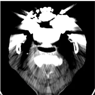
Fig. 3. Computed tomography (CT) artifact due to a dental prosthesis: artifacts in a CT scan can cause significant errors in proton therapy dose calculation.
Ninety-eight patients underwent definitive CCRT. Among them, 63 patients were treated with tomotherapy alone, while the others were treated with tomotherapy/IMPT combi- nation therapy. Before PSM, grade ≥2 mucositis (tomotherapy vs. tomotherapy/IMPT, 69.8% vs. 45.7%; P=0.019) was found to be less frequent in the tomotherapy/IMPT group than in the tomotherapy group. After PSM, it was still less frequent in the tomotherapy/IMPT group (tomotherapy vs.
tomotherapy/IMPT, 62.9% vs. 45.7%; P=0.150), but the dif- ference was not statistically significant. In the whole cohort, LR and distant failures occurred in 9 (9.2%) and 12 (12.2%) patients in the tomotherapy and tomotherapy/IMPT groups, respectively, with no difference between the groups. Our preliminary results showed comparable early therapeutic outcomes with more favorable acute toxicity profiles in the tomotherapy/IMPT combination group than in the tomother- apy group.
Pitfalls of proton therapy for HNC
During proton therapy planning, the exact location of the Bragg peak is crucial for precise delivery of the radiation dose. However, many factors affect the precise position of the Bragg peak. The range of the Bragg peak depends on the energy of the beam, as well as the density of the tissue that the beam passes. In HN sites, the path of the beam usually contains soft tissue, bone, air, and other materials, such as dental prostheses. Furthermore, changes in aeration (e.g., in nasal cavity and paranasal sinus), tumor volume, and body shape due to weight loss during the treatment occur in the majority of patients. These changes can shift the Bragg peak to a different position, which is calculated during the treatment-planning phase in a simulation. As a result, an overdose or underdose to the tumor and/or critical normal tissues can occur during treatment. To compensate for this uncertainty, additional margins and/or robust opti- mization methods are essential for optimal treatment planning.
In addition, adaptive planning, i.e., re-planning to compen- sate for changes during the course of RT, should be im- plemented if concerns for under- or overdosing arise.
Uncertainty is also related to the dose calculation method.
The treatment planning system (TPS) calculates the dose distribution in the body based on the interaction between the proton energy and matter (body) using the stopping
power acquired in simulation computed tomography (CT) scan. If dental or surgical implants are present in the treatment volume, artifacts in the CT scan can cause significant errors in the proton stopping powers used for dose calculation.52,53) (Fig. 3) In addition, a metal implant such as titanium within the beam path of proton therapy should be calibrated during CT density correction, which may result in a significant dose difference. In such cases, the use of proton therapy is currently not recommended. The Monte Carlo calculation method is known to be a more accurate algorithm for dose calculation; however, it is a time-consuming method. Currently, commercial TPSs used in the clinic facilitate Monte Carlo calculations.
Recent studies have shown an increase in RBE at the distal edge of the SOBP.54) This means that the biological effect of the proton beam can be enhanced at the distal edge of the beam. Thus, placing critical organs (e.g., the spinal cord and brainstem) at the distal edge of the proton beam should be avoided during treatment planning. Currently, dose calculation is performed based on a fixed RBE of 1.1.
Several investigators have reported unexpected brainstem necrosis in pediatric brain tumor patients treated with proton therapy, which may be associated with different RBE issues.
Currently, RBE-based dose optimization is under inves- tigation.55,56)
Proton therapy generally induces more severe radio- dermatitis than X-ray therapy does, especially when the
scattering technique is used. Multiple Bragg peaks of differ- ent energies are summed to create the SOBP. In addition, proton therapy uses a limited number of beams compared to X-ray therapy. This can lead to a substantial increase in the entrance dose to the skin. Several studies have demon- strated a higher risk of radiodermatitis in patients treated with proton therapy compared with that of patients treated with X-rays.36,43,57) Therefore, physicians should carefully monitor any skin reactions that occur during and after pro- ton therapy.
Future directions
Although many retrospective and case-matching analyses have shown that proton therapy is less toxic than IMRT, the use of proton therapy is still limited because of the high cost of construction and management of proton therapy facilities. In Korea, only two institutions use proton therapy.
Under these circumstances, clinicians should carefully select patients for whom proton therapy would be most beneficial.
A systematic review of cost-effective analyses showed that proton therapy is cost-effective for cases of pediatric brain tumors, well-selected breast cancers, locoregionally advanced non-small cel lung cancer (NSCLC), and high-risk HNCs.
However, this is not true for patients with prostate cancer and early stage NSCLC. The results and conclusions of such an economic analysis could be different in countries with other health systems. Future studies should investigate the utility of proton therapy in Korea. Many studies suggest that normal tissue complication probability (NTCP) model- ing could help select patients who are most suitable for proton therapy.58-60) This modeling technique allows estima- tion of the expected toxicity difference between modalities based on large validated data of irradiated dose and volume of normal tissue and clinical outcomes. For robust results of NTCP modeling, large datasets of dosimetric results of normal tissue and toxicity outcomes are essential. In addi- tion, data showing better efficacy of proton therapy com- pared to that of IMRT are scarce. Thus, large prospective studies, including randomized controlled studies, should be conducted to validate the clinical outcomes of proton ther- apy by providing confirmative level 1 evidence.
As mentioned above, there is a controversy around the use of a fixed RBE value of 1.1 during proton therapy
planning. RBE can vary depending on the fractionation, depth of protons, and properties of normal tissues or tumors.16,17) A recent study suggested that the brain-specific RBE is 1.18, based on the temporal lobe changes of 60 NPC cases treated with the scattering technique of proton therapy.61) An increase in application of proton therapy will help optimize normal tissue and tumor-specific RBE in the future. Furthermore, RBE-optimized planning is currently under investigation.
Proton therapy for HNC is generally delivered with two or three beams at fixed angles. Determining the optimal beam angle is a crucial component for obtaining the best dose distribution. In selected cases, arc therapy, which de- livers beams over 360 degrees, may achieve a significant reduction in high and moderate doses to normal tissues, even though arc beams inevitably increase the low dose area of normal tissues.62) The optimal beam arrangement, includ- ing use of arc therapy, requires further investigation. FLASH RT is a new cutting-edge technology. FLASH RT is an ultra-high dose rate RT with a mean dose rate of >40 Gy/s (c.f., the conventional usual dose rate of ≥0.01 Gy/s). Many cell- and animal-based studies have shown that FLASH RT using X-ray results in a significant decrease in normal tissue toxicity, making it an interesting treatment modality. Certainly, more research is needed to evaluate this technique in the clinical setting.
Conclusion
The current evidence of proton therapy utility is relatively weak; however, the technique is associated with lower tox- icity compared to those of standard treatments. The efficacy of proton therapy should be investigated in well-designed multi-center studies. The issues of cost-effectiveness and uncertainty will be solved with proper patient selection and rapid evolution of technology in the near future.
References
1) Veldeman L, Madani I, Hulstaert F, De Meerleer G, Mareel M, De Neve W. Evidence behind use of intensity-modulated radio- therapy: a systematic review of comparative clinical studies.
Lancet Oncol. 2008;9:367-375.
2) Luo MS, Huang GJ, Liu HB. Oncologic outcomes of IMRT versus CRT for nasopharyngeal carcinoma: A meta-analysis. Medicine
(Baltimore). 2019;98:e15951.
3) Gupta T, Kannan S, Ghosh-Laskar S, Agarwal JP. Systematic re- view and meta-analyses of intensity-modulated radiation therapy versus conventional two-dimensional and/or or three-dimen- sional radiotherapy in curative-intent management of head and neck squamous cell carcinoma. PLoS One. 2018;13:e0200137.
4) Zhang B, Mo Z, Du W, Wang Y, Liu L, Wei Y. Intensity-modu- lated radiation therapy versus 2D-RT or 3D-CRT for the treat- ment of nasopharyngeal carcinoma: A systematic review and meta-analysis. Oral Oncol. 2015;51:1041-1046.
5) Marta GN, Silva V, de Andrade Carvalho H, de Arruda FF, Hanna SA, Gadia R, et al. Intensity-modulated radiation therapy for head and neck cancer: systematic review and meta-analysis.
Radiother Oncol. 2014;110:9-15.
6) Rong Y, Welsh J. Basics of particle therapy II biologic and dosi- metric aspects of clinical hadron therapy. Am J Clin Oncol.
2010;33:646-649.
7) Palm A, Johansson KA. A review of the impact of photon and pro- ton external beam radiotherapy treatment modalities on the dose distribution in field and out-of-field; implications for the long-term morbidity of cancer survivors. Acta Oncol. 2007;
46:462-473.
8) PTCOG (http://www.ptcog.ch).
9) Alterio D, Marvaso G, Ferrari A, Volpe S, Orecchia R, Jereczek-Fossa BA. Modern radiotherapy for head and neck cancer. Semin Oncol. 2019;46:233-245.
10) Moreno AC, Frank SJ, Garden AS, Rosenthal DI, Fuller CD, Gunn GB, et al. Intensity modulated proton therapy (IMPT) - The future of IMRT for head and neck cancer. Oral Oncol.
2019;88:66-74.
11) Kim JK, Leeman JE, Riaz N, McBride S, Tsai CJ, Lee NY.
Proton Therapy for Head and Neck Cancer. Curr Treat Options Oncol. 2018;19:28.
12) Leeman JE, Romesser PB, Zhou Y, McBride S, Riaz N, Sherman E, et al. Proton therapy for head and neck cancer: expanding the therapeutic window. Lancet Oncol. 2017;18:e254-e265.
13) Li X, Lee A, Cohen MA, Sherman EJ, Lee NY. Past, present and future of proton therapy for head and neck cancer. Oral Oncol.
2020;110:104879.
14) Paganetti H, Niemierko A, Ancukiewicz M, Gerweck LE, Goitein M, Loeffler JS, et al. Relative biological effectiveness (RBE) values for proton beam therapy. Int J Radiat Oncol Biol Phys. 2002;53:407-421.
15) Paganetti H. Relative biological effectiveness (RBE) values for proton beam therapy. Variations as a function of biological end- point, dose, and linear energy transfer. Phys Med Biol. 2014;
59:R419-472.
16) Peeler CR, Mirkovic D, Titt U, Blanchard P, Gunther JR, Mahajan A, et al. Clinical evidence of variable proton biological effectiveness in pediatric patients treated for ependymoma.
Radiother Oncol. 2016;121:395-401.
17) Willers H, Allen A, Grosshans D, McMahon SJ, von Neubeck C, Wiese C, et al. Toward A variable RBE for proton beam therapy.
Radiother Oncol. 2018;128:68-75.
18) Mohan R, Grosshans D. Proton therapy - Present and future. Adv Drug Deliv Rev. 2017;109:26-44.
19) Mercado CE, Holtzman AL, Rotondo R, Rutenberg MS, Mendenhall WM. Proton therapy for skull base tumors: A review of clinical outcomes for chordomas and chondrosarcomas. Head Neck. 2019;41:536-541.
20) Munzenrider JE, Liebsch NJ. Proton therapy for tumors of the skull base. Strahlenther Onkol. 1999;175:57-63.
21) Weber DC, Rutz HP, Pedroni ES, Bolsi A, Timmermann B, Verwey J, et al. Results of spot-scanning proton radiation ther- apy for chordoma and chondrosarcoma of the skull base: the Paul Scherrer Institut experience. Int J Radiat Oncol Biol Phys.
2005;63:401-409.
22) Weber DC, Badiyan S, Malyapa R, Albertini F, Bolsi A, Lomax AJ, et al. Long-term outcomes and prognostic factors of skull-base chondrosarcoma patients treated with pencil-beam scanning proton therapy at the Paul Scherrer Institute. Neuro Oncol. 2016;18:236-243.
23) Kountouri M, Pica A, Walser M, Albertini F, Bolsi A, Kliebsch U, et al. Radiation-induced optic neuropathy after pencil beam scanning proton therapy for skull-base and head and neck tumours.
Br J Radiol. 2020;93:20190028.
24) Mody MD, Saba NF. Multimodal Therapy for Sinonasal Malignancies: Updates and Review of Current Treatment. Curr Treat Options Oncol. 2020;21:4.
25) Patel SH, Wang Z, Wong WW, Murad MH, Buckey CR, Mohammed K, et al. Charged particle therapy versus photon therapy for paranasal sinus and nasal cavity malignant diseases:
a systematic review and meta-analysis. Lancet Oncol. 2014;
15:1027-1038.
26) Yu NY, Gamez ME, Hartsell WF, Tsai HK, Laramore GE, Larson GL, et al. A Multi-Institutional Experience of Proton Beam Therapy for Sinonasal Tumors. Adv Radiat Oncol.
2019;4:689-698.
27) Pasalic D, Ludmir EB, Allen PK, Thaker NG, Chapman BV, Hanna EY, et al. Patient-reported outcomes, physician-reported toxicities, and treatment outcomes in a modern cohort of patients with sinonasal cancer treated using proton beam therapy.
Radiother Oncol. 2020;148:258-266.
28) Svajdova M, Dubinsky P, Kazda T. Radical external beam re-ir- radiation in the treatment of recurrent head and neck cancer:
Critical review. Head Neck. 2021;43:354-366.
29) Lee J, Shin IS, Kim WC, Yoon WS, Koom WS, Rim CH.
Reirradiation with intensity-modulated radiation therapy for re- current or secondary head and neck cancer: Meta-analysis and systematic review. Head Neck. 2020;42:2473-2485.
30) Foster CC, Fan M, Lee NY, Yom SS, Heaton CM, Deraniyagala R, et al. Is It Worth It? Consequences of Definitive Head and Neck Reirradiation. Semin Radiat Oncol. 2020;30:212-217.
31) Simone CB, Plastaras JP, Jabbour SK, Lee A, Lee NY, Choi JI, et al. Proton Reirradiation: Expert Recommendations for Reducing Toxicities and Offering New Chances of Cure in Patients With Challenging Recurrence Malignancies. Semin Radiat Oncol.
2020;30:253-261.
32) Phan J, Sio TT, Nguyen TP, Takiar V, Gunn GB, Garden AS, et al. Reirradiation of Head and Neck Cancers With Proton Therapy: Outcomes and Analyses. Int J Radiat Oncol Biol Phys.
2016;96:30-41.
33) McDonald MW, Zolali-Meybodi O, Lehnert SJ, Estabrook NC, Liu Y, Cohen-Gadol AA, et al. Reirradiation of Recurrent and Second Primary Head and Neck Cancer With Proton Therapy.
Int J Radiat Oncol Biol Phys. 2016;96:808-819.
34) Verma V, Rwigema JM, Malyapa RS, Regine WF, Simone CB, Systematic assessment of clinical outcomes and toxicities of pro- ton radiotherapy for reirradiation. Radiother Oncol. 2017;
125:21-30.
35) Gunn GB, Blanchard P, Garden AS, Zhu XR, Fuller CD, Mohamed AS, et al. Clinical Outcomes and Patterns of Disease Recurrence After Intensity Modulated Proton Therapy for Oropharyngeal Squamous Carcinoma. Int J Radiat Oncol Biol Phys. 2016;95:360-367.
36) Aljabab S, Liu A, Wong T, Liao JJ, Laramore GE, Parvathaneni U. Proton Therapy for Locally Advanced Oropharyngeal Cancer:
Initial Clinical Experience at the University of Washington. Int J Part Ther. 2020;6:1-12.
37) Slater JD, Yonemoto LT, Mantik DW, Bush DA, Preston W, Grove RI, et al. Proton radiation for treatment of cancer of the oropharynx: early experience at Loma Linda University Medical Center using a concomitant boost technique. Int J Radiat Oncol Biol Phys. 2005;62:494-500.
38) Sio TT, Lin HK, Shi Q, Gunn GB, Cleeland CS, Lee JJ, et al.
Intensity Modulated Proton Therapy Versus Intensity Modulated Photon Radiation Therapy for Oropharyngeal Cancer: First Comparative Results of Patient-Reported Outcomes. Int J Radiat Oncol Biol Phys. 2016;95:1107-1114.
39) Blanchard P, Garden AS, Gunn GB, Rosenthal DI, Morrison WH, Hernandez M, et al. Intensity-modulated proton beam ther- apy (IMPT) versus intensity-modulated photon therapy (IMRT) for patients with oropharynx cancer - A case matched analysis.
Radiother Oncol. 2016;120:48-55.
40) Cao J, Zhang X, Jiang B, Chen J, Wang X, Wang L, et al.
Intensity-modulated proton therapy for oropharyngeal cancer reduces rates of late xerostomia. Radiother Oncol. 2021;160:
32-39.
41) Yoon HG, Ahn YC, Oh D, Noh JM, Park SG, Nam H, et al. Early Clinical Outcomes of Intensity Modulated Radiation Therapy/
Intensity Modulated Proton Therapy Combination in Comparison with Intensity Modulated Radiation Therapy Alone in Oropharynx Cancer Patients. Cancers (Basel). 2021;13: 1549.
42) Stromberger C, Cozzi L, Budach V, Fogliata A, Ghadjar P, Wlodarczyk W, et al. Unilateral and bilateral neck SIB for head and neck cancer patients : Intensity-modulated proton therapy, tomotherapy, and RapidArc. Strahlenther Onkol. 2016;192:232-239.
43) Romesser PB, Cahlon O, Scher E, Zhou Y, Berry SL, Rybkin A, et al. Proton beam radiation therapy results in significantly re- duced toxicity compared with intensity-modulated radiation therapy for head and neck tumors that require ipsilateral radiation. Radiother Oncol. 2016;118:286-292.
44) Zakeri K, Wang H, Kang JJ, Lee A, Romesser P, Mohamed N, et al. Outcomes and prognostic factors of major salivary gland tu- mors treated with proton beam radiation therapy. Head Neck.
2021;43:1056-1062.
45) Moon SH, Cho KH, Lee CG, Keum KC, Kim YS, Wu HG, et al.
IMRT vs. 2D-radiotherapy or 3D-conformal radiotherapy of na- sopharyngeal carcinoma : Survival outcome in a Korean mul- ti-institutional retrospective study (KROG 11-06). Strahlenther Onkol. 2016;192:377-385.
46) Lewis GD, Holliday EB, Kocak-Uzel E, Hernandez M, Garden AS, Rosenthal DI, et al. Intensity-modulated proton therapy for nasopharyngeal carcinoma: Decreased radiation dose to normal structures and encouraging clinical outcomes. Head Neck.
2016;38:E1886-1895.
47) Liu SW, Li JM, Chang JY, Yu JM, Chen Q, Jiang QA, et al. A treatment planning comparison between proton beam therapy and intensity-modulated x-ray therapy for recurrent nasophar- yngeal carcinoma. J Xray Sci Technol. 2010;18:443-450.
48) Minatogawa H, Yasuda K, Dekura Y, Takao S, Matsuura T, Yoshimura T, et al. Potential benefits of adaptive intensity- modulated proton therapy in nasopharyngeal carcinomas. J Appl Clin Med Phys. 2021;22:174-183.
49) Holliday EB, Garden AS, Rosenthal DI, Fuller CD, Morrison WH, Gunn GB, et al. Proton Therapy Reduces Treatment- Related Toxicities for Patients with Nasopharyngeal Cancer: A Case-Match Control Study of Intensity-Modulated Proton Therapy and Intensity-Modulated Photon Therapy. International Journal of Particle Therapy. 2015;2:19-28.
50) Alterio D, D'Ippolito E, Vischioni B, Fossati P, Gandini S, Bonora M, et al. Mixed-beam approach in locally advanced na- sopharyngeal carcinoma: IMRT followed by proton therapy boost versus IMRT-only. Evaluation of toxicity and efficacy. Acta Oncol. 2020;59:541-548.
51) Park SG, Ahn YC, Oh D, Noh JM, Ju SG, Kwon D, et al. Early clinical outcomes of helical tomotherapy/intensity-modulated proton therapy combination in nasopharynx cancer. Cancer Sci.
2019;110:2867-2874.
52) Verburg JM, Seco J. Dosimetric accuracy of proton therapy for chordoma patients with titanium implants. Med Phys. 2013;
40:071727.
53) Richard P, Sandison G, Dang Q, Johnson B, Wong T, Parvathaneni U. Dental amalgam artifact: Adverse impact on tumor visual- ization and proton beam treatment planning in oral and orophar- yngeal cancers. Pract Radiat Oncol. 2015;5:e583-588.
54) Saager M, Peschke P, Brons S, Debus J, Karger CP. Determination of the proton RBE in the rat spinal cord: Is there an increase to- wards the end of the spread-out Bragg peak? Radiother Oncol.
2018;128:115-120.
55) Sanchez-Parcerisa D, Lopez-Aguirre M, Dolcet Llerena A, Udias JM. MultiRBE: Treatment planning for protons with selective ra- diobiological effectiveness. Med Phys. 2019;46:4276-4284.
56) Guan F, Geng C, Ma D, Bronk L, Kerr M, Li Y, et al. RBE Model- Based Biological Dose Optimization for Proton Radiobiology Studies. Int J Part Ther. 2018;5:160-171.
57) Kozak KR, Smith BL, Adams J, Kornmehl E, Katz A, Gadd M, et al. Accelerated partial-breast irradiation using proton beams:
initial clinical experience. Int J Radiat Oncol Biol Phys. 2006;
66:691-698.
58) Hansen CR, Friborg J, Jensen K, Samsoe E, Johnsen L, Zukauskaite R, et al. NTCP model validation method for DAHANCA patient selection of protons versus photons in head and neck cancer radiotherapy. Acta Oncol. 2019;58:1410-1415.
59) Rwigema JM, Langendijk JA, Paul van der Laan H, Lukens JN, Swisher-McClure SD, Lin A. A Model-Based Approach to Predict Short-Term Toxicity Benefits With Proton Therapy for Oropharyngeal Cancer. Int J Radiat Oncol Biol Phys. 2019;
104:553-562.
60) Brodin NP, Kabarriti R, Pankuch M, Schechter CB, Gondi V, Kalnicki S, et al. A Quantitative Clinical Decision-Support Strategy Identifying Which Patients With Oropharyngeal Head and Neck Cancer May Benefit the Most From Proton Radiation Therapy. Int J Radiat Oncol Biol Phys. 2019;104:540-552.
61) Zhang YY, Huo WL, Goldberg SI, Slater JM, Adams JA, Deng XW, et al. Brain-Specific Relative Biological Effectiveness of Protons Based on Long-term Outcome of Patients With Nasopharyngeal Carcinoma. Int J Radiat Oncol Biol Phys. 2021.
62) Apinorasethkul O, Kirk M, Teo K, Swisher-McClure S, Lukens JN, Lin A. Pencil beam scanning proton therapy vs rotational arc radiation therapy: A treatment planning comparison for post- operative oropharyngeal cancer. Med Dosim. 2017;42:7-11.