Ⅰ. INTRODUCTION
Impacted teeth are those with a delayed eruption time or that are not expected to erupt completely based on clinical and radiographic assessment
1). The maxillary canine is second next to the mandibular third molar in its frequency of impaction. The preva- lence of impacted maxillary canines is 12% in the general population
2,3). Management options for im- pacted maxillary canines can include (1) continued observation, (2) extraction of the primary canine to
aid spontaneous eruption, (3) uncovering and bond- ing of the impacted tooth and its eruption using or- thodontic traction4), (4) autotransplantation
5), and (5) extraction followed by prosthetic replacement
6). Autotransplantation is the process in which tooth, usually impacted, is surgically transferred to correct position or to replace another tooth in the same alve- olus
7). Autotransplantation should be considered when the degree of malposition is too severe to cor- rect by orthodontic alignment.
Medical Rapid Prototyping (MRP) is defined as the manufacture of dimensionally accurate physical mod- els of human anatomy derived from medical image da- ta using a variety of Rapid Prototyping (RP) technolo- gies. It has been applied to a range of medical special- ities, including oral and maxillofacial surgery
8-14), dental implantology
15), neurosurgery
16,17), and orthope-
Autotransplantation of an impacted maxillary canine using Rapid Prototyping : A case report
Nan-Ju Cho, Nan-Young Lee, Sang-Ho Lee
Department of Pediatric Dentistry, Collage of Dentistry, Chosun University
Management options for impacted maxillary canines can include (1) continued observation, (2) extrac- tion of the primary canine to aid spontaneous eruption, (3) uncovering and bonding of the impacted tooth and its eruption using orthodontic traction, (4) autotransplantation, and (5) extraction followed by pros- thetic replacement. Autotransplantation should be considered when the degree of malposition is too se- vere to correct by orthodontic alignment. The present report describes the management of an ectopic eruption of the left maxillary canine in an 10-year-old girl. The treatment included the extraction of pri- mary maxillary left canine and the autotransplantation using a Rapid Prototyping model. By using RP model to contour the recipient bone and check for fitting in the prepared socket, the extra-oral time can reduce. The autotransplanted canine showed mobility within normal limit, negative response to percus- sion and positive to electric pulp test after 6 months.
Key words : Impacted canine, Autotransplantation, Rapid Prototyping Abstract
교신저자 : 이 상 호
광주광역시 동구 서석동 375번지 조선대학교 치과대학 소아치과학교실
Tel: 062-220-3860 Fax: 062-225-8240 E-mail: [email protected]
※이 논문은 2007년도 조선대학교 학술연구비의 지원에 의해 연구되었음.
dics
18). Lee et a1.
19)reported a lower extra-oral time and improved contact between the donor tooth and recipient bone after performing autotransplantation using Rapid Prototyping for a similar model to the donor tooth.
This case report demonstrates an autotransplanta- tion of an impacted maxillary canine using Rapid Prototyping in a 10-year-old girl.
Ⅱ. CASE REPORT Clinical history
A 10-year-old girl came for her first visit with her parents. Their chief complaint was ectopic eruption of the left maxillary canine. Clinical examination showed a retained left maxillary deciduous canine and spacing of maxillary anteriors(Fig. 1). The radi-
ographic analysis revealed that the left maxillary ca- nine was impacted, and its crown was in buccal por- tion to the root of the left maxillary second premo- lar(Fig. 2). The location of the impacted canine was
Fig. 1. Initial clinical examination showing a retained left maxillary deciduous canine and spacing of maxillary anteriors.
Fig. 2. Radiographs revealing the left maxillay canine with open apex buccally impacted.
Fig. 3. The cusp of the impacted canine was located in sector 3 according to the criteria proposed by
Crescini et al.
20). The inclination of impacted maxillary canine was 113�, which is the angle between
the long axis of the impacted maxillary canine and midline. The distance between the cusp of the
canine and the occlusal plane was 17mm.
assessed by panoramic image using a criteria pro- posed by Crescini et al
20). The cusp of the impacted canine was located in sector 3. The inclination of the impacted maxillary canine was 113�, which is the angle between the long axis of the impacted maxil- lary canine and midline. The distance between the cusp of the canine and the occlusal plane was 17mm.
The left maxillary canine had an open apex(Fig. 3).
The patient’s medical history was noncontributory.
The canine was in a difficult malposition, so that orthodontic treatment was impossible. After discus- sion of treatment options, autotransplantation was selected. The treatment included the extraction of the left maxillary deciduous canine and orthodontic pre-treatment prior to autotransplantation.
Orthodontic pre-treatment with mesial and vertical traction of the impacted maxillary canine may bring the maxillary canine to a more favorable position and facilitate extraction and autotransplantation
21).
Orthodontic procedure
A full thickness flap was raised to expose the corti- cal plate, and the deciduous canine was removed.
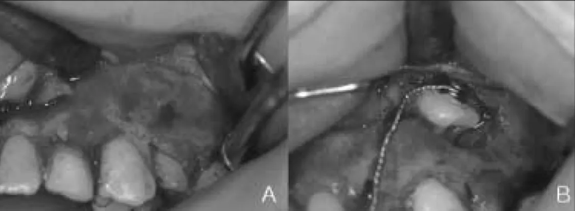
Cortical bone was removed to provide access to the crown, and follicular socket was eliminated. The chain was passed through guiding groove on cortical bone and fixed to impacted canine by a bonded but- ton(Fig. 4). The flap was then repositioned and su- tured in its original seat.
By means of fixed and removable appliances, a suf- ficient space was made in the maxillary arch for the impacted canine. At the same time orthodontic trac- tion was performed with a force of approximately 60
�100mg for 8 months. When the space opening was completed and the canine was located at buccal side of the left maxillary first premolar, the autotrans- plantation was performed(Fig. 5).
Fig. 4. Surgical exposure(A) and bonding of a button to the canine(B).
Fig. 5. By means of orthodontic appliances, a sufficient space was made for the impacted
canine and the canine was located at the buccal side of the left maxillary first premolar.
Autotransplantation
Computed tomography (CT) was taken for pre-ex- amination of the donor tooth and the recipient site.
The size of the donor tooth and the recipient site were measured. Three-dimensional data (DICOM format; digital imaging and communications in medi- cine) of the donor tooth using a CB MercuRay
�(Hitachi medical corp., Japan) were obtained . A slit thickness was 0.20-mm. DICOM CT files were im- ported into CT image processing software (Vwork 5.0, Cybermed, Seoul, Korea), which built up the image and constructed the virtual 3D model(Fig. 6).
The software sent the Structured Triangular Language (STL) file to the Rapid Prototyping ma- chine for fabrication of the donor tooth model by 3D printing.
250mg of systemic amoxillin (Ilsung Augumentin
�, Seoul, Korea) was administrated one hour before
surgery. After local anesthesia, a full thickness flap of the recipient site was reflected and the recipient bone was prepared using surgical bur with saline ir-
Fig. 7. Autotransplantation of the canine. A. RP model and the donor tooth. B. The donor tooth is placed into the prepared socket.
Fig. 8. 6 month after autotransplantation. A. Intra-oral view. B. Panoramic view.
Fig. 6. 3-D reconstructed image.
rigation until the model of the left maxillary canine fit well into recipient site. After preforming an intra- crevicular incision around the crown, the canine was extracted with minimal injury and placed into the re- cipient bone socket with slight sub-occlusion(Fig. 7).
Extra-oral time was about 50 seconds and the tooth was stored in Hank’s balanced salt solution (HBSS) during the extra-oral period. The transplanted tooth was stable enough for the fixation with modified an- chor suture and periodontal pack.
The patient was given systemic amoxillin (Ilsung Augumentin
�, Seoul, Korea), 750mg tid for 7 days, and mouth rinsing with 0.1 % chlorhexidine (Hexamedine
�, Bukwang Parmaceutical Co., Seoul, Korea) was also recommended. The sutures and peri- odontal pack were removed 1 week after surgery. No endodontic treatment was planned unless the check- up radiographs revealed complication because the donor tooth had an open apex.
The patient was recalled every 2 weeks for 3 months, and every 1 month thereafter. Registrations were made regarding tooth mobility, percussion, sen- sitivity upon electric stimulation, occlusal contact, periodontal condition and radiographic signs of root resorption. The autotransplanted canine showed mo- bility within normal limit, negative response to per- cussion and normal gingival condition with pocket depths below 3mm. The canine responded positively to electric pulp test after 6 months. The radiograph showed continuance of lamina dura and no root re- sorption(Fig. 8).
Ⅲ. DISCUSSION
The prognosis for successful autotransplantation is dependent on a number of factors, such as root de- velopment, position of the tooth, surgical technique, extra-oral time, type of splinting, fixation period and endodontic technique
22,23). The revasculation of the pulp appears more predictable in roots with open apices than in roots with closed apices. The pulp healing was the usual finding in teeth at stage 1�5 of root development and a diameter of the apical foramen above 1mm had a low risk of pulp necrosis
24). Continued root development after trans- plantation can also be expected if a donor tooth is immature and Hertwig’s epithelial sheath is pre-
served around the apices
25). However, the amount of root development to be expected cannot be predicted.
Therefore it is favorable for donor tooth to be its maximal length as possible. Tsukiboshi
23)recom- mended that considering pulp healing and continued root development, the ideal autotransplantation time of a tooth with incomplete root formation is at the stage 4�5 of root development. In this case report, the left maxillary canine had an open apex at the op- eration and responded positively to EPT after 6 month.
A vital periodontal ligament is essential for the long term survival of autotransplantation. Loss of vi- tality of periodontal ligament could lead to root re- sorption
26). Therefore it is important to minimize the extra-oral time and store the donor tooth in a physi- ologic storage medium during the extra-oral period in order to preserve the periodontal ligament. Lee et a1.
19)reported that the average extra-oral time was 7.4 min in the autotransplantation procedure using Rapid Prototyping. In this study, the extra-oral time was about 50 seconds and the tooth was stored in Hank’s balanced salt solution (HBSS) throughout the extra-oral time. No visible root resorption was observed on radiograph.
The gap between the recipient site and the root surface of the transplanted tooth is an important fac- tor in autotransplantation. When the root surface of the transplanted tooth is too close to recipient site, it is likely to occur ankylosis due to mechanical damage of periodontal ligament. On the other hand, when the recipient socket is too wide, the bone healing would be delayed
23). Optimal contact with the recipi- ent site can improve blood supply to periodontal liga- ment and provide better wound healing. Therefore the accuracy of RP model compared with real donor tooth may affect the prognosis of autotransplanta- tion. Lee et al.
27)evaluated the dimensional errors among the real tooth, the 3D CT image and RP mod- el and found that an average of absolute error was 0.291mm between the real teeth and the RP model.
In this case report, the RP model which was fabricat-
ed with 3D printing method was about 1mm larger
than real tooth. Several studies
19,28)have been report-
ed a successful rate of an autotransplantation when
the gap between the recipient site and the root sur-
face of the transplanted tooth was about 1�2mm
within the acceptable error. However the model in this case report did not show the fine dilaceration of the real donor tooth.
The type and period of splinting may have an effect on pulpal and periodontal healing of replanted and autotransplanted teeth. Bauss et al.
29)showed in a study of autotransplanted immature third molars that ankylosis and pulp necrosis increased signifi- cantly after rigid fixation for 4 weeks compared with suture splinting for a week and also prolonged rigid fixation group revealed a significantly shorter final root length and root length increment. In this case report, the fixation was made with modified anchor suture and periodontal pack for 1 week because the transplanted tooth was stable.
By using RP model to contour the recipient bone and check for fit in the prepared socket, the number of trials with the real donor tooth may decrease.
Therefore RP model can help to minimize the extra- socket time and the possible injury of transplanted tooth during the process of autotransplantation. Also it may improve the fitness between the donor tooth and recipient site. Especially in the case of impacted tooth, the use of a RP model seems to provide signif- icant advantages in the diagnosis, treatment plan- ning, and surgical procedure.
The present report describes the management of an impacted left maxillary canine in an 10-year-old girl.
The treatment included the extraction of primary maxillary left canine, the orthodontic pre-treatment and the autotransplantation using a Rapid Prototyping model. The autotransplanted canine showed mobility within normal limit, negative re- sponse to percussion and positive to electric pulp test after 6 months.
REFERENCES
1. Thilander B, Jakobsson SO : Local factors in im- paction of maxillary canines. Acta Odontol Scand, 26:145-168, 1968.
2. Bass TB : Observations on the misplaced upper canine tooth. Dent Pract Dent Rec, 18:25-33, 1967.
3. Rayne J : The unerupted maxillary canine. Dent Pract Dent Rec, 19:194-204, 1969.
4. 최형준, 이종은, 이제호 등 : 수평 매복된 상악 견치의
교정적 견인. 대한소아치과학회지, 30:600-604, 2003.
5. 김태완, 김현정, 김연진 등 : 매복된 상악 견치의 자가치 아이식을 통한 치험례. 대한소아치과학회지, 30:326- 333, 2003.
6. Saiar M, Rebellato J : Maxillary Impacted Canine with Congenitally Absent Premolars.
Angle Orthod, 74:568-575, 2004.
7. Morse DR : Plantation procedures: history, im- munology and clinical considerations. J Oral Implantol, 7:176-192 contd, 1977.
8. Anderl H, Zur Nedden D, Muhlbauer W, et al. : CT-guided stereolithography as a new tool in raniofacial surgery. Br J Plast Surg, 47:60-64, 1994.
9. Arvier JF, Barker TM, Yau YY, et al. : Maxillofacial biomodelling. Br J Oral Maxillofac Surg, 32:276-283, 1994.
10. D’Urso PS, Barker TM, Earwaker WJ, et al. : Stereolithographic biomodelling in cranio-maxillo- facial surgery: a prospective trial. J Cranioma- xillofac Surg, 27:30-37, 1999.
11. Eufinger H, Wehmoller M : Individual prefabri- cated titanium implants in reconstructive cranio- facial surgery: clinical and technical aspects of the first 22 cases. Plast Reconstr Surg, 102:300- 308, 1998.
12. Gateno J, Allen ME, Teichgraeber JF, et al. : An in vitro study of the accuracy of a new protocol for planning distraction osteogenesis of the mandible. J Oral Maxillofac Surg, 58:985-990, 2000.
13. Sailer HF, Haers PE, Zollikofer CP, et al. : The value of stereolithographic models for preopera- tive diagnosis of craniofacial deformities and planning of surgical corrections. Int J Oral Maxillofac Surg, 27:327-333, 1998.
14. Hughes CW, Page K, Bibb R, et al. : The cus- tom-made titanium orbital floor prosthesis in re- construction for orbital floor fractures. Br J Oral Maxillofac Surg, 41:50-53, 2003.
15. Heckmann SM, Winder W, Meyer M, et al. : Overdenture attachment selection and the load- ing of implant and denture-bearing area. Part 1:
In vivo verification of stereolithographic model.
Clin Oral Implants Res, 12:617-623, 2001.
16. Winder RJ, Cooke RS, Gray J, et al. : Medical
Rapid Prototyping and 3D CT in the manufacture
of custom made cranial titanium plates. J Med Eng Technol, 23:26-28, 1999.
17. Heissler E, Fischer F, Bolouri S, et al. : Custom- made cast titanium implants produced with CAD/CAM for the reconstruction of cranium de- fects. Int J Oral Maxillofac Surg, 27:334-338, 1998.
18. Minns RJ, Bibb R, Banks R, et al. : The use of a reconstructed three-dimensional solid model from CT to aid surgical management of a total knee arthroplasty: a case study. Med Eng Phys, 25:523-526, 2003.
19. Lee SJ, Jung IY, Lee CY, et al. : Clinical appli- cation of computer-aided Rapid Prototyping for tooth transplantation. Dent Traumatol, 17:114- 119, 2001.
20. Crescini A, Clauser C, Giorgetti R, et al. : Tunnel traction of infraosseous impacted maxil- lary canines. A three-year periodontal follow-up.
Am J Orthod Dentofacial Orthop, 105:61-72, 1994.
21. Berglund L, Kurol J, Kvint S : Orthodontic pre- treatment prior to autotransplantation of palatal- ly impacted maxillary canines: case reports on a new approach. Eur J Orthod, 18:449-456, 1996.
22. Norderam A : Autotransplantation of teeth. A clinical and experimental investigation. Acta Odontol Scand, 21:Suppl 33:7-76, 1963.
23. Tsukiboshi M : Autotransplantation of teeth: re- quirements for predictable success. Dent
Traumatol, 18:157-180, 2002.
24. Andreasen JO, Paulsen HU, Yu Z, et al. : A long-term study of 370 autotransplanted premo- lars. Part II. Tooth survival and pulp healing subsequent to transplantation. Eur J Orthod, 12:14-24, 1990.
25. Andreasen JO, Paulsen HU, Yu Z, et al. : A long-term study of 370 autotransplanted premo- lars. Part IV. Root development subsequent to transplantation. Eur J Orthod, 12:38-50, 1990.
26. Andreasen JO : Effect of extra-alveolar period and storage media upon periodontal and pulpal healing after replantation of mature permanent incisors in monkeys. Int J Oral Surg, 10:43-53, 1981.
27. Seong-Jae Lee, Eui-Seong Kim, Kee-Deog Kim, et al. : Accuracy of computer aided Rapid Prototyping model(CARP model) compared with real donor tooth in autogenous tooth transplanta- tion. Journal of Korean dental association, 44:115-122, 2006.
28. Nethander G : Periodontal conditions of teeth autogenously transplanted by a two-stage tech- nique. J Periodontal Res, 29:250-258, 1994.
29. Bauss O, SchilkeR, Fenske C, et al. : Autotransplantation of immature third molars:
influence of different splinting methods and fixa-
tion periods. Dent Traumatol, 18:322-328, 2002.
국문초록
Rapid Prototyping을 이용한 상악 매복 견치의 자가이식 치험례