十全大補湯 藥鍼液의 사람 피부아세포의 콜라게나제 활성 및 프로콜라겐 합성과 티로시나제 활성에 미치는 영향
Effects of Sipgeondaebo-tang Pharmacopuncture Extracts on the Collagenase Activity and Procollagen Synthesis in HS68 Human
Fibroblasts and Tyrosinase Activity
Original Articles
Sena Lee1), Myung-Gyou Kim1), Myoung-Hee Kim2), Hyung-Jun Kim1), Hak Jun Jo1), Kang-Hyun Leem1)*
1 : College of Oriental Medicine, Semyung University, Jechon 390-711, Korea, 2 : Department of Nursing, Semyung University, Jechon 390-711, Korea
ABSTRACT
Objectives This study was designed to investigate the collagen metabolism and tyrosinase activity of Sipgeondaebo-tang Pharmacopuncture extracts (SP).
Methods : The effect of SP on type I procollagen production and collagenase activity in human nor- mal fibroblasts HS68 after UVB (312 nm) irradiation was measured by ELISA method. The tyrosi- nase activity after treatment of SP was measured as well.
Results : Type I procollagen production was recovered by SP in UVB damaged HS68 cells. The increased collagenase activity after UVB damage was significantly recovered by SP. The tyrosinase activity was significantly reduced as well. However, the L-DOPA oxidation was not changed.
Conclusion : SP showed the anti-wrinkle effects and whitening effects in vitro. These results suggest that SP may be a potential pharmacopuncture as an anti-aging pharmacopuncture treatments.
Key Words:
Sipgeondaebo-tang, Shiquandabu-tang, pharmacopuncture extracts, type I procolla- gen, collagenase, tyrosi- nase
Received : 11. 02. 18 Revised : 11. 02. 25 Accepted : 11. 02. 25
The corresponding author : Kang-Hyun Leem. College of Oriental Medicine, Semyung University, Chungbuk 390-711, South Korea. Tel.:+82-43-649-1341; Fax:+82-43-649-1341; e-mail:
I. Introduction
Sipgeondaebo-tang pharmacopuncture extracts (Shiquandabu-tang in chinese, SP) are composed of ten herbal medicines. Its effects are to warm and tonify the qi and blood for qi and blood deficien- cy1,2).
Every man and woman has the desire to look and feel at least ten years younger than their chronolog- ical age. Therefore, in these days, aging seems to be treated as not a nature to accept but a disease or a disorder to overcome. Theories of aging fall into
two categories, (1) programmatic theory and (2) stochastic theory. The programmed theory proposes a clock in our bodies that controls not only our pro- cess of development but also triggers our self- destruction. The stochastic theory proposes that the cross-linking of proteins and other cellular macro- molecules leads to age-dependent diseases and dis- orders. Processes that are associated with cellular damage and aging are the production of free radi- cals (a process much enhanced after ultraviolet irra-
diation) and an increasing number of errors during DNA replication. Cellular manifestations of intrin- sic aging include decreased life span of cells, decreased responsiveness of cells to growth signals, which may reflect loss of cellular receptors to growth factors, and increased responsiveness to growth inhibitors. All these findings are more pro- nounced in cells derived from photo-damaged skin3). Aging process in skin (extrinsic aging) is generally referred to as photo-aging due to chronic exposure to short wavelength UV light (UVB) and is characterized by severe wrinkling and pigmen- tary changes, such as solar lentigo and mottled pig- mentation on exposed areas such as the face, neck, and forearm4). It has been shown that UV irradiation leads to the formation of reactive oxygen species (ROS) that activate the mitogen-activated protein (MAP) kinase pathway, which subsequently ind- uces the expression and activation of matrix metal- loproteinases (MMPs) in human skin in vivo5,6). MMPs including collagenase are considered key factors in the photo-aging process. Melanogenesis was induced after UV irradation as well. The key regulator in melanogenesis is well known as a type of enzyme, tyrosinase. Tyrosinase is a copper-con- taining enzyme present in animal tissues that cat- alyzes the production of melanin7).
In the present study, the effect of SP on type I procollagen production and collagenase activity in human normal fibroblasts HS68 after UVB (312 nm) irradiation were investigated. The tyrosinase activity after treatment of SP was measured as well.
II. Materials and methods
1. Sample preparation
The Ginseng Radix, Atractylodis Rhizoma Alba, Hoelen, Glycyrrhizae Radix, Angelicae Gigantis Radix, Paeonia Radix, Rehmanniae Radix Prepar- ata, Astragali Radix, and Cinnamomi Cortex were
purchased from Omniherb (Korea). SP was pre- pared as follow. The Ginseng Radix (12 g), Atractylodis Rhizoma Alba (12 g), Hoelen (12 g), Glycyrrhizae Radix (12 g), Angelicae Gigantis Ra- dix (12 g), Paeonia Radix (12 g), Rehmanniae Rad- ix Preparata (12 g), Astragali Radix (10 g) and Cin- namomi Cortex (10 g) in 2,000 ml distilled water was heated in a heating extractor for 3 hours. The extract was filtered and concentrated by using the rotary evaporator. The extracts were lyophilized by using freeze dryer (19.5 g). The extract was dissol- ved in water and filtered three times through micro- filter paper and syringe filter (Whatman #2, 0.45 m to 0.2 m). Filtered material was placed in the di- sinfected vial and was sealed for further study.
2. Reagents
All reagents were purchased from Sigma-Aldrich except as mentioned below (St. Louis, MO, USA).
3. Cell culture
HS68 human fibroblasts (Health Protection Agency Culture Collections, UK) were cultured in Dulbecco's Modified Eagle's medium (Gibco, USA) containing 10% fetal bovine serum, 1% antibiotics at 37 C in a humidified atmosphere of 5% CO2. When cells reached above confluency, subculture was conducted at a split ratio of 1:3.
4. UVB irradiation
A UVB lamp (Vilber Lourmat, France) was used as a UVB source. In brief, HS68 cells were rinsed twice with phosphate-buffered saline (PBS), and all irradiations were performed under a thin layer of PBS (200 l/well). Immediately after irradiation, fresh serum-free medium was added to the cells.
After 24 hours incubation period, responses were measured. Mock-irradiated blanks followed the same schedule of medium changes without UVB irradiation.
5. Cell viability
General viability of cultured cells was deter- mined by reduction of 3-(4,5-dimethylthiazol-2-yl)- 2,5-diphenyltetrazolium bromide (MTT) to for- mazan. The human fibroblast cells (HS68) were seeded in 24-well plates at a density of 2 105/ml per well and cultured at 37 C in 5% CO2. Cells we- re pretreated with the sample at a concentration of 100, 10, 1 g/ml for 24 hours prior to UVB irradia- tion. After UVB irradiation, cells were retreated with the sample and incubated for additional 24 hours, before being treated with 0.05 mg/ml (final concentration) of MTT. The blank and control group was cultivated without sample treatment. The cells were then incubated at 37 C for additional 4 hours. The medium containing MTT was discarded, and MTT formazan that had been produced was extracted with 200 l of DMSO. The absorbance was read at 595 nm with a reference wavelength of 690 nm. The cell viability was calculated as fol- lows:
Cell viability (%)
=[(OD595 of sample)/(OD595 of control)] 100
6. Assays of collagen type I synthesis and colla- genase inhibition
HS68 human fibroblasts were inoculated into 24- well plate (2 105 cells/well) and cultured at 37 C in 5% CO2. Cells were pretreated with the sample at a concentration of 10, 30, and 100 g/ml for 24 ho- urs prior to UVB irradiation. After UVB irradiation, cells were retreated with the sample and incubated
for additional 24 hours. The blank and control group was cultivated without sample treatment.
After culturing, the supernatant was collected from each well, and the amount of pro-collagen type I was measured with a procollagen type I C-peptide assay kit (Takara Bio, Japan). The activity of colla- genase was measured with a matrix metallopro- teinase-1 (MMP-1) human biotrak ELISA system (Amersham life science, USA)
7. Tyrosinase inhibition assay
Tyrosinase activity was determined essentially as previously described8). The reation mixtures were prepared by adding 40 U of mushroom tyrosinase to 20 l of SP dissolved in distilled water, and then adding 40 l of 1.5 mM L-tyrosine and 220 l of 0.1 M sodium phosphate buffer (pH 6.5). The resul ting mixture (300 l) was incubated for 10 min at 37 C and then absorbance at 490 nm was mea- sured. The same mixture, but without SP extract, was used as a control.
8. Inhibition of L-DOPA oxidation
The inhibitory effect of SP on L-DOPA oxidation was determined according to the method of Joshi with a slight modification9). 50 l of SP dissolved in 0.1 M sodium phosphate buffer was added to 40 U of mushroom tyrosinase in 900 l of 0.1 M sodium phosphate buffer (pH 6.5). After 6 min of incuba- tion at 37 C, 3 mM of L-DOPA was added. Then the mixture was incubated at 37 C for 15 min.
Activities were quantified by measuring absorbance at 475 nm. The same mixture, but without SP extract, was used as a control.
9. Statistical analysis
The results were expressed as means standard error of the mean (SEM). Significances of changes were determined using the one-way ANOVA with a Dunnett's post hoc test. Values of p 0.05 were con- sidered significant.
III. Results
1. Cytotoxicity on HS68 human fibroblasts
In order to evaluate the cytotoxicity of SP, sam- ples were prepared at various concentrations and used to treat human fibroblasts (HS68). The results of this evaluation are shown in Figure 1 at concen- trations of 10, 30, 100 g/ml. The cell viability was recalculated into 100% of control group. The cell viabilities of SP 10 g/ml treated, SP 30 g/ml treated, SP 100 g/ml treated are 105.2 2.0%, 104.5 0.7%, and 106.2 0.5%, respectively. SP showed no cytotoxicity up to the effective concen- tration for anti-wrinkle activity (less than 100 g/ml).
2. Assay of collagen type I synthesis
To evaluate the amount of collagen type I synthe- sis that occurred upon exposure to the sample, col- lagen type I was quantitatively detected by using the previously described procollagen type I C-pep- tide assay kit. Collagens are synthesized as precur- sor molecules, called procollagens. These molecules contain additional peptide sequences, usually referred to as 'propeptides', at both the amino-terminal end and the carboxy-terminal end.
These propeptides are cleaved from the collagen triple-helix molecule during its secretion, after which the triple-helix collagens are polymerized into extracellular collagen fibrils. Thus, the amount of free propeptide stoichiometrically reflects the amount of collagen molecules synthesized10). The
amounts of type I collagen synthesis of SP were shown in Figure 2. SP did not increase the expres- sion of type I collagen at all concentrations of 10, 30, and 100 g/ml (11.5 2.3 ng/ml, 12.1 2.4 ng/ml, and 13.9 2.3 ng/ml) compared with con- trol group (15.3 1.6 ng/ml, Fig. 2).
3. Assay of collagenase activity
To evaluate the collagenase activity, matrix met- alloproteinase-1 (MMP-1) activity was quantita- tively measured by using the previously described matrix metalloproteinase-1 assay kit. The activities of MMP-1 of SP treatment were recalculated into 100% of control group (Figure 3). SP significantly reduced the MMP-1 activity at concentrations of 10 g/ml (59.1 1.9%, p < 0.05). Both 30 g/ml, and 100 g/ml showed the tendency of reducing pat- terns. However, there were no significances (62.8
9.1%, and 55.9 16.2% respectively, Fig. 3).
4. Tyrosinase activity assay
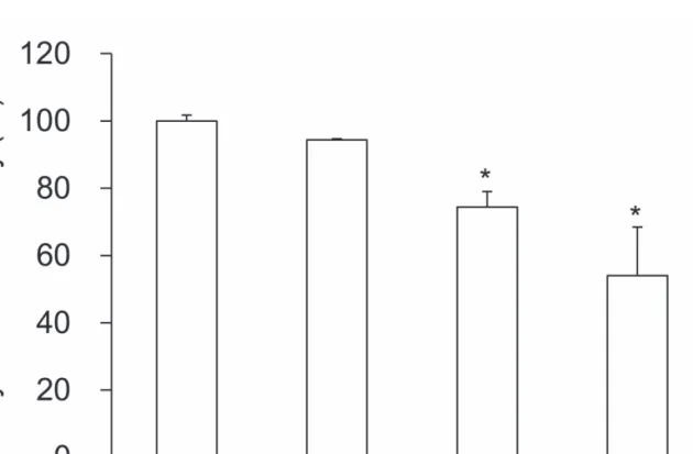
The activities of SP on tyrosinase activity were recalculated into 100% of control group (Fig. 4). SP significantly reduced the tyrosinase activity at con- centrations of 1 and 10 mg/ml (74.4 4.6% and 54.0 14.4%, p < 0.05) in a dose dependent man- ner. The tyrosinase activity of SP 0.1 mg/ml treated group did not show any significance (94.4 0.3%).
5. L-DOPA oxidation
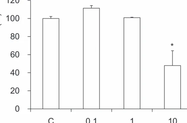
The activities of SP on L-DOPA oxidation were recalculated into 100% of control group (Figure 5).
SP significantly reduced the L-DOPA oxidation activity at concentrations of 10 mg/ml (47.9 16.5%, p < 0.05). SP 0.1 and 1 mg/ml treated
groups did not show any activity (111.3 2.9%
and 100.9 0.5% respectively).
IV. Discussion and conclusion
Sipgeondaebo-tang pharmacopuncture is com- posed of ten herbal medicines; Ginseng Radix, Atractylodis Rhizoma Alba, Hoelen, Glycyrrhizae Radix, Angelicae Gigantis Radix, Paeonia Radix, Rehmanniae Radix Preparata, Astragali Radix, and Cinnamomi Cortex1). Its effects are to warm and tonify the qi and blood for qi and blood deficiency in consumptive disorders with coughing, reduced appetite, spermatorrhea, and weakness of the lower extremities. The qi and blood deficiency is com- monly showed in aging-process. This decoction is a very commonly used formula for qi and blood defi- ciency with a predominance of deficient qi tending toward cold1,2).
The skin aging is one of the most obvious evi- dence of aging. The skin is increasingly exposed to ambient UV-irradiation thus increasing risks for photo-oxidative damage with long-term detrimental effects like photo-aging, characterized by wrinkles, loss of skin tone and resilience. Photo-aged skin displays alterations in the cellular component and extracellular matrix with accumulation of disorga- nized elastin and its microfibrillar component fib- rilin in the deep dermis and a severe loss of intersti- tial collagens, the major structural proteins of the dermal connective tissue. MMPs are known to be upexpressed in human fibroblasts within hours after exposure to UV irradiation and are, therefore, con- sidered key factors in the photo-aging process.
Therefore, agents with the ability to elevate ECM protein levels or inhibit the major collagen-degrad- ing enzymes like MMPs would prove to be useful in the development of effective anti-aging agents.
Collagen is a group of naturally occurring proteins.
In nature, it is found exclusively in animals, espe- cially in the flesh and connective tissues of mam-
mals11). It is the main component of connective tis- sue, and is the most abundant protein in mammals, making up about 25% to 35% of the whole-body protein content12). Collagen, in the form of elongat- ed fibrils, is mostly found in fibrous tissues such as tendon, ligament and skin, and is also abundant in cornea, cartilage, bone, blood vessels, the gut, and intervertebral disc. In muscle tissue it serves as a major component of endomysium. Collagen consti- tutes 1% to 2% of muscle tissue, and accounts for 6% of the weight of strong, tendinous muscles13). Collagen occurs in many places throughout the body. So far, only 29 types of collagen have been identified and described. Over 90% of the collagen in the body, however, is of type I, II, III, and IV.
Among them, collagen type I is placed at skin, ten- don, vascular, ligature, organs, and bone (main component of bone). Collagen-related diseases most commonly arise from genetic defects or nutri- tional deficiencies that affect the biosynthesis, assembly, postranslational modification, secretion, or other processes involved in normal collagen pro- duction .
In this study, we evaluate the cytotoxicity of SP on human fibroblasts (HS68) at various concentra- tions. There was no cytotoxicity in all SP-treated concentrations. However, the amount of collagen type I was not increased at all concentration of SP treatments.
To evaluate the collagenase activity, matrix met- alloproteinase-1 (MMP-1) activity was quantitati- vely measured. SP significantly reduced the MMP- 1 activity at a concentration of 10 g/ml. However, 30 g/ml and 100 g/ml treatments showed the almost same effects as a 10 g/ml treatment. The reason of the insignificance is thought to due to the variances of each trial.
Tyrosinase is one of the important enzymes that has a key role in pigmentation process14). L-DOPA oxidation was also undertaken by tyrosinase. The effects of SP on tyrosinase activity and L-DOPA oxidation were significantly effective at both 1 and
10 mg/ml treatments, and 10 mg/ml treatment, resp- ectively.
In conclusion, SP showed the anti-wrinkle and whitening effects. These results suggest that SP may be a potential pharmacopuncture as an anti- aging pharmacopuncture treatments. We think fur- ther studies will be needed to unravel exactly under the effects in vivo and clinical experiments and the molecular mechanisms of the effects.
V. Acknowledgement
This work was supported by a grant from the Ministry of Knowledge Economy of the Republic of Korea (RIC-07-06-01).
VI. References
1. The Bangje-hak textbook committee. Bangje- hak. Seoul : Young-Lim Press. 1999 : 294.
2. Bensky D, Barolet R. Chinese Herbal Medici- ne Formulas & Strategies. Washington : Eastland Press. 1990 : 260.
3. Yaar M, Gilchrest BA. Cellular and molecular mechanisms of cutaneous aging. J Dermatol Surg Oncol. 1990 ; 16(10) : 915-22.
4. Gelse K, Poschl E, Aigner T. Collagens--struc- ture, function, and biosynthesis. Adv Drug Deliv Rev. 2003 ; 55(12) : 1531-46.
5. Fisher GJ, Datta SC, Talwar HS, Wang ZQ, Varani J, Kang S, Voorhees JJ. Molecular basis of sun-induced premature skin ageing and retinoid antagonism. Nature.
1996 ; 379(6563) : 335-9.
6. Shin JY, Hur W, Wang JS, Jang JW, Kim CW, Bae SH, Jang SK, Yang SH, Sung YC, Kwon OJ, Yoon SK. HCV core protein promotes liv- er fibrogenesis via up-regulation of CTGF with TGF-beta1. Exp Mol Med.
2005 ; 37(2) : 138-45.
7. Wei X, Liu Y, Xiao J, Wang Y. Protective eff- ects of tea polysaccharides and polyphenols on skin. J Agric Food Chem.
2009 ; 57(17) : 7757-62.
8. Vanni A, Gastaldi D, Giunata G. Kinetic inves- tigations on the double enzyme activity of the tyrosinase mushroom. Ann Chim.
1990 ; 80 : 35-60.
9. Joshi PC, Carraro C, Pathak MA. Involvement of reactive oxygen species in the oxidation of tyrosine and dopa to melanin and in skin tan- ning. Biochem Biophys Res Commun.
1987 ; 142(1) : 265-74.
10. Kim YH, Chung CB, Kim JG, Ko KI, Park SH, Kim JH, Eom SY, Kim YS, Hwang YI, Kim KH. Anti-wrinkle activity of ziyuglyco- side I isolated from a Sanguisorba officinalis root extract and its application as a cosmeceu- tical ingredient. Biosci Biotechnol Biochem.
2008 ; 72(2) : 303-11.
11. Mu..
ller WEG. The Origin of Metazoan Comp- lexity: Porifera as Integrated Animals. Integr.
Comp. Biol. 2003 ; 43(1) : 3-10.
12. Di Lullo GA, Sweeney SM, Korkko J, Ala- Kokko L, San Antonio JD. Mapping the lig- and-binding sites and disease-associated muta- tions on the most abundant protein in the human, type I collagen. J Biol Chem.
2002 ; 277(6) : 4223-31.
13. Sikorski ZE. Chemical and Functional Prope- rties of Food Proteins. Boca Raton : CRC Press. 2001 : 242.
14. Lei TC, Virador VM, Vieira WD, Hearing VJ.
A melanocyte-keratinocyte coculture model to assess regulators of pigmentation in vitro. Anal Biochem. 2002 ; 305(2) : 260-8.
Figure 1. Cell viability of SP on HS68 human fibroblasts. B: blank, distilled water treated group without UVB irradiation. C:
control, distilled water treated group with UVB irradiation. 10, 30, and 100: Sipgeondaebo-tang pharmacopuncture extracts (SP 10, 30, and 100 g/ml) treated group. Data are expressed as the mean SEM of three experiments.
Figure 2. Effect of SP on collagen type I synthesis in human fibroblast cells. B: blank, distilled water treated group without UVB irradiation. C: control, distilled water treated group with UVB irradiation. 10, 30, and 100: Sipgeondaebo-tang pharmacopuncture extracts (SP 10, 30, and 100 g/ml) treated group. Data are expressed as the mean SEM of three experiments.
Figure 3. Effect of SP on collagenase activity in human fibroblast cells. B: blank, distilled water treated group without UVB irradiation. C: control, distilled water treated group with UVB irradiation. 10, 30, and 100: Sipgeondaebo-tang pharmacopuncture extracts (SP 10, 30, and 100 g/ml) treated group. Data are expressed as the mean SEM of three experiments. *: significantly different from the control, p 0.05.
Figure 4. Effect of SP on tyrosinase activity. C: control, distilled water treated group. 0.1, 1, and 10: Sipgeondaebo-tang pharmacopuncture extracts (SP 0.1, 1, and 10 mg/ml) treated group. Data are expressed as the mean SEM of three experiments. *: significantly different from the control, p 0.05.
Figure 5. Effect of SP on L-DOPA oxidation. C: control, distilled water treated group. 0.1, 1, and 10: Sipgeondaebo-tang pharmacopuncture extracts (SP 0.1, 1, and 10 mg/ml) treated group. Data are expressed as the mean SEM of three experiments.