Calcineurin inhibitors such as cyclosporine and FK506 (tacrolimus) are potent immunosuppressive agents that are widely used in organ transplantation of the kidney.
However, these agents can induce neurotoxicity. The spectrum of neurological disturbances caused by cal- cineurin inhibitors ranges from very mild symptoms such as paresthesia, tremor, headache and flushing to severe changes that may cause a lethal outcome (1, 2).
The neurotoxicity of these agents in the central nervous system has been increasingly reported with the wide- spread use of magnetic resonance imaging (MRI). FK506 leukoencephalopathy has been included within posteri-
or reversible leukoencephalopathy syndrome (PRES), and this has also been caused by cyclosporine, eclamp- sia, malignant hypertension, renal failure and drug-in- duced thrombotic thrombocytopenic purpura. The MR finding of FK506-induced PRES are well documented and these include increased signal intensities in both parieto-occipital lobes on the T2-weighted images (T2WI), the diffusion-weighted images (DWI) and the apparent diffusion coefficient (ADC) mapping. These findings indicate vasogenic edema rather than cytotoxic edema (3, 4). However, reports about FK506-induced cerebral infarction are not common.
We report here on a case of acute cerebral infarction after FK506 administration in a kidney transplantation recipient.
Case Report
An 11-year-old girl with chronic renal failure due to
J Korean Soc Radiol 2011;64:109-112
─ 109 ─
Acute Cerebral Infarction after FK 506 Administration in a Kidney Transplantation Recipient: A Case Report1
Ji Kyung Lim, M.D., Woo Mok Byun, M.D., Jae Woon Kim, M.D.
1Department of Diagnostic Radiology, College of Medicine, Yeungnam University
Received May 4, 2010 ; Accepted November 11, 2010
Address reprint requests to : Woo Mok Byun, M.D., Department of Diagnostic Radiology, College of Medicine, Yeungnam University, 317-1, Daemyung-dong, Nam-gu, Daegu 705-717, Korea.
Tel. 82-
53-620-3046
Fax. 82-53-653-5484
E-mail:[email protected]
FK506 is widely used as a potent immunosuppressive agent following organ trans- plantation. However, the use of FK506 is associated with a wide spectrum of neurotox- icity. FK506-induced cerebral infarctions have rarely been reported. We report here on a case of the acute cerebral infarction caused by vasospasm after FK506 administra- tion in a kidney transplantation recipient. There were areas with increased signal in- tensity on the diffusion-weighted image. The areas showing increased signal intensity on the diffusion- and T
2-weighted images demonstrated decreased signal intensity on the apparent diffusion coefficient mapping. MR angiography showed diffuse stenosis in both the anterior and middle cerebral arteries.
Index words : Tacrolimus
Neurotoxicity Syndrome Stroke
Kidney Transplantation
Neurotoxins
congenital renal hypoplasia underwent kidney trans- plantation from her mother. This patient was born pre- maturily. She had suffered from renal failure for 11 years. After renal transplantation, she was receiving FK506 to prevent graft versus host disease. The dosage of FK506 (approximately 0.2 mg/kg per day) was in- creased to attain the targeted blood level. The serum concentration of FK506 was maintained between 10-22 ng/mL for 11 post-operative days. Twelve days after re- nal transplantation, she showed symptoms of left hemi- paresis and headache. The serum concentration of FK506 was 10.5 ng/mL and the patient’s blood pressure was 160/100 mmHg. Brain MRI was performed.
Increased signal intensity on the DWI and decreased sig- nal intensity on the ADC mapping in the right frontal
lobe were demonstrated and these findings indicated acute infarction. MR angiography (MRA) showed dif- fuse stenosis in both the anterior and middle cerebral ar- teries. Follow-up MRA was obtained one month after the discontinuation of FK506 administration, and the stenosis of both anterior and middle cerebral arteries was improved with a normal caliber of the vessels. The clinical symptoms were also improved.
Discussion
FK506 (tacrolimus) is a macrolide antibiotic and it has immunosuppressant activity similar to that of cy- closporine. However, it is between 10 and 100 times more potent than cyclosporine in terms of its immuno-
Ji Kyung Lim,
et al : Acute Cerebral Infarction after FK 506 Administration in a Kidney Transplantation Recipient
─ 110 ─
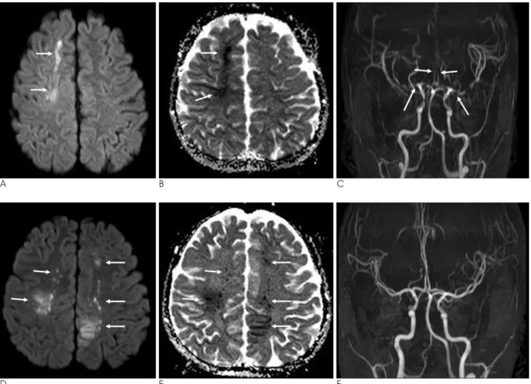
A B C
D E F
Fig. 1. Acute infarction with vasospasm caused by FK506 administration.
A, B. Increased signal intensity on DWI (A) and decreased signal intensity on ADC mapping (B) in the right frontal lobe are demon- strated (arrows). The findings indicate acute infarction.
C. MRA shows diffuse stenosis in both the anterior and middle cerebral arteries (arrows).
D, E. Follow-up DWI (D) and ADC mapping (E) after one weak reveal another new acute infarction in both the fronto-parietal lobes (arrows).
F. Improvement of vasospasm is seen on the follow-up MRA one month after the discontinuation of FK506.
suppressive properties (5). Widespread use of FK506 as an immunosuppressive agent following organ transplan- tation has led to a number of neurological symptoms.
The common clinical features of FK506 neurotoxicity in- clude the sudden onset of seizures, headache, an altered mental status, visual abnormalities, aphasia and hemi- paresis. The onset of neurological symptom may be acute or subacute. In solid organ transplant recipients, these major neurological toxicities have been observed in approximately 5% of the patients receiving FK506 and they have occurred at median of 10-13 days after its initial administration (6).
The pathophysiology of both cyclosporine and FK506 neurotoxicity currently remain unclear. Two pathogenic mechanisms have been suggested. One is that the acute hypertension in posterior leukoencephalopathy induces a loss of autoregulation with passive dilatation of the cerebral arterioles; the hydrostatic pressure results in the extravasation of proteins and fluid into the intersti- tium. Multiple reports of reversible T2 hyperintense white matter lesions with a predominance in the posteri- or circulation as well as increased perfusion seen on SPECT scanning in patients with posterior leukoen- cephalopathy support this theory (3). Another theory is vasculopathy. Diffuse vessel injury can lead to cerebral vasoconstriction and related ischemia. FK506-induced leukoencephalopathy was been increasingly reported with the widespread use of the MRI. However, reports about FK506-induced cerebral infarction are rare. We think that the FK506-induced cerebral infarction is caused by vasospasm and this vasospasm may have im- proved after discontinuing the administration of FK506.
It has been postulated that FK506 neurotoxicity may be similar to hypertensive encephalopathy. However, not all the patients with immunosuppressive induced leukoencephalopathy have hypertension. Hypertensive encephalopathy is the most widely recognized cause of posterior reversible leukoencephalopathy syndrome (PRES), which is a condition that is characterized by the rapid development of vasogenic edema in the posterior head regions (7). Ahn et al. (3) reported the DWI and ADC mapping findings of a FK506 neurotoxicity patient who showed increased signal intensities in both parieto- occipital lobes on the T2WI, DWI and ADC mapping.
They suggested that vasogenic edema rather than cy- totoxic edema may play a pivotal role in the pathogene- sis of FK506 neurotoxicity. Kilinc et al. (8) reported that a 18-year-old male with chronic renal failure underwent uncomplicated renal transplantation from his mother
using an immunosuppression protocol of mycopheno- late mofetil, tacrolimus and prednisolone. Brain MRI on the second day of hospitalization showed signal change in the deep bilateral temporal and occipital regions as well as in the right frontal and right posterior parietal re- gions. The DWI revealed vasogenic edema in the right frontal, right occipital, and left temporal regions. An au- topsy was performed 6 hours after death. The principal pathologic diagnosis was multiple cerebral hemispheric infarctions that were possibly due to vasculitis.
Although reversible vasogenic edema due to cere- brovascular autoregulatory dysfunction is the underly- ing pathophysiologic mechanism, irreversible lesions re- sulting from cytotoxic edema can be associated with FK506 neurotoxicity. Kinoshita et al. (4) mentioned that the MR abnormalities related to the FK506 neurotoxici- ty included in PRES are similar to those observed in hy- pertensive encephalopathy. However, the cortical lami- nar necrosis in their other case could have developed af- ter treatment with FK506. The initial images show cy- totoxic edema in the gray and white matter. Follow-up studies demonstrate cortical hyperintensity in the boundary zone on the T1-weighted images, and this is consistent with cortical laminar necrosis. They suggest- ed this is probably due to a transient hypoxic-ischemic process. Curro et al. (9) reported the case of a male liver transplant recipient who developed de novo migraine while on FK506 therapy. Brain MRI showed a single fo- cal ischemic lesion in the occipital lobe with no abnor- malities in the cortex other than those in the white mat- ter in the remnant brain. MRA suggested vasospasm, which was supported by the findings of a reduction of the lumina of the left anterior and middle cerebral arter- ies with normal contralateral arteries. They suggest that the combination of rizatriptan and tacrolimus can poten- tially lead to an excessive risk of clinically evident cere- bral vasospasm.
In conclusion, we report here on a case of cerebral acute infarction caused by vasospasm in a patient with FK506 neurotoxicity. On DWI, there were areas with in- creased signal intensity in the cerebral hemispheres.
The areas showing increased signal intensity on DWI and T2WI demonstrated decreased signal intensity on the ADC map image. Follow-up MRA was obtained af- ter the discontinuation of FK506, and the stenosis of both anterior and middle cerebral arteries was im- proved with a normal caliber of the vessels. These find- ings suggest that cytotoxic edema is the main cause of the increased signal intensities seen on T2WI, and va-
J Korean Soc Radiol 2011;64:109-112
─ 111 ─
sospasm-induced infarction rather than breakdown of autoregulation played a pivotal role in the pathogenesis of FK506 neurotoxicity. Discontinuing FK506 adminis- tration may result in improvement of the vasospasm.
References
1. Gijtenbeek JM, van den Bent MJ, Vecht CJ. Cyclosporine neuro- toxicity: a review. J Neurol 1999;246:339-346
2. Basic-Jukic N, Basic-Kes V, Kes P, Furic-Cunko V, Bacic-Baronica K. Neurological complications in renal transplant recipients. Acta
Med Croatica 2008;62 Suppl 1:76-813. Ahn KJ, Lee JW, Hahn ST, Yang DW, Kim PS, Kim HJ, et al.
Diffusion-weighted MRI and ADC mapping in FK506 neurotoxici- ty. Br J Radiol 2003;76:916-919
4. Kinoshita T, Moritani T, Shrier DA, Hiwatashi A, Wang HZ, Numaguchi Y, et al. Diffusion-weighted MR imaging of posterior
reversible leukoencephalopathy syndrome: a pictorial essay. Clin
Imaging 2003;27:307-3155. Keenan RJ, Konishi H, Kawai A, Paradis IL, Nunley DR, Iacono AT, et al. Clinical trial of tacrolimus versus cyclosporine in lung transplantation. Ann Thorac Surg 1995;60:580-584
6. Eidelman BH, Abu-Elmagd K, Wilson J, Fung JJ, Alessiani M, Jain A, et al. Neurologic complications of FK 506. Transplant Proc 1991;23:3175-3178
7. Junna MR, Rabinstein AA. Tacrolimus induced leukoencephalopa- thy presenting with status epilepticus and prolonged coma. J
Neurol Neurosurg Psychiatry 2007;78:1410-14118. Kilinc M, Benli S, Can U, Yilmaz A, Karakayali H, Colak T, et al.
FK 506-induced fulminant leukoencephalopathy after kidney transplantation: case report. Transplant Proc 2002;34:1182-1184 9. Curro G, Baccarani U, Adani GL, Lorenzin D, Bresadola F.
Transient ischemic attack after rizatriptan administration in a liver transplant recipient: a case report. Transplant Proc 2006;38:3138- 3139
Ji Kyung Lim, et al : Acute Cerebral Infarction after FK 506 Administration in a Kidney Transplantation Recipient
─ 112 ─
대한영상의학회지 2011;64:109-112
신장 이식 환자에서 FK506 투여 후 발생한 급성 뇌 경색:
증례 보고11