• 검색 결과가 없습니다.

Cerebral infarction caused by endocarditis in a patient with COVID-19

N/A
N/A
Protected

Academic year: 2021

Share "Cerebral infarction caused by endocarditis in a patient with COVID-19"

Copied!
4
0
0

로드 중.... (전체 텍스트 보기)

전체 글

(1)

INTRODUCTION

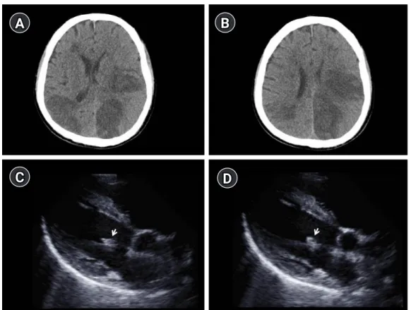
Coronavirus disease 2019 (COVID-19) is a newly identified in- fectious disease with rapid human to human transmission capaci- ty. COVID-19 can result in several fatal complications such as acute respiratory distress syndrome, RNAaemia, acute cardiac in- jury, acute kidney injury, secondary infection, and septic shock [1,2]. Since February 21, 2020, the authors of this paper have been working at a hospital designated for treating COVID-19 pa- tients and by June 15, 2020, 1,027 COVID-19 patients had been

Cerebral infarction caused by

endocarditis in a patient with COVID-19

Doo Hyuk Kwon, MD 1 ; Jae-Seok Park, MD 2 ; Hyung Jong Park, MD 1 ; Sung-Il Sohn, MD 1 ; Jeong-Ho Hong, MD, PhD 1

1

Department of Neurology, Keimyung University School of Medicine, Daegu, Republic of Korea

2

Department of Internal Medicine, Keimyung University School of Medicine, Daegu, Republic of Korea

CASE REPORT

Received: August 6, 2020 Revised: August 19, 2020 Accepted: August 20, 2020 Corresponding Author:

Jeong-Ho Hong, MD, PhD

Department of Neurology, Keimyung University School of Medicine, 1035 Dalgubeol-daero, Dalseo-gu, Daegu 42601, Korea

Tel: +82-53-258-7839 Fax: +82-53-258-4380

E-mail: neurohong79@gmail.com

Background: Coronavirus disease 2019 (COVID-19) has become a worldwide health threat due to its highly contagious nature and se- vere complications. The authors experienced a case of cerebral infarction caused by endocarditis in a patient infected with severe acute respiratory syndrome coronavirus 2 (SARS-CoV-2).

Case Report: A 65-year-old male patient with COVID-19 was admitted to our hospital for cohort isolation. On day 11 after admission, the patient suddenly presented right-sided hemiparesis. Computed tomography of the brain showed multiple hypodense lesions, which confirmed acute cerebral infarction. Transthoracic echocardiogram revealed a vegetation of about 5 mm on the mitral valve and en- docarditis was diagnosed as the cause of cerebral infarction.

Conclusion: Cerebral infarction is one of the complications of COVID-19 and can occur at any time during the course of the disease.

Hence, a neurologist has an important role in the medical team involved in COVID-19 treatment.

Keywords: COVID-19; Heart injury; Endocarditis; Cerebral infarction; Neurologist J Neurocrit Care 2020;13(2):101-104

https://doi.org/10.18700/jnc.200019

hospitalized. Our critical care team implemented a collaborative approach for intensive care unit patients with severe acute respira- tory syndrome coronavirus 2 (SARS-CoV-2) infection enabling simultaneous co-management of pulmonological, neurocritical, and nutritional services. Here, we report a case of multiple cere- bral infarctions in a patient hospitalized with COVID-19.

CASE REPORT

A 65-year-old South Korean male patient with COVID-19 was eISSN 2508-1349

101 www.e-jnc.org

© 2020 The Korean Neurocritical Care Society

This is an Open Access article distributed under the terms of the Creative Commons Attribution Non-Commercial License (http://creativecommons.org/licenses/by-nc/4.0/)

which permits unrestricted noncommercial use, distribution, and reproduction in any medium, provided the original work is properly cited.

(2)

admitted to our hospital for cohort isolation. The patient had no underlying conditions and developed cough and sputum 5 days pri- or to diagnosis. On admission, the patient’s vital signs were recorded as a temperature of 38.0°C, blood pressure of 135/88 mm Hg, heart rate of 78 beats per minute, respiratory rate of 22 breaths per min- ute, and oxygen saturation of 95% on room air. Chest computed tomography (CT) showed pneumonia in the lower lobes of both lungs. We started treatment with lopinavir/ritonavir and hydroxy- chloroquine, which are known to be effective against COVID-19 [1,2]. Additionally, ceftriaxone and azithromycin were simultane- ously administered as empirical antibiotics to treat pneumonia.

Six days later, pneumonia gradually worsened, and the patient was put on mechanical ventilation in the intensive care unit as his blood pressure had dropped (90/60 mm Hg), heart and respiratory rate had increased (120 beats per minute and 30 breaths per minute, respectively), and oxygen saturation levels were below 85%. Based on the results of blood culture (positive for Pseudomonas aerugino- sa) and drug sensitivity test, antibiotic treatment was changed to in- clude piperacillin/tazobactam and levofloxacin. Inotropes were ad- ministered to raise the patient’s blood pressure, and 0.4–0.6 µg/kg/

hr of dexmedetomidine and 0.1–0.2 µg/kg/min of remifentanil were additionally administered during mechanical ventilation.

On day 11 after admission, when the patient’s vital signs stabi- lized, we gradually reduced sedative administration, and the pa- tient began to recover consciousness. However, the patient sud- denly developed right-sided hemiparesis (Medical Research Council grade 3). He had a National Institute of Health Stroke Scale score of 7 (partial hemianopia, 1; facial palsy, 1; right arm motor drift, 2; right leg motor drift, 2; and dysarthria, 1). Emer- gency brain CT with intracranial CT angiography was performed.

The brain CT showed multiple hypodense lesions with surround- ing edema in different areas of the brain (Fig. 1A and B), which confirmed acute cerebral infarction. Since the intracranial CT an- giography was normal, cardioembolic stroke was suspected and transthoracic echocardiogram (TTE) was performed to assess the cause of the stroke. TTE showed a vegetation of approximately 5 mm on the mitral valve (Fig. 1C and D) and endocarditis was di- agnosed as the cause of cerebral infarction. Aspirin (100 mg) that was administered at the time of diagnosis of cerebral infarction was discontinued on suspicion of infectious endocarditis. Antimi- crobial therapy with piperacillin/tazobactam and levofloxacin was administered based on blood culture (positive for P. aeruginosa) and antibiotic sensitivity test. Two weeks later, the patient was re- leased from quarantine after two consecutive negative findings on

B

D A

C

Fig. 1. Findings of brain computed tomography (CT) and transthoracic echocardiogram (TTE). (A, B) Brain CT demonstrates multiple hypodense lesions with surrounding edema in different areas of the brain. (C, D) In the TTE (supine view), a mobile and irregularly shaped echodense mass of approximately 1.0×0.5 cm is attached to the anterior mitral valve (white arrows).

https://doi.org/10.18700/jnc.200019 102

Doo Hyuk Kwon, et al. • Cerebral infarction in COVID-19 patients

(3)

COVID-19 real-time reverse transcription polymerase chain reac- tion testing and was transferred to another tertiary hospital (not a COVID-19 designated hospital) for further treatment.

DISCUSSION

Recently published studies report that COVID-19 could be ac- companied by acute cardiac injury (defined as blood levels of hy- persensitive troponin I above the 99th percentile upper reference limit (> 28 pg/mL) or new abnormalities shown on electrocardi- ography and echocardiography), secondary infection, and sepsis [1,2]. The vegetation of endocarditis is initiated through bactere- mia, which then binds and attaches to the damaged endothelium.

Thromboplastin, which is secreted by tissue factors in the dam- aged endothelium, causes platelet aggregation and cleavage of fi- brinogen to fibrin. Acute cardiac injury, secondary infections, and sepsis can cause endocarditis [3,4]. In our study, P. aeruginosa was reported in blood culture after the patient was confirmed to have COVID-19. Hence, endocarditis can occur due to complications of COVID-19 accompanied by secondary bacterial infection. De- spite improved management, endocarditis remains associated with high mortality and serious complications [3]. Symptomatic neurological complications appear in 15%–30% of endocarditis patients and occur primarily due to embolism from vegetations [4]. Neurologic symptoms most occur often before or during en- docarditis diagnosis, but new or recurrent events may occur after endocarditis diagnosis. Clinical presentation is variable and may include multiple symptoms or signs in the same patient. Typically, focal signs are diagnosed due to ischemic stroke. Further, transient ischemic attack, intracerebral or subarachnoid hemorrhage, brain abscess, meningitis, and toxic encephalopathy can be present, and there is evidence that 35%–60% of patients with endocarditis de- velop additional silent cerebral embolisms [5].

We believe that this is the first reported case of cerebral infarc- tion caused by endocarditis in a patient with COVID-19. Several complications can result from COVID-19 and acute cerebral in- farction can further potentially occur during the course of this dis- ease [1,2,6]. Due to the lack of equipment required for neurologi- cal disease assessment and inaccessibility of medical staff in our designated hospital optimized for COVID-19 treatment, it was difficult to perform diagnosis of stroke by physical examination and evaluation. However, it is clear that neurologists have an im- portant role besides respiratory and infectious physicians in the treatment of COVID-19.

ARTICLE INFORMATION

Ethics statement

This case was reviewed and approved by the Institutional Review Board of Keimyung University Dongsan Hospital (IRB No. 2020- 09-054). Informed consent was waived by the Board.

Conflict of interest

Dr. JH Hong is an editorial board member of the journal but was not involved in the peer reviewer selection, evaluation, or decision process of this article. There are no other potential conflicts of in- terest relevant to this article to declare.

Funding

This work was supported by the Research Program of Medicity Dae- gu Council funded by Daegu Metropolitan City (No. COVID19_

DM08).

ORCID

Doo Hyuk Kwon https://orcid.org/0000-0001-7541-9226 Jae-Seok Park https://orcid.org/0000-0002-5751-7209 Hyung Jong Park https://orcid.org/0000-0002-6112-2939 Sung-Il Sohn https://orcid.org/0000-0002-6900-1242 Jeong-Ho Hong https://orcid.org/0000-0002-8235-9855 Author contributions

Conceptualization: DHK, JHH. Data curation: DHK, JSP, HJP, JHH. Formal analysis: all authors. Funding acquisition: DHK. In- vestigation: HJP, SIS. Supervision: JSP, HJP, SIS. Visualization:

DHK, HJP, JHH. Writing–original draft: DHK, JHH. Writing–

review & editing: DHK, JHH.

REFERENCES

1. Huang C, Wang Y, Li X, Ren L, Zhao J, Hu Y, et al. Clinical fea- tures of patients infected with 2019 novel coronavirus in Wu- han, China. Lancet 2020;395:497-506.

2. Chen N, Zhou M, Dong X, Qu J, Gong F, Han Y, et al. Epidemi- ological and clinical characteristics of 99 cases of 2019 novel coronavirus pneumonia in Wuhan, China: a descriptive study.

Lancet 2020;395:507-13.

3. Thuny F, Grisoli D, Collart F, Habib G, Raoult D. Management of infective endocarditis: challenges and perspectives. Lancet 2012;379:965-75.

4. Murdoch DR, Corey GR, Hoen B, Miró JM, Fowler VG Jr, Bay-

er AS, et al. Clinical presentation, etiology, and outcome of in-

fective endocarditis in the 21st century: the International Col-

103

https://doi.org/10.18700/jnc.200019

(4)

laboration on Endocarditis-Prospective Cohort Study. Arch In- tern Med 2009;169:463-73.

5. Snygg-Martin U, Gustafsson L, Rosengren L, Alsiö A, Acker- holm P, Andersson R, et al. Cerebrovascular complications in patients with left-sided infective endocarditis are common: a prospective study using magnetic resonance imaging and neu-

rochemical brain damage markers. Clin Infect Dis 2008;47:23- 30.

6. Mao L, Jin H, Wang M, Hu Y, Chen S, He Q, et al. Neurologic manifestations of hospitalized patients with coronavirus disease 2019 in Wuhan, China. JAMA Neurol 2020;77:1-9.

https://doi.org/10.18700/jnc.200019 104

Doo Hyuk Kwon, et al. • Cerebral infarction in COVID-19 patients

수치

Fig. 1. Findings of brain computed tomography (CT) and transthoracic echocardiogram (TTE)

참조

관련 문서

2.World Health Organization: Clinical management of severe acute respiratory infection when Novel coronavirus (nCoV) infection is suspected: Interim Guidance.

In this group of high-risk patients with severe COVID-19 pneumonia, treatment with lenzilumab was associated with a significantly shorter time to clinical improvement compared

자료원 : 벨라루스 통계청, 벨라루스 관세청, https://connect.ihs.com(Global Trade Atlas), www.belta.ru, www.news.tut.by,

In addition to the problem of this bias, the problem caused by using the time variable with a weighted wage rate may be a multicollinearity between time value (=time x

Time series of vertical cross section of potential vorticity and wind vector calculated by Case 1 along the A-A' line indicated at Fig.. Same

Haemorragic fever with renal syndrome (HFRS) caused by Hantanvirus has been one of the principal acute febrile disease in Korea. Hantaviruses are carried by numerous

Therefore, cTnT is a recommended biomarker for use in the detection of myocardial infarction (MI) and in acute coronary syndromes.[8] Indeed, several authors

The locations of aneurysms were middle cerebral artery in 15 patients, cerebral artery in 15 patients, cerebral artery in 15 patients, cerebral artery in