Acute allograft dysfunction mimicking thrombotic microangiopathy in kidney transplant recipient with renal infarction:
case report and review of literatures
Sua Lee
1, Lo-Yi Ho
2, Byung Ha Chung
3,4, Sun Cheol Park
5, Chul Woo Yang
3,41Division of Nephrology, Department of Internal Medicine, Seoul National University Hospital, Seoul, Korea
2Department of Medicine and Geriatrics, Kwong Wah Hospital, Hong Kong, China
3 Transplant Research Center, Department of Internal Medicine, Seoul St. Mary’s Hospital, College of Medicine, The Catholic University of Korea, Seoul, Korea
4 Division of Nephrology, Department of Internal Medicine, College of Medicine, The Catholic University of Korea, Seoul, Korea
5 Division of Vascular and Transplant Surgery, Department of Surgery, College of Medicine, The Catholic University of Korea, Seoul, Korea
Acute allograft dysfunction is rarely observed in kidney transplantation (KT). We report an unusual case of acute allograft dysfunction mimicking thrombotic microangiopa- thy (TMA) in recipient with renal infarction. A 65-year-old man underwent KT from his 39-year-old son. Pre-transplant donor evaluation was normal except for the branches of the upper and lower pole renal arteries originating from the aorta in renal computed topo- graphic angiography, respectively. The immediate post-transplant clinical course was un- eventful, but serum creatinine (SCr) increased from 2.2 to 4.5 mg/dL, anemia and throm- bocytopenia were shown, and serum lactate dehydrogenase increased to 919 U/L on the third day after transplantation. We suspected TMA, because of no evidence of acute bleeding. The laboratory parameters associated with TMA were within normal ranges.
Renal magnetic resonance angiography revealed a focal wedge-shaped perfusion defect in the upper pole of the graft and renal Doppler ultrasonography showed decreased per- fusion of the lower pole of the graft. Graft function improved with conservative therapy.
The patient was discharged with SCr of 1.21 mg/dL. Graft function has been stable after discharge. Acute allograft infarction should be considered in the differential diagnosis of acute allograft dysfunction mimicking TMA in recipients with grafts supplied by multiple renal arteries.
Keywords: Kidney transplantation; Primary graft dysfunction; Thrombotic microangiopathy
Received July 30, 2020 Revised October 15, 2020 Accepted October 26, 2020
Corresponding author: Chul Woo Yang Department of Internal Medicine, Seoul St. Mary’s Hospital, College of Medicine, The Catholic University of Korea, 222 Banpo-daero, Seocho-gu, Seoul 06591, Korea
Tel: +82-2-2258-6851 Fax: +82-2-2258-6917 E-mail: [email protected]
© The Korean Society for Transplantation This is an Open Access article distributed under the terms of the Creative Commons Attribution Non-Commercial License (http://creativecommons.org/licenses/
by-nc/4.0/) which permits unrestricted non-commercial use, distribution, and reproduction in any medium, provided the original work is properly cited.
pISSN 2671-8790
eISSN 2671-8804
INTRODUCTION
Renal infarction is a rare cause of acute kidney injury that results from inadequate blood flow to the kidney [1]. The incidence of acute infarction in native kidneys is reported between 0.004% and 0.1% [2,3]. From autopsy study, the incidence was reported as 0.48–1.4% [4]. The incidence of infarction in renal allografts is not confirmed, but it is con- sidered to be less than that in native kidneys. The etiology of acute allograft dysfunction in the early posttransplant period is varied. Some of the causes of acute allograft dysfunction have clinical manifestations that are similar to those of thrombotic microangiopathy (TMA), and they should be considered during differential diagnosis. The main clinical manifestations of renal infarction include abdominal pain, hematuria, and elevated serum lactate dehydrogenase (LDH) level, but since these also occur in TMA, diagnosis of renal infarction in the early posttrans- plantation period is more difficult. Here, we report an un- usual case of renal infarction mimicking TMA in a kidney transplant (KT) recipient with a graft that was supplied by multiple renal arteries.
CASE REPORT
This study was approved by the Institutional Review Boards (IRB) of the Catholic University of Korea (KC19ZESE0699) and an informed consent was received from the patient and a caregiver. The images are published under agree- ment of the patient.
A 65-year-old man visited the department of transplan- tation of our hospital for KT. He was diagnosed with end- stage renal disease of unknown origin. Renal ultrasonog- raphy (US) and magnetic resonance imaging performed 4 years ago revealed a 2 cm renal mass at the lower pole of the kidney suspected to be renal cell carcinoma, and radiofrequency ablation was used to treat the renal mass.
Follow-up radiological evaluations did not show tumor re- currence. The demographic characteristics and underlying diseases of patient were presented in Table 1. His 39-year- HIGHLIGHTS
• Renal infarction is a rare complication of allograft and is associated with high rates of allograft loss.
• The clinical manifestations of renal infarction are simi- lar to those of thrombotic microangiopathy.
• Multiple renal arteries supplying the allograft are one of causes in renal infarction.
Table 1. Demographic characteristics of patient
Clinical feature Result
Body weight (kg) 69
Height (m) 1.76
Body mass index (kg/m
2) 22.3
Systolic and diastolic blood pressure (mmHg) 140/90 Underlying disease
Hypertension +
Diabetes –
Cardiovascular disease –
Valvular heart disease –
Atrial fibrillation –
Current smoking Non-smoker
Current alcohol use (-)
A B
Fig. 1. Renal computed tomography an-
giogram of the donor. A small lower polar
branch arises from the aorta (arrow), supply-
ing the lower pole (A) and a tiny upper polar
branch arises from the aorta (arrow), supply-
ing the upper pole of the left kidney (B).
old son was a kidney donor. The results of pretransplant donor evaluation were within normal ranges except for a lower polar renal artery branch 2.7 mm in diameter found on renal computed tomography (CT) angiogram, which originated from the aorta and separately supplied the low- er pole of the left kidney (Fig. 1A).
The transplantation was an ABO-compatible transplan- tation. The patient’s human leukocyte antigen (HLA) type was A02/33, B35/44, DR12/13 and the donor’s HLA type was A02/33, B35/67, DR12/-. There was only one HLA mismatch. The results of panel reactive antibody and HLA crossmatch tests were negative. The patient underwent living donor KT on a scheduled day. He was administered basiliximab (anti-cluster of differentiation 25 monoclonal antibody) for induction and tacrolimus, methylpredniso- lone, and mycophenolic acid for maintenance immuno- suppressive therapy. The transplantation was completed successfully with no complications. The total ischemia time was 60 minutes and the warm ischemia time was 2 minutes 43 seconds. The lower polar renal artery branch seen on renal CT angiogram was reconstructed using the end-to-side anastomosis technique with the middle renal artery. The total volume of blood lost during the operation was estimated to be about 250 mL. On the 1st day after transplantation, serum creatinine (SCr) level fell to 2.19 mg/dL, hourly urine output maintained at 100–300 mL, and the tacrolimus trough level was 4.3 ng/mL.
However, on the 2nd day after transplantation, SCr level increased to 2.61 mg/dL and hourly urine output decreased to 60–130 mL. The patient complained of no symptoms such as abdominal pain, nausea, vomiting and fever. At first, we suspected acute T-cell mediated allograft rejection and started the patient on 1.25 mg/kg of anti-thy-
mocyte immunoglobulin (ATG).
SCr level increased to 4.53 mg/dL on the 3rd day after transplantation despite ATG administration, and within 3 days after transplantation, serum hemoglobin concentration decreased from 10.9 g/dL to 7.0 g/dL (ref- erence, 13.0–18.0 g/dL) and platelet count decreased from 159,000/mm
3to 65,000/mm
3(reference, 150,000–
450,000/mm
3). Additionally, serum LDH level suddenly increased to 919 U/L (reference, 250–450 U/L). The rapid progression of allograft dysfunction, the sudden progres- sion of anemia and thrombocytopenia, and the increase in serum LDH level informed our suspicion of TMA in accor- dance with the diagnostic criteria of TMA [5].
On the 4th day after transplantation, SCr level eventual- ly rose to 5.59 mg/dL with worsening anemia and throm- bocytopenia at 6.1 g/dL and 65,000/mm
3, respectively.
However, the laboratory parameters associated with TMA were within normal range (corrected reticulocyte count, 0.8%; absolute reticulocyte count, 37/mm
3; haptoglobin concentration, 147 mg/dL [reference, 30–200 mg/dL], Coombs test was negative, and no schistocytes were found). We excluded TMA and considered other causes.
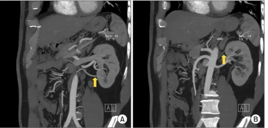
On the 6th day after transplantation, renal magnetic reso- nance angiography (MRA) revealed a focal wedge-shaped perfusion defect in the upper pole of the graft suggestive of infarction without evidence of acute bleeding such as hematoma, renal arterial bleeding (Fig. 2A).
We reviewed the pretransplant examinations and renal CT angiogram showed an upper polar renal artery branch about 1 mm in diameter originating from the aorta and supplying the left kidney, but this finding was not previ- ously mentioned by the radiologist (Fig. 1B). A surgeon in- formed us of an additional small-caliber polar renal artery
A B
Fig. 2. Radiologic findings of the recipient. Focal wedge shaped perfusion defect in the upper pole of the transplanted kidney on flair images (arrow),
coronal view of renal magnetic resonance angiography (A) and slightly decreased vascularity and perfusion in inferior pole of graft kidney on kidney Dop-
pler ultrasonography (B). Peak systolic velocity of lower inter-lobar artery reduced to 14.3 cm/sec.
branch, which supplied the upper pole of the graft kidney and appeared damaged after nephrectomy, and it was therefore ligated before transplantation. Renal Doppler US revealed decreased perfusion of the lower pole of the graft, which was suggestive of infarction (Fig. 2B).
Therefore, the patient was diagnosed with renal infarc- tion and acute kidney injury caused by ligation of the upper polar branch and reconstruction of the lower polar branch.
Renal function improved with conservative therapy, and on the 21st day after transplantation, the patient was dis- charged with a SCr level of 1.21 mg/dL. The SCr level re- mained at 1.28 mg/dL 3 months after transplantation.
The change of laboratory findings after KT is shown in Fig. 3.
DISCUSSION
Allograft dysfunction calls for immediate attention and ac- tion to transplant nephrologists and surgeons. In general, recovery of allograft function takes 7 to 14 days and there are several important causes of early allograft dysfunc- tion. Hyperacute rejection, renal vascular complications, delayed allograft function, and TMA are possible causes of allograft dysfunction in the early posttransplant period [6].
TMA of renal allografts is a serious complication in KT recipients and can either be recurrent or de novo. It mostly occurs within the first month of transplantation at a report- ed incidence of more than 60% [5]. TMA may be suspected when the following are present: anemia, thrombocytope- nia, schistocytes in peripheral blood smears, low hapto- globin levels, elevated serum LDH levels, low hematocrit, and acute allograft dysfunction. This is because TMA has
OD 1 1,000
875 750 625 500 375 250 125
LDH(U/L)
0
SCr(mg/dL)
8 7 6 5 4 3 2 1 0 OD POD 1 POD 2 POD 3 POD 4 POD 6 POD 7 POD 21 POD 90 LDH
SCr 5.29
566 544
2.77 2.19
411
2.61 504
919
4.53
533 5.59
4.4
420 2.46
697
1.21 861
652
1.28
OD 1 200 180 160 140 120 100 80 60 40 20 PLT(10/L)3
0
Hb(g/dL)
20 18 16 14 12 10 8 6 4 2 0 OD POD 1 POD 2 POD 3 POD 4 POD 6 POD 7 POD 21 POD 90 PLT
Hb 168
12.2 159
10.9
8.1 107
7.6 104
7 65
33
6.1 60
6.4 7.4 132
158
9 177
13.6
A
B
Fig. 3. Change of laboratory findings after kidney transplantation. (A) On the 1st day after transplantation, serum creatinine (SCr) decreased to 2.19