INTRODUCTION
Improved treatment modalities for childhood cancers have in- creased 5-year survival rates to nearly 80%.1,2 However, com- plications may arise later in the lives of childhood cancer sur- vivors who received cytotoxic drugs, radiation, or other treat- ments. Due to the increasing number of long-term survivors,
these late effects have become an important issue in childhood cancer management.3 Endocrine-related late effects are com- monly reported, of which thyroid disorders are the most fre- quent.4
Thyroid hormones play an important role in growth and de- velopment during childhood. The thyroid is especially radio- sensitive, and irradiation of the neck, head, or brain can cause thyroid dysfunction.4,5 Recent reports demonstrate that child- hood cancer survivors who received total body irradiation, mantle irradiation for Hodgkin disease (HD), craniospinal irra- diation for brain tumors, or other conditioning regimens are at risk for thyroid disease.4-6
The most common thyroid disease of childhood cancer sur- vivors is hypothyroidism.5,7 There are two types of hypothyroid- ism, characterized as follows: primary hypothyroidism, with el- evated levels of serum thyroid-stimulating hormone (TSH); and secondary or central hypothyroidism, with reduced levels of
Subclinical Hypothyroidism in Childhood Cancer Survivors
Hyun Joo Lee
1,2, Seung Min Hahn
1,2, Song Lee Jin
1,2, Yoon Jung Shin
1,2, Sun Hee Kim
1,2, Yoon Sun Lee
3, Hyo Sun Kim
1,2, Chuhl Joo Lyu
1,2, and Jung Woo Han
1,21Division of Pediatric Hematology and Oncology, Department of Pediatrics, Yonsei University College of Medicine, Yonsei University Health System, Seoul, Korea;
Departments of 2Pediatric Hemato-Oncology and 3Pharmacy, Yonsei Cancer Center, Yonsei University Health System, Seoul, Korea.
Purpose: In childhood cancer survivors, the most common late effect is thyroid dysfunction, most notably subclinical hypothy- roidism (SCH). Our study evaluated the risk factors for persistent SCH in survivors.
Materials and Methods: Survivors (n=423) were defined as patients who survived at least 2 years after cancer treatment completion.
Thyroid function was assessed at this time and several years thereafter. Two groups of survivors with SCH were compared: those who regained normal thyroid function during the follow-up period (normalized group) and those who did not (persistent group).
Results: Overall, 104 of the 423 survivors had SCH. SCH was observed in 26% of brain or nasopharyngeal cancer survivors (11 of 43) and 21.6% of leukemia survivors (35 of 162). Sixty-two survivors regained normal thyroid function, 30 remained as persistent SCH, and 12 were lost to follow-up. The follow-up duration was 4.03 (2.15–5.78) years. Brain or nasopharyngeal cancer and Hodg- kin disease were more common in the persistent group than in the normalized group (p=0.002). More patients in the persistent group received radiation (p=0.008). Radiation to the head region was higher in this group (2394±2469 cGy) than in the normalized group (894±1591 cGy; p=0.003). On multivariable analysis, lymphoma (p=0.011), brain or nasopharyngeal cancer (p=0.039), and head radiation dose ≥1800 cGy (p=0.039) were significant risk factors for persistent SCH.
Conclusion: SCH was common in childhood cancer survivors. Brain or nasopharyngeal cancer, lymphoma, and head radiation
≥1800 cGy were significant risk factors for persistent SCH.
Key Words: Hypothyroidism, neoplasm, survivor, child
pISSN: 0513-5796 · eISSN: 1976-2437
Received: May 29, 2015 Revised: August 30, 2015 Accepted: November 1, 2015
Corresponding author: Dr. Jung Woo Han, Division of Pediatric Hematology and Oncology, Department of Pediatrics, Yonsei University College of Medicine, Yonsei University Health System, 50-1 Yonsei-ro, Seodaemun-gu, Seoul 03722, Korea.
Tel: 82-2-2228-2060, Fax: 82-2-393-9118, E-mail: [email protected]
•The authors have no financial conflicts of interest.
© Copyright: Yonsei University College of Medicine 2016
This is an Open Access article distributed under the terms of the Creative Com- mons Attribution Non-Commercial License (http://creativecommons.org/licenses/
by-nc/3.0) which permits unrestricted non-commercial use, distribution, and repro- duction in any medium, provided the original work is properly cited.
Yonsei Med J 2016 Jul;57(4):915-922 http://dx.doi.org/10.3349/ymj.2016.57.4.915
circulating TSH. Primary hypothyroidism can be overt [reduc- ed levels of free thyroxine (fT4) and triiodothyronine (T3)] or subclinical (normal fT4 and T3 levels). In central hypothyroid- ism, the reduced thyroid hormone levels result from inade- quate stimulation of the thyroid gland due to reduced TSH in cases where hypothalamic-pituitary disturbance is present.8-10 Overt hypothyroidism is usually treated via daily administra- tion of thyroxine pills,8-10 and the distinction between it and subclinical hypothyroidism (SCH) is determined via biochem- ical measurement of fT4 and T3 concentrations in serum. There is no standard management for patients with SCH and mildly high serum TSH concentrations, although treatment of patients with concentrations of 10 mU/L or more is recommended.8-10
Pediatric endocrinologists are undecided as to how to treat children with SCH, as controlled pediatric SCH studies are rare. Two recent studies reported that untreated patients with SCH had normal TSH levels at follow-up.10,11 However, eluci- dation of the natural history of SCH as a late effect in childhood cancer survivors is lacking, despite its frequent occurrence in such survivors.12,13
The aim of this study was to determine which children are at risk for persistent SCH after cancer treatment and who there- fore should be treated early during the follow-up period.
MATERIALS AND METHODS
Study population
Starting in 2005, we established a long-term follow-up clinic at the Yonsei Cancer Center, Yonsei University Health System, Ko- rea to evaluate cancer survivors once or twice a year.7 In accor- dance with published guidelines, survivors were defined as individuals with no evidence of cancer for 2 or more years after the completion of cancer treatment.14 Among the more than 700 registered survivors in the clinic, 423 had been tested for thy- roid function, and seven showed hyperthyroidism (three overt and four subclinical) at 2 years after cancer treatment. The re- mainder (n=416) included survivors of leukemia (162, 38.9%);
non-Hodgkin lymphoma (NHL; 47, 11.3%); HD (12, 2.9%); brain or nasopharyngeal cancer (brain/naso; 43, 10.3%); Wilms tu- mor (WT), neuroblastoma (NB), or abdominal malignancies including hepatoblastoma (WT/NB/abdomen; 79, 19.0%); and sarcoma or other non-abdominal malignancies (sarcoma/oth- ers; 73, 17.5%). Two years after completion of cancer treatment, 300 survivors (72.1%) had normal thyroid function, 12 (2.9%) had overt hypothyroidism, and 104 (25.0%) had SCH. Twelve of the survivors with SCH were lost to follow-up; therefore, 92 survivors with SCH were ultimately included in this study. This study was approved by the Institutional Review Board of Sever- ance Hospital, Yonsei University Health System (4-2015-0016).
Thyroid function test and follow-ups
Serum T3, fT4, and TSH levels were monitored in the survivors;
the reference values were 80–200 ng/dL, 0.95–2.23 ng/dL, and 0.3–4.0 mU/L, respectively, as defined by our institution. SCH was defined as a condition in which TSH levels were higher than the reference value and T3 and T4 levels were within the range of the reference values. To detect thyroid disorders in sur- vivors, the thyroid function test (TFT) was performed 2 years after cancer treatment completion as a baseline test.7 Survivors with no thyroid disease-related symptoms received no addi- tional TFTs as routine surveillance to comply with the Korean health insurance policy. Survivors with abnormal thyroid func- tion were followed-up annually or biannually. If thyroid func- tion became normal, they were designated as “normalized.” If it did not, they were designated as “persistent.”
As recommended by an endocrinologist, thyroid hormone (levothyroxine) was prescribed for survivors whose TSH levels were >8–10 mU/L, with consideration of their clinical symp- toms, signs, and developmental stage. Thyroid hormone re- placement was tapered out after 6–12 months. If the TSH level increased after tapering or discontinuing the treatment, the treatment was reinitiated. Thyroid function was monitored ev- ery 1–2 years after short-term discontinuation of replacement therapy. If the therapy was permanently discontinued, the sur- vivor was considered to be normalized.
Definitions and variables
The six cancer diagnosis groups were leukemia, NHL, HD, br- ain/naso, WT/NB/abdomen, and sarcoma/others. We review- ed the treatment modalities, including chemotherapy, radia- tion therapy, and hematopoietic stem cell transplantation (HSCT). The types of chemotherapeutic agents used were also reviewed, as were radiation body sites and doses. The total ra- diation dose per survivor was defined as the maximum total dose of radiation at a specific radiation site. If a survivor had received craniospinal irradiation long after prophylactic brain radiation, we summed the doses at each site and calculated the maximum dose for each site.
The time variables were as follows: age at diagnosis, age at cancer treatment completion, age at the initial TFT (the age at which thyroid function was first categorized after treatment completion), age at the final TFT, and follow-up period (time from the initial TFT to the final TFT).
Statistical analysis
Categorical variables including diagnoses and treatment mo- dalities were evaluated by using the chi-square test or Fisher’s exact test. Continuous variables including time variables and radiation doses were assessed by using Student’s t-test or an analysis of variance for parametric tests and the Mann-Whit- ney U-test or Kruskal Wallis test for non-parametric tests. Pair- ed variables were tested using the Wilcoxon signed rank test.
The cumulative incidence of thyroid function normalization was assessed in each cancer diagnosis group by using a Kaplan- Meier analysis and the log-rank test. For the Kaplan-Meier an-
alysis and regression analysis, diagnosis groups were categoriz- ed into three groups: brain/naso, lymphoma, and other tumors.
Variables that were significant in a univariate analysis, includ- ing treatment modalities and age at diagnosis, were included in the multivariable model. We calculated the odds ratios us- ing logistic regression after adjustment for the risk factors of persistent SCH including age, gender, HSCT, head radiation, neck radiation, and chemotherapeutic agents such as enzymes and epipodophyllotoxins. All statistical analyses were perform- ed by using SPSS version 21 for Windows (SPSS Inc., Chicago, IL, USA).
RESULTS
Patient demographics
Two years after cancer treatment completion, 116 of the 423 childhood cancer survivors initially surveyed (27.4%) had hy- pothyroidism; 112 (26.5%) had primary hypothyroidism, and four (0.94%) had secondary hypothyroidism. The four survivors with secondary hypothyroidism were brain tumor survivors, and all were cases of overt hypothyroidism. Eight of the 112 (7.1%) survivors with primary hypothyroidism had overt hy- pothyroidism, whereas 104 (92.9%) had SCH. Six of the eight (75%) survivors with overt hypothyroidism were brain tumor
survivors (data not shown). The percentage of survivors with SCH was similar for all diagnoses (p=0.322, data not shown):
leukemia, 35 of 162 (21.6%); NHL, 12 of 47 (25.5%); HD, 6 of 12 (50.0%); brain/naso, 11 of 43 (25.6%); WT/NB/abdomen, 23 of 79 (29.1%); and sarcoma/others, 17 of 73 (32.3%, data not shown).
Our findings showed that SCH was the most common thy- roid hormone disturbance after cancer treatment (n=104). Ow- ing to losses (n=12) during follow-up, our study group ultimate- ly comprised 92 childhood cancer survivors with SCH; their characteristics are shown in Table 1. The number of male sur- vivors was 61 (66.3%). The median ages at cancer diagnosis, cancer treatment completion, initial TFT, and final TFT were 3.97 (1.80–8.11), 6.00 (2.95–10.50), 9.75 (6.92–14.59), and 14.42 (10.82–19.15) years, respectively. The interval from comple- tion of treatment to enrollment was 2.97 (2.33–4.95) years. The follow-up duration (initial TFT to final TFT) was 4.03 (2.15–
5.78) years. The most common diagnosis was leukemia (n=32, 34.8%), followed by WT/NB/abdomen (n=21, 22.8%). Most survivors underwent chemotherapy (n=91, 98.9%), and 43 sur- vivors received HSCT (46.7%).
Risk factors for persistent SCH: demographic findings Sixty-two survivors with SCH regained normal thyroid function more than 2 years after cancer treatment completion (the nor-
Table 1. Demographic Characteristics of Patients with Persistent Subclinical Hypothyroidism (SCH)
Characteristic Normalized SCH (n=62, 67.4%) Persistent SCH (n=30, 32.6%) Total (n=92) p value
Sex (male:female) 39 (62.9):23 (37.1) 22 (73.3):8 (26.7) 61 (66.3):31 (33.7) 0.321
Age at diagnosis (yr) 3.88 (1.66–7.96) 4.70 (1.80–11.47) 3.97 (1.80–8.11) 0.355
Age at completion (yr) 5.95 (2.73–9.76) 6.11 (3.20–12.10) 6.00 (2.95–10.50) 0.373
Age at initial study (yr) 9.75 (7.30–13.92) 9.62 (6.18–18.83) 9.75 (6.92–14.59) 0.748
Time from completion to initial study (yr) 3.48 (2.37–5.18) 2.47 (2.18–3.56) 2.97 (2.33–4.95) 0.051
Age at final study (yr) 14.24 (10.83–17.22) 15.54 (10.55–21.72) 14.42 (10.82–19.15) 0.283
Follow-up period (yr) 3.80 (2.01–5.51) 4.42 (3.10–6.54) 4.03 (2.15–5.78) 0.142
Diagnosis, n (%) 0.002
Leukemia 24 (75.0) 8 (25.0) 32 (34.8)
NHL 6 (60.0) 4 (40.0) 10 (10.9)
HD 2 (33.3) 4 (66.7) 6 (6.5)
Brain/naso 1 (12.5) 7 (87.5) 8 (8.7)
WT/NB/abdomen 18 (85.7) 3 (14.3) 21 (22.8)
Sarcoma/others 11 (73.3) 4 (26.7) 15 (16.3)
Treatment modality, n (%)
HSCT 0.378
Yes 27 (62.8) 16 (37.2) 43 (46.7)
No 35 (71.4) 14 (28.6) 49 (53.3)
Radiation 0.017
Yes 27 (56.3) 21 (43.8) 48 (52.2)
No 35 (79.5) 9 (20.5) 44 (47.8)
Chemotherapy 0.326
Yes 62 (68.1) 29 (31.9) 91 (98.9)
No 0 (0.0) 1 (100) 1 (1.1)
NHL, non-Hodgkin leukemia; HD, Hodgkin disease; naso, nasopharyngeal; WT, Wilms tumor; NB, neuroblastoma; HSCT, hematopoietic stem cell transplantation.
malized group), whereas 30 did not (the persistent group).
There were no differences in sex or any time variables (age at diagnosis or age at cancer treatment completion) between the normalized and persistent groups. There were, however, sig- nificant differences in terms of cancer type: leukemia (75.0%), WT/NB/abdomen (85.7%), and sarcoma/others (73.3%) were more common in the normalized group, whereas brain/naso (87.5%) and HD (66.7%) were more common in the persistent group. The fT4 levels were statistically similar in all survivors regardless of cancer type (p=0.176) (Table 2). TSH levels were significantly higher in survivors of brain/naso cancer than in survivors of the other cancer types (p=0.010). As for the treat- ment modality, fT4 was significantly higher in patients treated with HSCT than in those without HSCT (p=0.021), and fT4 was significantly lower in patients treated with radiation (p=0.044);
however, the fT4 levels in all groups were within the normal range.
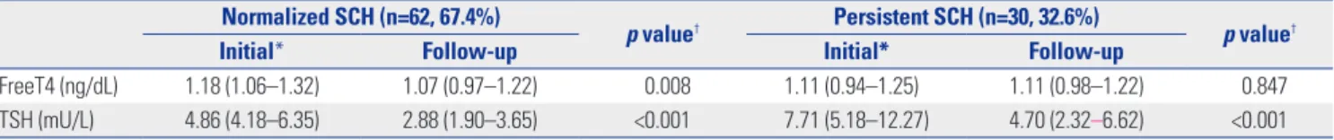
The initial TSH level was significantly higher in the persistent group than in the normalized group (7.71 mU/L vs. 4.86 mU/
L; p=0.001 via Mann-Whitney U-test, data not shown). In the normalized group, the TSH level was significantly lower at the final follow-up than at the initial follow-up (2.88 mU/L vs. 4.86 mU/L; p<0.001 via Wilcoxon signed rank test) (Table 3). The fT4 level was significantly higher at the initial follow-up than at the final follow-up (1.18 ng/dL vs. 1.07 ng/dL; p=0.008 via Wil- coxon signed rank test); however, all were in the normal range.
In the persistent group, the TSH level was also lower at the fi- nal follow-up than at the initial follow-up (4.70 mU/L vs. 7.71 mU/L; p<0.001 via Wilcoxon signed rank test).
In the persistent SCH group, 23 (76.7%) survivors received thyroid hormone replacement therapy. The mean TSH levels tended to be higher in the replacement group than in the non- replacement group, though not significant [8.79 (5.46–12.71) mU/L vs. 6.27 (4.90–7.12) mU/L; p=0.149 via Mann-Whitney U- test], and fT4 levels were not different [1.11 (0.95–1.28) ng/dL vs. 1.09 (0.94–1.20) ng/dL; p=0.756 via Mann-Whitney U-test, data not shown]. Survivors with good clinical status did not re- ceive hormone replacement therapy, and their TSH level did
Table 2. Thyroid Function at 2 Years after Cancer Treatment Completion
Characteristic FreeT4 (ng/dL) p value* TSH (mU/L) p value*
Sex (n) 0.650 0.617
Male (61) 1.15 (1.03–1.31) 5.36 (4.00–8.25)
Female (31) 1.19 (0.98–1.31) 5.44 (4.51–7.91)
Diagnosis (n) 0.176 0.010
Leukemia (32) 1.18 (0.94–1.35) 5.45 (4.19–7.71)
NHL (10) 1.20 (1.11–1.39) 4.60 (2.86–5.62)
HD (6) 1.03 (0.93–1.36) 4.82 (3.55–6.68)
Brain/naso (8) 0.96 (0.92–1.11) 12.27 (9.16–14.22)
WT/NB/abdomen (21) 1.18 (1.10–1.37) 5.44 (4.69–10.02)
Sarcoma/others (15) 1.14 (1.04–1.26) 5.29 (4.14–6.52)
Treatment modality
HSCT (n) 0.021 0.837
Yes (43) 1.20 (1.06–1.38) 5.48 (4.20–8.35)
No (49) 1.11 (0.98–1.25) 5.26 (4.30–7.62)
Radiation 0.044 0.275
Yes (48) 1.11 (0.96–1.21) 5.68 (4.05–9.09)
No (44) 1.23 (1.07–1.35) 4.90 (4.26–7.63)
Chemotherapy - -
Yes (91) 1.17 (1.02–1.31) 5.35 (4.24–7.88)
No (1) - -
TSH, thyroid-stimulating hormone; NHL, non-Hodgkin leukemia; HD, Hodgkin disease; naso, nasopharyngeal; WT, Wilms tumor; NB, neuroblastoma; HSCT, he- matopoietic stem cell transplantation.
*Tested by Mann-Whitney U-test or Kruskal Wallis test.
Table 3. Change of Thyroid Function during Follow-Up Normalized SCH (n=62, 67.4%)
p value† Persistent SCH (n=30, 32.6%)
p value†
Initial* Follow-up Initial* Follow-up
FreeT4 (ng/dL) 1.18 (1.06–1.32) 1.07 (0.97–1.22) 0.008 1.11 (0.94–1.25) 1.11 (0.98–1.22) 0.847
TSH (mU/L) 4.86 (4.18–6.35) 2.88 (1.90–3.65) <0.001 7.71 (5.18–12.27) 4.70 (2.32–6.62) <0.001 SCH, subclinical hypothyroidism; TSH, thyroid-stimulating hormone.
*Initial means the time point of 2 years after cancer treatment completion, †Tested via Wilcoxon signed rank test.
not exceed 10 mU/L throughout the follow-up period.
Risk factors for persistent SCH: treatment factors More survivors treated with radiation than without radiation showed persistent SCH (21 of 48, 43.8% vs. 9 of 44, 20.5%; p=
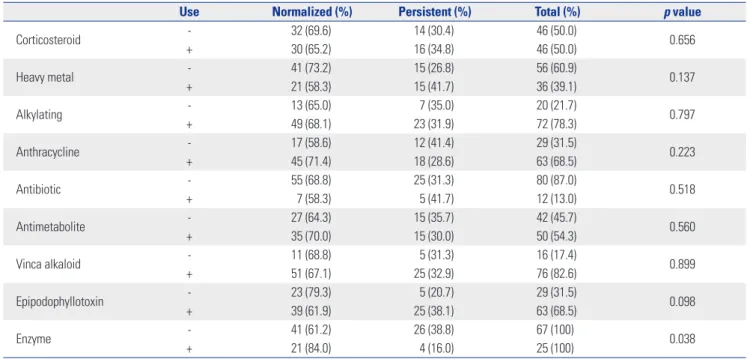
0.017); the numbers of survivors previously receiving HSCT (p=0.378) or chemotherapy (p=0.326) were similar in both gr- oups (Table 1). There were no group differences in the types of chemotherapeutic agents used with two exceptions: more sur- vivors showed normalized SCH when treated with enzyme (e.g., L-asparaginase) than those treated without enzyme [21 of 25 (84.0%) vs. 41 of 67 (61.2%); p=0.038], whereas epipodophyl- lotoxin usage tended to be higher in the persistent group [25 of 63 (38.1%) vs. 5 of 29 (20.7%); p=0.098] (Table 4).
Radiation doses to the head region were significantly higher in the persistent group (2394±2469 cGy) than in the normalized group (894±1591 cGy; p=0.003), as were doses to the spine region (1319±1603 cGy vs. 462±921 cGy; p=0.001) (Table 5). Sixteen of the 37 (43.2%) survivors who had received head radiation
≥1800 cGy still had SCH at the final follow-up. In contrast, all four survivors (100%) who had received head radiation of <1800 cGy returned to normal at the final follow-up (p=0.031, data not shown).
Radiation doses to the neck region were also higher in the persistent group than in the normalized group, although the difference was not significant (626±1468 cGy vs. 115±357 cGy;
p=0.086) (Table 5). All of the four survivors who had received neck radiation ≥1500 cGy still showed SCH at the final follow- up. Seven of the nine survivors (77.8%) who had received neck radiation <1500 cGy recovered normal thyroid function (p=
0.009, data not shown). The total radiation dose per survivor was higher in the persistent group than in the normalized group
(2883±2304 cGy vs. 1266±1707 cGy; p=0.001) (Table 5).
Time trend of thyroid function normalization
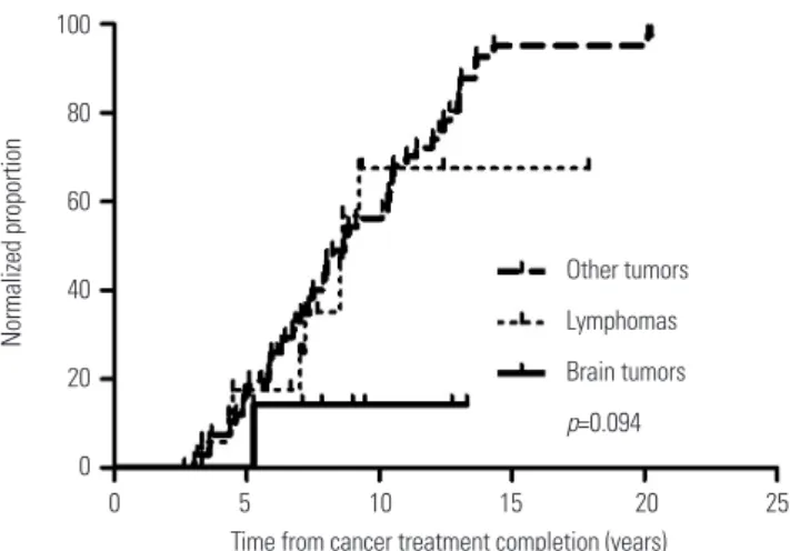
The median interval from time of cancer treatment completion to time of normalization of thyroid function was 8.84 years (95% confidence interval, 7.10–10.58 years). When the survi- vors were split into three cancer type groups (brain tumor sur- vivors, lymphoma survivors, and survivors of other cancers), there was a tendency of difference in the normalized propor- tion of SCH among the three groups (p=0.094) (Fig. 1). Brain tu- mor survivors did not regain normal thyroid function during the follow-up compared to the survivors in the other groups (p=0.039).
Multivariable analysis of the risk factors for persistent SCH
Lymphoma (odds ratio, 6.71; p=0.011), and brain/naso (odds Table 4. Thyroid Function According to Chemotherapeutic Agent
Use Normalized (%) Persistent (%) Total (%) p value
Corticosteroid - 32 (69.6) 14 (30.4) 46 (50.0)
0.656
+ 30 (65.2) 16 (34.8) 46 (50.0)
Heavy metal - 41 (73.2) 15 (26.8) 56 (60.9)
0.137
+ 21 (58.3) 15 (41.7) 36 (39.1)
Alkylating - 13 (65.0) 7 (35.0) 20 (21.7)
0.797
+ 49 (68.1) 23 (31.9) 72 (78.3)
Anthracycline - 17 (58.6) 12 (41.4) 29 (31.5)
0.223
+ 45 (71.4) 18 (28.6) 63 (68.5)
Antibiotic - 55 (68.8) 25 (31.3) 80 (87.0)
0.518
+ 7 (58.3) 5 (41.7) 12 (13.0)
Antimetabolite - 27 (64.3) 15 (35.7) 42 (45.7)
0.560
+ 35 (70.0) 15 (30.0) 50 (54.3)
Vinca alkaloid - 11 (68.8) 5 (31.3) 16 (17.4)
0.899
+ 51 (67.1) 25 (32.9) 76 (82.6)
Epipodophyllotoxin - 23 (79.3) 5 (20.7) 29 (31.5)
0.098
+ 39 (61.9) 25 (38.1) 63 (68.5)
Enzyme - 41 (61.2) 26 (38.8) 67 (100)
0.038
+ 21 (84.0) 4 (16.0) 25 (100)
Table 5. Thyroid Function According to Radiation Site and Dose Radiation site
Radiation dose (cGy, mean±SD)
p value Normalized SCH
(n=62)
Persistent SCH (n=30)
Head 893.5±1591.1 2394±2468.8 0.003
Neck 115.2±357.4 626±1467.7 0.086
Chest 196±563.9 260±869.2 0.841
Abdomen 302.6±836.9 323±776.6 0.535
Extremity 220±621.6 110±338.7 0.882
Testis 115.2±357.4 110±338.7 >0.999
Spine 462.1±920.7 1319±1603 0.001
Total radiation
dose per survivor 1266.3±1707 2883±2303.8 0.001 SCH, subclinical hypothyroidism.
ratio, 15.35; p=0.039) were significant risk factors for persistent SCH when compared to other tumors as a reference (Table 6).
A head radiation dose of ≥1800 cGy was also a significant risk factor (odds ratio, 3.78; p=0.039). Sex, HSCT, type of chemo- therapeutic agent, and neck radiation dose ≥1500 were not sig- nificant risk factors.
DISCUSSION
Primary hypothyroidism is caused by direct damage to the thy-
roid gland and is categorized as SCH or, if more severe, overt hypothyroidism. Central hypothyroidism is caused by damage to the hypothalamus-pituitary gland axis and can result in loss of endocrine function. In our study, SCH was the most com- mon thyroid disease after treatment of childhood cancer (104 of 112 survivors, 92.9%). Development of SCH during the fol- low-up period was unrelated to the previous cancer type. How- ever, the percentage of survivors who regained a euthyroid sta- tus differed according to cancer type and treatment. Our aim was to identify survivors at risk for persistent SCH and to sug- gest a watchful waiting strategy for the normalized group and an early therapy for the persistent group.
Diagnosis of SCH is based on laboratory findings. Most pa- tients with SCH show no symptoms or signs. Some present with non-specific symptoms such as poor quality of life, anxi- ety, depression, fatigue, or constipation.15 The prevalence of SCH ranges from 1% to 12.4% in the general population.10 Other estimates are 4.3% (the National Health and Examination Sur- vey III) and 5.4–17.3% (large studies of thyroid dysfunctions in Koreans 17 years of age or older).16,17 Prevalence tends to in- crease as age increases.17 There are no studies on SCH preva- lence in Korean children.
TSH levels return to normal in 15–65% of untreated individ- uals 1–6 years after one-time elevations18,19 and in >50% of indi- viduals with increased or decreased levels.19 However, a signi- ficant proportion of adult patients with SCH progress to overt hypothyroidism. The rate of progression to overt hypothyroid- ism was 1–4% per year in one study and 2.9% per 5 years in an- other.15,19 In a prospective study with a mean observation time of 9 years, 30–50% of female patients with SCH developed overt hypothyroidism, depending on clinical risk factors.20
The clinical consequences of SCH are controversial. In a me- ta-analysis of 55287 subjects, the risk of coronary heart disease and mortality increased as TSH concentration increased.21 The hazard ratio in the meta-analysis was 1.89 (95% confidence interval, 1.28–2.80) at 10 mU/L TSH. Conversely, two prospec- tive studies found no association between SCH and the out- comes of patients with cardiovascular disease.22,23 Other con- sequences of SCH include non-alcoholic fatty liver disease24 and, although not observed in all studies, psychiatric manifes- tations such as depression, anxiety, and cognitive dysfunction.25
Endocrinologists recommend thyroid hormone replacement for patients with TSH concentrations of >10 mU/L and routine surveillance for patients with TSH concentrations of ≤10 mU/L.8,9 Replacement therapy is sometimes advised for people aged less than 65 years, as their cardiovascular risks are greater than those of people aged 65 years or more.15
SCH is less prevalent in children than in adults. Although in- sufficient, data thus far suggest that SCH occurs in 1.7–9.5% of otherwise normal children.10 The natural course of SCH in ch- ildhood has not been well studied. In a recent meta-analysis, only nine relevant studies could be evaluated,6 most of which included only a small number of subjects. An exception was a Table 6. Multivariable Analysis of Risk Factors for Persistent Subclinical
Hypothyroidism
Odds ratio (95% CI) p value Sex
Female 0.80 (0.24–2.70) 0.716
HSCT
Yes 3.38 (0.93–12.36) 0.065
Chemotherapy
Enzymes 0.26 (0.054–1.22) 0.087
Epipodophyllotoxins 0.92 (0.21–3.92) 0.905
Age at diagnosis
>10 years 0.85 (0.19–3.87) 0.838
Diagnosis
Other tumors* 1
Lymphoma 6.71 (1.16–28.92) 0.011
Brain/naso 15.35 (1.15–204.65) 0.039
Head radiation
Dose ≥1800 cGy 3.78 (1.07–13.30) 0.039
Neck radiation
Dose ≥1500 cGy - 0.999
HSCT, hematopoietic stem cell transplantation; naso, nasopharyngeal; CI, confidence interval.
*Other tumors included Wilms tumor, neuroblastoma, abdomen tumors, sar- coma, and other cancer types.
Fig. 1. Cumulative incidence of normalization of thyroid function accord- ing to cancer type. Other tumors included Wilms tumor, neuroblastoma, abdomen tumors, sarcoma, and other cancer types.
100 80 60 40 20
0
0 5 10 15 20 25 Time from cancer treatment completion (years)
Normalized proportion
Other tumors Lymphomas Brain tumors p=0.094
large-scale retrospective study conducted in Israel; however, most of its subjects had autoimmune thyroiditis.11 The rate of recovery in the meta-analysis was 21.9–50%, and progression to overt hypothyroidism was 0–28.8%.6 In the Israeli study, SCH prevalence was 3.3% (n=3938), and 70.1% of patients with SCH had normal TSH levels in subsequent tests.11
Thyroid dysfunction in childhood cancer survivors is a well- known late effect.3,7,26 Primary hypothyroidism is the most com- mon abnormal thyroid-related response to thyroid irradiation.4 Risk factors for hypothyroidism include neck, craniospinal, and total body irradiation3,4 yet apparently not chemotherapy.4,13 Compared with primary overt hypothyroidism, SCH in child- hood cancer survivors is less understood. The percentage of long-term survivors of Hodgkin lymphoma (n=55) who devel- op SCH is 20%.
Data on the natural history of SCH in childhood cancer sur- vivors are limited. A previous study showed impaired thyroid function in 24% of survivors (71 of 291) during a median follow- up period of 6.1 years13 yet did not address SCH status after this time. Ishiguro, et al.12 found that 26.5% of patients (39 of 147) who underwent bone marrow transplantation developed SCH.
Their TSH levels decreased from the upper normal range 8 years after bone marrow transplantation to the mid to normal range thereafter. However, the proportion of patients who recov- ered from SCH was not presented.
In our study, 12 (2.8%) of the 423 childhood cancer survivors had overt hypothyroidism, mainly the brain/naso survivors.
SCH was the most common thyroid dysfunction in the 423 sur- vivors (104, 24.6%), as well as in the 123 survivors with thyroid abnormalities (104, 84.5%). Among the study cohort (n=92), 67.4% of patients with SCH regained normal thyroid function.
This result is similar to those in other pediatric studies on pa- tients with autoimmune disease or healthy thyroids.6,11 In our study, 60–85% of leukemia, NHL, and WT/NB/abdomen sur- vivors regained normal thyroid function, whereas only 12.5–
33.3% of brain/naso and HD survivors did so; this difference was significant (p=0.002) (Table 2). Although the proportion of leukemia survivors with SCH was substantial (21.6%, which was much higher than in a normal pediatric cohort), most ex- hibited normalized thyroid function at the final follow-up. The percentages of brain/naso survivors with SCH or overt hypo- thyroidism were 25.6% (11 of 43) and 20.9% (9 of 43), respec- tively; however, most of them did not recover a euthyroid sta- tus. Collectively, these data show that SCH is frequently found in childhood cancer survivors regardless of cancer type. Leu- kemia and WT/NB/abdomen survivors can be managed with- out further treatment, whereas brain/naso survivors should re- ceive thyroid hormone replacement as soon as possible, even if they have SCH rather than overt hypothyroidism.
The risk factors for persistent SCH are similar to those for overt hypothyroidism as reported in previous studies.4,5 How- ever, the data on the dose-volume effect for the development of SCH are generally lacking. Our multivariable analysis con-
firmed that brain/naso tumors, lymphoma, and head radiation doses ≥1800 cGy were all significant risk factors for SCH. Brain tumor survivors have an increased risk of endocrine, psychiat- ric, cognitive, neurological, and developmental disorders ow- ing to previous radiotherapy.3,4 The threshold dose for pituitary- hypothalamus hypothyroidism was 5000 cGy in the studies on the cranial irradiation.27-29 When the radiation dose to the pitu- itary gland was increased from 2000 to 4500 cGy, the risk of hy- pothyroidism increased from 9% to 52%.29 As for direct thyroid radiation, the thresholds are less clear; however, radiation dos- es greater than 3000–4000 cGy are generally accepted as a th- reshold.29,30 Nevertheless, the threshold for SCH has not yet been established. A large study on the general health status of cancer survivors reported that exposure of the hypothalamus- pituitary axis to ≥1800 cGy disrupted this axis in >50% of sur- vivors.31 However, it regarded all physical and laboratory find- ings including growth, pubertal progress, menstrual history, insulin-like growth factor 1 (IGF-1), cortisol, estradiol, TSH, and fT4 by screening studies as outcome measurements and did not specify the kind, proportion, or severity of axis disruption.31 Therefore, the radiation dose for inducing SCH could not be confirmed. In this study, radiation exposure of 1800 cGy or more to the head region posed the risk of persistent SCH, and this dose was lower than the dose causing overt hypothyroidism.
Whether cancer survivors who develop SCH during follow- up should receive treatment is an unanswered question. Our study provides suggestions based on the presence of risk fac- tors. However, it had several limitations. First, the study cohort was prospective, whereas the analysis was retrospective. Sec- ond, follow-ups regularly occurred every 6 months; however, several survivors were lost during follow-up. Third, the dura- tion of the follow-up was 4.2 years, yet the range was variable.
Fourth, certain cancer types had an insufficient number of sur- vivors for analysis. Fifth, survivors with normal thyroid function at the initial follow-up did not receive additional TFTs in com- pliance with the Korean health insurance policy. Sixth, thyroid autoantibodies as a risk factor for progression to thyroid dis- ease were not examined owing to the retrospective nature of our analysis. Despite these limitations, our study is one of the few studies on SCH in childhood cancer survivors, and it pro- vides new data for Korean survivors. We showed that a sub- stantial number of leukemia and WT/NB/abdomen survivors developed SCH, and our follow-up data described the natural course of SCH in survivors.
In conclusion, SCH is a frequent late effect in childhood can- cer survivors. Most leukemia and WT/NB/abdomen survivors with SCH recovered spontaneously. There was an increased risk of persistent SCH in survivors with brain/naso, lymphoma, or head radiation at doses ≥1800 cGy.
REFERENCES
1. Hewitt M, Weiner SL, Simone JV. Childhood cancer survivorship:
improving care and quality of life. Washington, DC: National Acad- emies Press; 2003.
2. Pui CH, Pei D, Pappo AS, Howard SC, Cheng C, Sandlund JT, et al.
Treatment outcomes in black and white children with cancer: re- sults from the SEER database and St Jude Children’s Research Hos- pital, 1992 through 2007. J Clin Oncol 2012;30:2005-12.
3. Kurt BA, Armstrong GT, Cash DK, Krasin MJ, Morris EB, Spunt SL, et al. Primary care management of the childhood cancer survivor.
J Pediatr 2008;152:458-66.
4. Chemaitilly W, Sklar CA. Endocrine complications in long-term survivors of childhood cancers. Endocr Relat Cancer 2010;17:
R141-59.
5. Nandagopal R, Laverdière C, Mulrooney D, Hudson MM, Meacham L. Endocrine late effects of childhood cancer therapy: a report from the Children’s Oncology Group. Horm Res 2008;69:65-74.
6. Monzani A, Prodam F, Rapa A, Moia S, Agarla V, Bellone S, et al.
Endocrine disorders in childhood and adolescence. Natural history of subclinical hypothyroidism in children and adolescents and po- tential effects of replacement therapy: a review. Eur J Endocrinol 2012;168:R1-11.
7. Han JW, Kwon SY, Won SC, Shin YJ, Ko JH, Lyu CJ. Comprehensive clinical follow-up of late effects in childhood cancer survivors shows the need for early and well-timed intervention. Ann Oncol 2009;
20:1170-7.
8. Gharib H, Tuttle RM, Baskin HJ, Fish LH, Singer PA, McDermott MT. Subclinical thyroid dysfunction: a joint statement on manage- ment from the American Association of Clinical Endocrinologists, the American Thyroid Association, and the Endocrine Society. J Clin Endocrinol Metab 2005;90:581-5.
9. Garber JR, Cobin RH, Gharib H, Hennessey JV, Klein I, Mechanick JI, et al. Clinical practice guidelines for hypothyroidism in adults:
cosponsored by the American Association of Clinical Endocrinol- ogists and the American Thyroid Association. Thyroid 2012;22:
1200-35.
10. Catli G, Abaci A, Büyükgebiz A, Bober E. Subclinical hypothyroid- ism in childhood and adolescense. J Pediatr Endocrinol Metab 2014;27:1049-57.
11. Lazar L, Frumkin RB, Battat E, Lebenthal Y, Phillip M, Meyerovitch J. Natural history of thyroid function tests over 5 years in a large pe- diatric cohort. J Clin Endocrinol Metab 2009;94:1678-82.
12. Ishiguro H, Yasuda Y, Tomita Y, Shinagawa T, Shimizu T, Morimoto T, et al. Long-term follow-up of thyroid function in patients who re- ceived bone marrow transplantation during childhood and adoles- cence. J Clin Endocrinol Metab 2004;89:5981-6.
13. Madanat LM, Lähteenmäki PM, Alin J, Salmi TT. The natural histo- ry of thyroid function abnormalities after treatment for childhood cancer. Eur J Cancer 2007;43:1161-70.
14. Landier W, Bhatia S, Eshelman DA, Forte KJ, Sweeney T, Hester AL, et al. Development of risk-based guidelines for pediatric cancer survivors: the Children’s Oncology Group Long-Term Follow-Up Guidelines from the Children’s Oncology Group Late Effects Com- mittee and Nursing Discipline. J Clin Oncol 2004;22:4979-90.
15. Cooper DS, Biondi B. Subclinical thyroid disease. Lancet 2012;379:
1142-54.
16. Kim TH, Kim KW, Ahn HY, Choi HS, Won H, Choi Y, et al. Effect of seasonal changes on the transition between subclinical hypothy- roid and euthyroid status. J Clin Endocrinol Metab 2013;98:3420-9.
17. Kim YA, Park YJ. Prevalence and risk factors of subclinical thyroid disease. Endocrinol Metab (Seoul) 2014;29:20-9.
18. Somwaru LL, Rariy CM, Arnold AM, Cappola AR. The natural his- tory of subclinical hypothyroidism in the elderly: the cardiovas- cular health study. J Clin Endocrinol Metab 2012;97:1962-9.
19. Meyerovitch J, Rotman-Pikielny P, Sherf M, Battat E, Levy Y, Surks MI. Serum thyrotropin measurements in the community: five-year follow-up in a large network of primary care physicians. Arch In- tern Med 2007;167:1533-8.
20. Huber G, Staub JJ, Meier C, Mitrache C, Guglielmetti M, Huber P, et al. Prospective study of the spontaneous course of subclinical hy- pothyroidism: prognostic value of thyrotropin, thyroid reserve, and thyroid antibodies. J Clin Endocrinol Metab 2002;87:3221-6.
21. Rodondi N, den Elzen WP, Bauer DC, Cappola AR, Razvi S, Walsh JP, et al. Subclinical hypothyroidism and the risk of coronary heart disease and mortality. JAMA 2010;304:1365-74.
22. Boekholdt SM, Titan SM, Wiersinga WM, Chatterjee K, Basart DC, Luben R, et al. Initial thyroid status and cardiovascular risk factors:
the EPIC-Norfolk prospective population study. Clin Endocrinol (Oxf) 2010;72:404-10.
23. Rodondi N, Newman AB, Vittinghoff E, de Rekeneire N, Satterfield S, Harris TB, et al. Subclinical hypothyroidism and the risk of heart failure, other cardiovascular events, and death. Arch Intern Med 2005;165:2460-6.
24. Chung GE, Kim D, Kim W, Yim JY, Park MJ, Kim YJ, et al. Non-alco- holic fatty liver disease across the spectrum of hypothyroidism. J Hepatol 2012;57:150-6.
25. Baldini IM, Vita A, Mauri MC, Amodei V, Carrisi M, Bravin S, et al.
Psychopathological and cognitive features in subclinical hypothy- roidism. Prog Neuropsychopharmacol Biol Psychiatry 1997;21:
925-35.
26. Han JW, Kim HS, Kim BS, Kwon SY, Shin YJ, Kim SH, et al. Increas- ing and worsening late effects in childhood cancer survivors during follow-up. J Korean Med Sci 2013;28:755-62.
27. Vogelius IR, Bentzen SM, Maraldo MV, Petersen PM, Specht L.
Risk factors for radiation-induced hypothyroidism: a literature- based meta-analysis. Cancer 2011;117:5250-60.
28. Jereczek-Fossa BA, Alterio D, Jassem J, Gibelli B, Tradati N, Orec- chia R. Radiotherapy-induced thyroid disorders. Cancer Treat Rev 2004;30:369-84.
29. Littley MD, Shalet SM, Beardwell CG, Robinson EL, Sutton ML. Ra- diation-induced hypopituitarism is dose-dependent. Clin Endo- crinol (Oxf) 1989;31:363-73.
30. Boomsma MJ, Bijl HP, Langendijk JA. Radiation-induced hypothy- roidism in head and neck cancer patients: a systematic review. Ra- diother Oncol 2011;99:1-5.
31. Hudson MM, Ness KK, Gurney JG, Mulrooney DA, Chemaitilly W, Krull KR, et al. Clinical ascertainment of health outcomes among adults treated for childhood cancer. JAMA 2013;309:2371-81.