Novel Mutations in the UNC13D Gene Carried by a Chinese Neonate with Hemophagocytic Lymphohistiocytosis
Yuanyuan Chen, Zhujun Wang, Yuping Cheng, and Yongmin Tang
Division of Hematology-Oncology, Children’s Hospital of Zhejiang University School of Medicine, Hangzhou, PR China.
Received: December 7, 2011 Revised: July 1, 2012 Accepted: September 7, 2012
Corresponding author: Dr. Yongmin Tang, Division of Hematology-Oncology, Children’s Hospital of Zhejiang University School of Medicine, #57, Zhuganxiang Road, Yan-an Street, Hangzhou 310003, PR China.
Tel: 0086 571 87061007 ext. 32460, Fax: 0086 571 87033296 E-mail: [email protected]
∙ The authors have no financial conflicts of interest.
© Copyright:
Yonsei University College of Medicine 2013 This is an Open Access article distributed under the terms of the Creative Commons Attribution Non- Commercial License (http://creativecommons.org/
licenses/by-nc/3.0) which permits unrestricted non- commercial use, distribution, and reproduction in any medium, provided the original work is properly cited.
Hemophagocytic lymphohistiocytosis (HLH) in different ethnicities has been de- scribed in the literature, but few cases in patients of Chinese descent have been re- ported. Here, we describe the case of a Chinese neonate presenting with HLH car- rying novel, compound heterozygous mutations of the UNC13D gene, including [c.2295_2298delGCAG, p.Glu765Aspfs*27] in exon 23, c.-250C>T, c.1+30G>A, c.279C>T, c.888G>C, c.18+36A>G, c.20-48T>C, c.1977C>T, c.2296C>T, c.24- 46C>T, c.26-9_26-8insC, c.2599A>G, c.28+48C>T and c.3198A>G, some of which have not been reported in the literature. Cytokine profile analyses were per- formed in this patient, and the results were consistent with our previous findings in HLH patients. Cytokine profile monitoring may be helpful in differentiating among various clinical phases of HLH.
Key Words: FHL, hemophagocytic lymphohistiocytosis, UNC13D, CD107a, cy- tokine monitoring
INTRODUCTION
Hemophagocytic lymphohistiocytosis (HLH) comprises the inherited form, prima- ry HLH, and secondary HLH, which usually is virus or malignancy associated.
Primary HLH often occurs in infancy and includes familial hemophagocytic lym- phohistiocytosis (FHL) and the immune deficiency syndromes associated with HLH (Chediak-Higashi syndrome, Griscelli syndrome, and X-linked lymphopro- liferative syndrome).1 FHL is a potentially lethal disorder of immune dysregulation that requires prompt and accurate diagnosis to initiate life-saving immunosuppres- sive therapy and to prepare for hematopoietic stem cell transplantation.2 Five sub- types of FHL have been identified so far, with mutations in the following genes:
unknown (FHL1), PRF1 (FHL2), UNC13D (FHL3), STX11 (FHL4), and STXBP2 (FHL5), respectively.3-6 Functional tests with resting NK cells and/or CTL cells, including perforin detection and degranulation assay quantifying CD107a surface expression with flow cytometry, can direct genetic analyses. Perforin expression is constantly reduced or absent in most FHL2 patients, while patients with FHL3, FHL4 or FHL5 can be screened by a degranulation assay result below 5%.7 In this paper, we describe the case of a Chinese male neonate who presented with HLH and had one identical, novel, heterozygous mutation in the UNC13D gene.
sive thrombocytopenia and was admitted to our hospital on February 15, 2011. His body temperature fluctuated around 38.5°C. At the time of admission, his liver and spleen had enlarged 3 cm and 4 cm below his costal margins, respec- tively. No rashes were noticed on his skin. Auscultation on his lungs and heart was normal. No CNS symptoms and signs were found. The following laboratory data were re- corded: WBC of 7.65×10⁹/L; Hb of 129g/L; platelet count of 18×109/L; feritin >1500 ng/mL; bilirubin total/direct 120.3 μmol/L/32.6 μmol/L; AST of 76 IU/L; ALT of 33 IU/L; fi- brinogen of 1.24 g/L; PT/APTT of 12.8/41.8 sec; thrombin time of 17.7 sec; D-dimer of 563 μg/L; triglyceride of 2.24 mmol/L and an LDH of 617 IU/L. Abdominal B-mode ul- trasound examination showed massive hepatosplenomega- ly. A bone marrow biopsy revealed numerous hemophago- cytic histiocytes consistent with a diagnosis of HLH.8 The parents of this patient also had given birth to a daughter two years before, who died several hours after her birth because of “suspected infection”.
FHL-related gene analysis using genomic DNA showed that the patient had novel, compound heterozygous muta- tions in the UNC13D gene, including c.-250C>T, c.1+30G>
A, c.279C>T, c.888G>C9, c.18+36A>G, c.20-48T>C, c.1977C>T, c.2296C>T (Fig. 1A), c.24-46C>T, c.26-9_26- 8insC, c.2599A>G, c.28+48C>T and c.3198A>G. Gene
CASE REPORT
An eight-day old Chinese neonate presented with progres-
A
A
B
B
Father
Father
Father 86.24%
9.04%
50.22%
4.76%
14.81%
0.51%
55.72%
0.45%
M1
M1
M1
M1
M1
M1
M1
M1
CountsCounts
Father Mother
Mother
Mother Mother
Patient
Patient
Patient Acute phase of HLH
Acute phase of HLH
Relieved status of HLH
Relieved status of HLH
IgG2b-PEPerforin-PE Before K562 cell stimulationAfter K562 cell stimulation
Perforin
CD107a Splicing site shifted
UNC13D-Genomic DNA
c.2296C>T UNC13D-cDNA
c.2295_2298delGCAG Patient Deleted
Fig. 2. The perforin concentration (A) and CD107a positivity before and after K562 cell stimulation (B) in NK cells of this patient and his parents. HLH, hemo- phagocytic lymphohistiocytosis; PE, phycoerythrin.
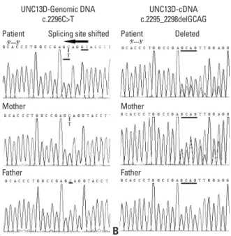
Fig. 1. Sequencing results of UNC13D gene in a Chinese male neonate and his parents. Genomic DNA sequencing results showed the patient and his mother had heterozygous point mutation c.2296C>T (A), which caused shifted splicing site in the transcription process and in mRNA level mani- fested as a frameshift mutation c.2295_2298delGCAG (B). In protein level, it lead to a premature stop codon p.Glu765Aspfs*27 and encoded truncated protein munc13-4 (1-792).
analysis using cDNA with a pair of primers for UNC13D- Sequence Coding for Amino Acids in Protein (CDS).
showed a frameshift mutation of (c.2295_2298del GCAG, p.Glu765Aspfs*27) (Fig. 1B) in UNC13D, which was con- sistent with the genomic DNA sequencing results for c.2296C>T, as the splicing site shifted in the transcription process based on the “GT-AG” splicing law. These genom- ic DNA and cDNA sequencing results for the patient were confirmed by the sequencing results of his parents. We also sequenced other FHL-related genes for this patient and his parents, including PRF1, STX11 and STXBP2, but no mu- tation was found except for one synonymous change in PRF1 gene (c.900C>T, p.His300His). In the acute phase of HLH, the perforin expression percentage with resting NK cell by flow cytometry of this patient was 14.81% (normal range 70.57±21.32%) (Fig. 2A) and surface CD107a level after K562 stimulation was 0.51% (normal range 14.1±
7.2%) (Fig. 2B), both of which were significantly reduced.
However, when his condition of HLH was controlled, his level of perforin expression returned to normal (55.72%), whereas his damaged function of degranulation never re- covered on repeated testing (never over 1%). Th1/Th2 cy- tokine levels, quantitatively determined by the cytometric bead array (CBA) kit-BDTM CBA Human Th1/Th2 Cyto- kine Kit II (BD Biosciences, San Jose, CA, USA), were successively monitored during the course of the disease in this patient. We also detected bacteria and viruses in his blood, with almost all negative results except for EBV-VCA IgG (Table 1). Bacteria and viruses were also detected in his parents, and all were showed negative results.
He received HLH-2004 chemotherapy, including etopo- side (VP-16), dexamethasone, cyclosporine A and intrathe- cal methotrexate, and responded well. Four days after initial administration, his platelets began to rise steadily and the spleen gradually shrank back to its normal position. During the non-hospitalized period, he was followed-up mainly by telephone. Although an intensive search for a HLA-matched donor among candidates in the Chinese Hematopoietic Stem Cell Bank with over 1.3 million donors was conduct- ed, no proper donor was found. The patient remained on the immunosuppressive therapy until his death in October 2011 because of gastrointestinal hemorrhage.
DISCUSSION
HLH in different ethnicities has been described in the litera- Table 1.h1/Th2 Cytokine Peak Levels in This Patient (pg/mL) T IL-2IL-4IL-6IL-10 TNF-α IFN-γ Testing dateLaboratory examinationClinical conditionsSpecific Th1/Th2 cytokine patterns (1.1-9.8)(0.1-3.0)(1.7-16.6)(2.6-4.9)(0.1-5.2)(1.6-17.3) 18/02/20111.71.117.1133.11.6 8.7Blood culture (-)Acute phase of HLHIncreased levels of IL-10 and/or IFN-γ Exaggerated status of Higher IFN-γ and IL-10 levels than those 24/02/20111.61.920.1693.02.051.0CMV DNA (-) HLH at initial diagnosis Exaggerated status of Accompanied by slightly elevated level 25/02/20112.11.229.3355.72.232.6EBV-VCA IgG: 127.30 IU/mL HLH of IL-6 IFN-γ and IL-10 levels are significantly 04/03/20112.72.2 6.7235.42.3 8.5EBV-DNA: (-)Relieved status of HLH decreased IL-10 and/or IFN-γ levels continued to 09/03/20111.90.917.1135.71.312.4CMV IgG: (+)Relieved status of HLH decline Exaggerated status of Declined IL-10 and IFN-γ levels, with 16/03/20111.81.020.0317.91.834.0EBV-VCA IgG: 120.80 IU/mL HLH fluctuations related to clinical changes 19/04/20112.62.0 3.8 10.32.6 3.4Blood culture (-)Controlled HLHAll the cytokines returned back to normal HLH, hemophagocytic lymphohistiocytosis. EBV-VCA IgG normal range: 0-22.0 IU/mL; -: negative; +: positive.
cessively monitor Th1/Th2 cytokine levels in the present patient during the course of HLH, e.g. acute phase of HLH, exaggerated status of HLH, relieved status of HLH and con- trolled HLH, indicating that the cytokine patterns were con- sistent with HLH clinical phases. Cytokine profile monitor- ing could be an important biomarker for monitoring HLH status and allow us to make an earlier diagnosis through which to administer more effective treatment.
ACKNOWLEDGEMENTS
This work was supported in part by the grants from the Na- tional Natural Science Foundation of China, No. 81170502, No.30971283 and the Zhejiang Provincial Natural Science Foundation of China, No. LZ12H08001. The authors would like to thank Mr. Ning Zhao, Ms. Baiqin Qian, Ping Chen and Mr. Hongqiang Shen for their excellent technical assistance.
REFERENCES
1. Zhizhuo H, Junmei X, Yuelin S, Qiang Q, Chunyan L, Zhengde X, et al. Screening the PRF1, UNC13D, STX11, SH2D1A, XIAP, and ITK gene mutations in Chinese children with Epstein-Barr vi- rus-associated hemophagocytic lymphohistiocytosis. Pediatr Blood Cancer 2012;58:410-4.
2. Murata Y, Yasumi T, Shirakawa R, Izawa K, Sakai H, Abe J, et al.
Rapid diagnosis of FHL3 by flow cytometric detection of intra- platelet Munc13-4 protein. Blood 2011;118:1225-30.
3. Stepp SE, Dufourcq-Lagelouse R, Le Deist F, Bhawan S, Certain S, Mathew PA, et al. Perforin gene defects in familial hemophago- cytic lymphohistiocytosis. Science 1999;286:1957-9.
4. Feldmann J, Callebaut I, Raposo G, Certain S, Bacq D, Dumont C, et al. Munc13-4 is essential for cytolytic granules fusion and is mutated in a form of familial hemophagocytic lymphohistiocyto- sis (FHL3). Cell 2003;115:461-73.
5. zur Stadt U, Schmidt S, Kasper B, Beutel K, Diler AS, Henter JI, et al. Linkage of familial hemophagocytic lymphohistiocytosis (FHL) type-4 to chromosome 6q24 and identification of mutations in syntaxin 11. Hum Mol Genet 2005;14:827-34.
6. zur Stadt U, Rohr J, Seifert W, Koch F, Grieve S, Pagel J, et al.
Familial hemophagocytic lymphohistiocytosis type 5 (FHL-5) is caused by mutations in Munc18-2 and impaired binding to syn- taxin 11. Am J Hum Genet 2009;85:482-92.
7. Bryceson YT, Pende D, Maul-Pavicic A, Gilmour KC, Ufheil H, Vraetz T, et al. A prospective evaluation of degranulation assays in the rapid diagnosis of familial hemophagocytic syndromes. Blood 2012;119:2754-63.
8. Henter JI, Horne A, Aricó M, Egeler RM, Filipovich AH, Imashu- ku S, et al. HLH-2004: diagnostic and therapeutic guidelines for hemophagocytic lymphohistiocytosis. Pediatr Blood Cancer 2007;48:124-31.
9. Zhang K, Biroschak J, Glass DN, Thompson SD, Finkel T, Passo
ture, but few patients of Chinese descent have been report- ed.1 Yoon, et al.10 reported that FHL3 accounted for 89% of cases of FHL in Korea, and approximately 20% to 25% in Japan, indicating that UNC13D mutations may be responsi- ble for a large part of HLH patients in Asian countries.
Here, we clearly showed a neonate with HLH harboring compound heterozygous mutations (c.2295_2298delGCAG, p.Glu765Aspfs*27) in exon 23 of the UNC13D gene by bi- directional sequencing. So far as we know, such mutation in UNC13D has never been reported in the literature. In many published reports, compound heterozygous mutations were common in FHL patients. Although FHL belongs to the autosomal recessive form of the disease, whether an au- tosomal dominant trait in FHL patients exists or not remains to be defined. Our patient showed a clear heterozygous mu- tation, and family history of a sister who died previously may be partial evidence for the possibility of such a genetic defect.11 Results on degranulation assay constantly below 5% also suggested that the patient had a diagnosis of FHL.7 The mutations in the UNC13D gene of this patient were all inherited from his mother, yet the mother had no presenta- tions of HLH with a mild reduction of degranulation func- tion (4.76%), indicating that EBV infection plays an impor- tant role in FHL onset. However, due to the fact that our gene analysis was carried out using conventional sequenc- ing of exons and splice-sites, possibilities of deep intronic mutations and other genetic aberrations cannot be definitely excluded. Another interesting question is that the neonate presented with a low level of perforin expression with NK cells in the acute phase of his disease. However, when his condition of HLH was controlled, his level of perforin ex- pression returned to normal. These results indicate that the abnormality of perforin expression in NK cells is not a de novo defect, but rather a temporary phenomenon.
A specific Th1/Th2 cytokine pattern to diagnose HLH was first reported by our group with excellent clinical applica- tion.12,13 During the acute phase of HLH without complicat- ing bacterial infection, both IFN-γ and IL-10 levels should be increased, while IL-6 remains normal or only slightly ele- vated. When HLH patients enters remission, the levels of IFN-γ and IL-10 both decline, usually with IFN-γ declining faster than IL-10. According to our previous studies, IL-6 levels are much higher in microbiologically documented infection patients than in patients with non-sepsis.13 When IFN-γ, IL-10 and IL-6 levels return to normal or close to nor- mal, the HLH related conditions of the patient can be con- sidered to be in control. In this regard, we were able to suc-
magnetic resonance imaging. Neuropediatrics 2011;42:191-3.
12. Tang Y, Xu X, Song H, Yang S, Shi S, Wei J, et al. Early diagnos- tic and prognostic significance of a specific Th1/Th2 cytokine pat- tern in children with haemophagocytic syndrome. Br J Haematol 2008;143:84-91.
13. Tang Y, Liao C, Xu X, Song H, Shi S, Yang S, et al. Evaluation of Th1/Th2 cytokines as a rapid diagnostic tool for severe infection in paediatric haematology/oncology patients by the use of cyto- metric bead array technology. Clin Microbiol Infect 2011;17:
1666-73.
MH, et al. Macrophage activation syndrome in patients with sys- temic juvenile idiopathic arthritis is associated with MUNC13-4 polymorphisms. Arthritis Rheum 2008;58:2892-6.
10. Yoon HS, Kim HJ, Yoo KH, Sung KW, Koo HH, Kang HJ, et al.
UNC13D is the predominant causative gene with recurrent splic- ing mutations in Korean patients with familial hemophagocytic lymphohistiocytosis. Haematologica 2010;95:622-6.
11. van Egmond ME, Vermeulen RJ, Peeters-Scholte CM, Augoustides- Savvopoulou P, Abbink F, Boelens JJ, et al. Familial hemophago- cytic lymphohistiocytosis in a pediatric patient diagnosed by brain