Original Article
Lower Eyelid Epiblepharon Associated with Lower Eyelid Retraction
Mi Sun Sung
1, Min Joung Lee
2, Ho-Kyung Choung
3, Nam Ju Kim
4, Sang In Khwarg
21
Department of Ophthalmology, Inje University Sanggye Paik Hospital, Seoul, Korea
2
Department of Ophthalmology, Seoul National University Hospital, Seoul, Korea
3
Department of Ophthalmology, Seoul National University Boramae Hospital, Seoul, Korea
4
Department of Ophthalmology, Seoul National University Bundang Hospital, Seongnam, Korea
Purpose: To describe a series of patients with lower eyelid epiblepharon associated with lower eyelid retraction.
Methods: We retrospectively reviewed the medical records of patients who underwent surgery for lower eyelid retraction, epiblepharon, or thyroid-associated ophthalmopathy (TAO) between October 1999 and March 2007.
Patients with both lower eyelid retraction and epiblepharon on preoperative examination were included in this study.
Results: Twenty-seven eyelids of 20 patients with both lower eyelid retraction and epiblepharon were enrolled. The underlying causes of lower eyelid retraction included congenital retraction (seven eyelids), congenital fibrosis of the extraocular muscles (CFEOM; seven eyelids), TAO (seven eyelids), post-operative cicatricial retraction (five eyelids), and facial nerve palsy (one eyelid). Eight of 27 eyelids were successfully corrected after the repair of retraction without the repair of epiblepharon, regardless of the cause of lower eyelid retraction. Another four eyelids with epiblepharon associated with TAO resolved after only orbital decompression. Cilia-everting sutures were additionally applied for epiblepharon in another 14 eyelids, 12 of which did not require the excision of a skin fold or the orbicularis muscles. Only one eyelid with mild retraction and epiblepharon underwent simple epiblepharon repair. Recurrence of retraction or epiblepharon developed in three eyelids during follow-up.
Conclusions: In cases with both lower eyelid retraction and epiblepharon, the retraction should be repaired first, and then the epiblepharon can be corrected selectively according to the severity of the case.
Key Words: Congenital fibrosis syndrome of the extraocular muscles, Congenital retraction, Epiblepharon, Lower eyelid retraction, Thyroid-associated ophthalmopathy
Received:August 20, 2009 Accepted: January 7, 2010
Reprint requests to Sang In Khwarg. Department of Ophthalmology, Seoul National University Hospital, #101 Daehangno, Jongno-gu, Seoul 110-744, Korea. Tel: 82-2-2072-2879, Fax: 82-2-741-3187, Email:
* This study was presented at the 98th Meeting of the Korean Ophtha- lmological Society, November 2007, Ilsan, Korea.
ⓒ 2010 The Korean Ophthalmological Society
This is an Open Access article distributed under the terms of the Creative Commons Attribution Non-Commercial License (http://creativecommons.org/licenses /by-nc/3.0/) which permits unrestricted non-commercial use, distribution, and reproduction in any medium, provided the original work is properly cited.
Lower eyelid retraction describes an abnormality in which the resting position of the lower eyelid is too low. The normal position of the lower eyelid is at the level of the inferior limbus. Although there are many normal variations, lower eyelid retraction is clinically diagnosed when any sclera is visible between the inferior corneoscleral limbus and the eyelid margin or when the lower eyelid margins of the two
eyes are at different levels [1].
Lagophthalmos, exposure keratopathy, conjunctivitis, and unnatural appearance are well known complaints and indications for surgical intervention in patients with lower eyelid re- traction [2]. Inward rotation of the lower eyelashes, namely lower eyelid epiblepharon, can be found in the presence of lower eyelid retraction and can cause functional problems.
However, this consideration has not been adequately addressed in the literature. To the best of the author’s knowledge, only a few cases of acquired lower eyelid ep- iblepharon have been described in patients with thyroid eyelid retraction [3].
In this study, we report the clinical features, management,
and outcomes in patients with lower eyelid epiblepharon
associated with lower eyelid retraction.
Materials and Methods
We reviewed the medical records of all patients who underwent surgery for lower eyelid retraction or lower eyelid epiblepharon under the care of one surgeon (SIK) at Seoul National University Hospital between October 1999 and March 2007. We also reviewed the records of patients who were surgically treated for thyroid-associated ophthalmopathy (TAO) during the same period, regardless of the surgical procedure performed. We reviewed all TAO patients who underwent surgery because there is a possibility that eyelid retraction or epiblepharon in these patients can be resolved after orbital wall decompression without the need for lid surgery.
Patients with both lower eyelid retraction and epi- blepharon on the preoperative examination were included as subjects of special interest to the authors. Patients with entropion, in which the eyelid margins were in-turning, were excluded. Patients with anophthalmic retraction were also excluded, because their cornea-touching cilia were thought to be attributable to entropion related to hor- izontal laxity and the effect of gravity of the prosthesis [4].
The protocol of this study was approved by the Institutional Review Board of Seoul National University Hospital.
We retrospectively studied each patient’s past history, including trauma or surgery, findings on preoperative exami- nations, diagnosis, surgical procedures and intraoperative findings, and surgical outcomes and complications.
Results
From October 1999 to March 2007, 60 patients with lower eyelid retraction and 497 patients with lower eyelid epiblepharon were surgically treated at Seoul National University Hospital. Among these, 23 eyelids of 18 patients exhibiting both lower eyelid retraction and epiblepharon were enrolled in the study, representing 30% of lower eye- lid retraction cases and 3.6% of epiblepharon cases. Two patients (three eyelids) had been previously diagnosed with TAO. On the other hand, 50 patients with TAO were surgically treated during the same period. The operative
indications varied for the cases, including eyelid retraction, strabismus, proptosis, and compressive optic neuropathy.
Using this approach, two patients (four eyelids) were ad- ditionally found to have both lower eyelid retraction and epiblepharon on preoperative examination. These patients underwent orbital wall decompression in both eyes.
Patients ranged in age from 3 to 49 years (mean age, 17.3 years; sex ratio, M:F=8:12). The mean postoperative follow-up period was 20.05 months (range, 1 to 102 months).
The underlying causes of lower eyelid retraction included congenital retraction, congenital fibrosis of the extraocular muscles (CFEOM), post-operative cicatricial retraction, facial nerve palsy, and TAO (Table 1).
Congenital retraction was diagnosed only after other causes of eyelid retraction were excluded. There was no history of uneventful delivery or other trauma including surgery. No patients had abnormalities of the skin or con- junctiva of the lower eyelid, nor did they have proptosis.
Seventh nerve and oculomotor nerve function were normal, and the findings of full ophthalmic examinations were normal, with the exception of bilateral congenital blepharoptosis and nasolacrimal duct obstruction in one patient and uni- lateral congenital nasolacrimal duct obstruction in one patient. No systemic abnormalities were detected, with the exception of one patient with Marfan syndrome, a clinical entity previously unknown to be associated with eyelid retraction [5].
Patients with CFEOM were diagnosed on the basis of downward fixation of one or both eyes and marked ble- pharoptosis with or without chin elevation. They were subsequently divided into five subgroups according to the classification of Harley et al. [6] (Table 1). The diagnosis of cicatricial retraction was made if there was a definite scar or symblepharon in the lower eyelid with a history of previous operation in the lower eyelid.
All patients underwent eyelid surgery under general (12
patients aged under 20 years of age) or local (six patients
aged over 20 years of age) anesthesia. When surgery for
lower eyelid epiblepharon associated with lower eyelid
retraction was performed in the non-TAO group, correction
of lower eyelid retraction with spacer grafts was initially
A B
C D
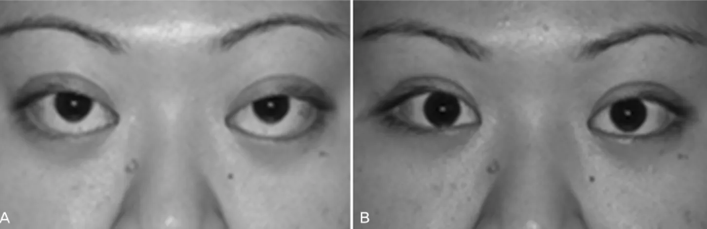
Fig. 1. Congenital lower eyelid retraction and epiblepharon. (A) Lower eyelid retraction and epiblepharon were observed in the preoperative photograph of a nine-yr-old boy. (B) Bilateral autogenous ear cartilage grafting and cilia-everting suturing were performed. After one month, the retraction and epiblepharon were resolved. (C) An eight-yr-old girl had unilateral retraction in the right lower eyelid and bilateral epiblepharon. (D) She underwent autogenous ear cartilage grafting in her right lower eyelid. Epiblepharon repair was performed on both eye- lids during the operation. The postoperative results were good.
performed. After that, epiblepharon procedures were added intraoperatively if the cornea-touching cilia were not suffi- ciently resolved. In the TAO group, there were two surgical options. If there was a possibility that orbital decompression would be requested functionally or cosmetically, orbital surgery was performed before any kind of lid surgery. If no orbital decompression was required, the lower eyelid retraction surgery was planned. Two patients who required orbital decompression received general anesthesia. The surgical procedures are described in Table 2. The grafts used for correcting lower eyelid retraction included 16 cases of autogenous ear cartilage, two Medpor
®sheets (Porex Surgical Inc., College Park, GA, USA), and one preserved homologous sclera. However, in one case exhibiting a very mild degree of retraction, correction was performed only for the epiblepharon. Epiblepharon repair included cilia-everting suture techniques with or without the excision of skin and muscle [7, 8]. A horizontal skin incision was made 2 mm below the lower lid margin, after which dissection was performed between the lower tarsus and the pretarsal orbicularis oculi. Fixation of subciliary subcutaneous tissue to the inferior margin of the lower tarsus was accomplished with 8-0 nylon (approximately five interrupted sutures), and the skin was repaired.
In all but one patient with congenital retraction (seven eyelids in six patients), lower eyelid retraction repair was accomplished with an autogenous ear cartilage graft, and epiblepharon repair was performed (mean age, 11.7 years;
sex ratio, M:F=3:3). The exception was a patient who had a very mild degree of retraction who underwent only epiblepharon repair with skin excision. The mean post- operative follow-up period was 19.8 months. All of the results were positive, and there were no complications, including recurrence (Fig. 1).
Congenital fibrosis of the extraocular muscles (seven
eyelids in six patients) patients underwent one of the following
treatments: lower eyelid retraction repair with autogenous
ear cartilage grafts and without epiblepharon repair (three
eyelids in three patients); lower eyelid retraction repair
with autogenous ear cartilage grafts and epiblepharon repair
(three eyelids in two patients); and lower eyelid retraction
repair using a scleral graft (one eyelid) (mean age, 12.7
years; sex ratio, M:F=4:2). The mean postoperative follow-up
period was 30.3 months. Epiblepharon was well corrected
immediately after the procedure. However, lower eyelid
retraction remained in one case where a preserved scleral
graft was used to correct lower eyelid retraction and epi-
blepharon, and scleral grafting was repeated two weeks
A B C
Fig. 2. Congenital fibrosis of the extraocular muscles. (A) A five-yr-old boy had lower eyelid retraction and epiblepharon, hypotropia and exotropia, and blepharoptosis in the left eyelid. He first underwent lower eyelid retraction repair using an autogenous ear cartilage graft. The surgeon then concluded that epiblepharon procedures were necessary. Cilia-everting tarsal fixation sutures were placed. (B) Epiblepharon recurred at four months postoperatively, although the lower eyelid was well positioned. (C) The patient underwent repeat epiblepharon repair on his left eyelid, including skin and orbicularis muscle excision. The photograph, taken only one week post-operatively, noted well-everted cilia and a prominent incision line along the lower eyelid margin. However, the line faded during follow-up.
Cicatricial
retraction 9 5 2 + (Medpor® sheets) + - 2 displacements of Medpor® sheets
2 + (ear cartilage) + - -
1
*+ (ear cartilage) - - -
Facial nerve palsy 48 1 1 + (ear cartilage) + - -
Thyroid-associated
ophthalmopathy 6.25 7 4
*-
†- - -
3
*+ (ear cartilage) - - 1 overcorrection of retraction
Total 20.05 27 eyelids 7 eyelids
*