http://dx.doi.org/10.20307/nps.2016.22.1.53
53
Dihydrobenzofuran Neolignans Isolated from Euonymus alatus Leaves and Twigs Attenuated Inflammatory Responses
in the Activated RAW264.7 Macrophage Cells
Na-Hyun Kim
1, Min Hye Yang
2, Jeong-Doo Heo
1, Sang Hyun Sung
3, and Eun Ju Jeong
4,*
1Gyeongnam Department of Environment & Toxicology, Korea Institute of Toxicology, 17 Jegok-gil, Munsan-eup, Gyeongnam 660-844, Republic of Korea
2College of Pharmacy, Pusan National University, Busan 609-735, Republic of Korea
3College of Pharmacy and Research Institute of Pharmaceutical Science, Seoul National University, Seoul 151-742, Republic of Korea
4Department of Agronomy & Medicinal Plant Resources, College of Life Sciences and Natural Resources, Gyeongnam National University of Science and Technology, Jinju 660-758, Republic of Korea
Abstract − Anti-inflammatory effects of dihydrobenzofuran neolignans isolated from Euonymus alatus leaves and twigs were evaluated in lipopolysaccharide (LPS)-stimulated RAW264.7 macrophage cells. Six neolignans, (+)- simulanol (1), (+)-dehydrodiconiferyl alcohol (2), (−)-simulanol (3), (−)-dehydrodiconiferyl alcohol (4), (+)- dihydrodehyrodiconiferyl alcohol (5), threo-buddlenol B (6) effectively inhibited the production of nitric oxide (NO) induced by LPS, and the activity of iNOS. (−)-dehydrodiconiferyl alcohol (4), which showed the most potent inhibitory activity, attenuated the activity of iNOS enzyme and also the expression of iNOS and COX-2 proteins. The subsequent production of pro-inflammatory cytokines, interleukin-1β, interleukin-6, tumor necrosis factor-α and prostaglandin E2 were also inhibited by the pretreatment of RAW264.7 cells with (−)- dehydrodiconiferyl alcohol (4). These neolignans are thought to contribute to anti-inflammatory effects of E.
alatus, and expected to be potential candidates to prevent/treat inflammation-related diseases.
Keywords − Euonymus alatus, Neolignan, Anti-inflammation, RAW264.7
Introduction
Inflammation is one of the most important host defense systems against tissue injuries and invading pathogens.
Although acute inflammation is a beneficial process particularly in response to infectious challenges, chronic inflammation is an undesirable phenomenon that can ultimately lead to development of inflammatory diseases, such as rheumatoid arthritis, bronchitis, gastritis, multiple sclerosis, and inflammatory bowel disease.
1The pathogenesis of inflammation is a complex process which is regulated by cytokine networks and the induction of many pro- inflammatory genes. Inflammatory response is characteri- zed by the abundant production of nitric oxide (NO), prostaglandin E
2(PGE2), and cytokines such as interleukin-
1 β (IL-1β), interleukin-6 (IL-6) and tumor necrosis factors- α (TNF-α).
Macrophage plays an important role in the regulation of inflammation and the immune response. Upon activation, macrophage releases various growth factors, cytokines, and lipid mediators that promote inflammatory process by directing cellular migration to the site of inflammation. Macrophage activation is normally mediated by an exposure to cytokines such as interferon-u (IFN-7) and interleukin-2 (IL-2), and also activated by the exposure to bacteria, bacterial products and particulates.
2RAW264.7 cells, a murine macrophage-like cell line, retain most of the morphological, phenotypical and functional properties of macrophage, such as lipopoly- saccharide (LPS)-induced iNOS and COX-2 expressions and the secretion of inflammatory cytokines. In this reason, LPS-stimulated RAW264.7 cells has been used as an excellent model for screening and subsequent evaluating the effects of candidate drugs for inflammation-related diseases.
*Author for correspondence
Eun Ju Jeong, Department of Agronomy & Medicinal Plant Resources, College of Life Sciences and Natural Resources, Gyeo- ngnam National University of Science and Technology, Jinju 660- 758, Republic of Korea
Tel: +82-55-751-3224; E-mail: [email protected]
Based on such background studies, we previously reported anti-inflammatory compounds isolated from the leaves and twigs of Euonymus alatus leaves and twigs which showed the significant inhibitory activities on nitric oxide production in LPS-stimulated RAW264.7 macrophage cells and BV2 microglia cells.
3,4Euonymus alatus (Thunb.) Sieb. (Celastraceae) is a deciduous tree which is commonly known as winged euonymus. The cork cambium on the twigs is called ‘gui-jun woo’ and has been used traditionally to regulate blood circulation, to relieve pain, to eliminate stagnant blood and to treat dysmenorrheal. Sesquiterpenes, sesquiterpene alkaloids, triterpenes, flavonoids and phenolic compounds have reported as major constituents of E. alatus.
5-7In the present study, in order to extend the understanding of anti- inflammatory effects of E. alatus leaves and twigs, the inhibitory effects of dihydrobenzofuran neolignans isolated from E. alatus, (+)-simulanol (1),
8(+)-dehy- drodiconiferyl alcohol (2),
9( −)-simulanol (3),
8( −)-dehy- drodiconiferyl alcohol (4),
9(+)-dihydrodehyrodiconiferyl alcohol (5),
10,11threo-buddlenol B (6)
12on the expression of inflammatory proteins, iNOS and COX-2, and the production of pro-inflammatory cytokines, IL-1 β, IL-6 and TNF- α were evaluated in LPS-stimulated RAW264.7 cells.
Experimental
Plant Material − The leaves and twigs of E. alatus were collected in Nambu forest of Seoul National University, Beagwoon Mountain, Gwangyang city, Jeolla- nam-do, Korea, in September 2007 and authenticated by Dr. Jong Hee Park, professor of Pusan National University.
A voucher specimen (CS-99) has been deposited in Herbarium of the Medicinal Plant Garden, College of Pharmacy, Seoul National University, Koyang, Korea.
Extraction and Isolation − The air-dried plant material (15 kg) was extracted three times with 80% MeOH in an ultrasonic apparatus. Removal of the solvent in vacuo yielded a methanolic extract (1.2 kg). The methanolic extract was then suspended in distilled water and partitioned successively with CHCl
3, EtOAc, and n- BuOH. The CHCl
3fraction (112 g) was subjected to column chromatography on a silica gel column using mixtures of CHCl
3-MeOH-Water of increasing polarity as eluents to give 9 fractions (CI~VIIII). CVI was subjected to ODS gel column chromatography with a gradient elution of MeOH-H
2O to give nine fractions (CVI-1~9).
CVI-1 was chromatographed on silica gel column eluted with n-hexane-EtOAc (2:1 → 1:10) to yield four fractions
(CVI-1-1~4). Compound 4 was obtained from CVI-1-2 by C
18RP HPLC (AcCN-H
2O 30:70, 2.0 ml/min, 210 nm). CVI-1-3 was applied to C
18RP HPLC (AcCN-H
2O 30:70, 2.0 ml/min, 210 nm) to yield compounds 1 (10.2 mg). CVI-3 was subjected to ODS gel column chromato- graphy with a gradient elution of MeOH-H
2O to give three fractions (CVI-1~3). Compounds 2 (6.8 mg), 3 (6.9 mg), 5 (6.3 mg), 6 (10.7 mg) were obtained from CVI-2 by additional purification on C
18RP HPLC.
Reagents for cell cultures − Dulbecco’s modified Eagle’s media (DMEM), Hank’s balanced salt solution (HBSS), penicillin/streptomycin, HEPES, L-glutamine, sodium bicarbonate, trypsin for cultures of RAW264.7 cells were purchased from Sigma Chemical Co. (St.
Louis, MO, USA). LPS (from Salmonella enteritidis), sodium nitrite, 3-(4,5-dimethylthiazol-2-yl)-2,5-dipheny- ltetrazolium (MTT), and reagents for Griess assay (sulfanilamide, N-1-naphtylethylenediamine dihydrochloride and phosphoric acid) were also obtained from Sigma Chemical Co (St. Louis, MO, USA). Fetal bovine serum (FBS) was obtained from Hyclone Co. (Logan, UT, USA).
Multi well culture plate and cell culture dishes were purchased from Corning (New York, NY, USA).
Cell cultures of RAW264.7 cells − RAW264.7 macro- phage cells were obtained from Korea Cell Line Bank (Seoul, Korea). The cell line was maintained in DMEM containing 20 mM HEPES, 2 mM L-Glutamine, 10%
FBS with penicillin (100 IU/ml) and streptomycin (10 mg/ml) at 37
oC in a humidified atmosphere of 95% air- 5% CO
2.
Estimation of NO production − RAW264.7 cells (1 × 10
5cells/well in 48 well plates) were treated with test samples for 1 h before exposure to 100 ng/ml of LPS.
After 24 h incubation, nitrite in culture media was measured to assess NO production in RAW264.7 cells using Griess reagent. To remove any trace of phenol red, the cell culture was washed and the medium was replaced with the medium containing Griess reagent and further incubated with the samples to be tested and LPS. After 24 h incubation, 100 μl of sample aliquots was mixed with 100 μl of Griess reagent in a 96 well plate and incubated at room temperature for 15 min. The absorbance at 550 nm was measured on a microplate reader. The concen- tration was determined using nitrite standard curve.
13Estimation of cell viability − After 100 μl of sample
aliquot was collected for Griess assay, MTT (0.2 mg/ml)
was directly added to cultures, followed by incubation at
37
oC for 2 h. The supernatant was then aspirated and 100
μl of DMSO was added to dissolve the formazan. After
insoluble crystals were completely dissolved, absorbance
at 540 nm was measured using a microplate reader. Data were expressed as percent cell viability relative to control cultures.
Determination of iNOS activity − RAW264.7 cells were plated overnight in 96-well plates at a density of 5 × 10
3cells per well. The activity of iNOS in RAW264.7 cells was measured using Fluorometric Cell-Associated Nitric Oxide Synthase Detection System (Sigma-Aldrich, St. Louis, MO, USA) According to the manufacturer's protocol, RAW264.7 cells were treated with the samples to be tested before exposure to 100 ng/ml of LPS. After 24 h incubation, the medium was removed and replaced with a reaction mixture containing two substrates, arginine for iNOS and the fluorescent probe, diacetate derivative of 4,5-diaminofluorescein (DAF-2 DA) for NO. The plate was incubated at room temperature, protected from light, for 2 h. The reaction of DAF-2 DA with NO, produced via iNOS activity, yielded green- fluorescent triazolofluoresceins. The fluorescent product was quantified at excitation wavelength 492 nm and emission wavelength 515 nm.
Determination of IL-1β, IL-6, TNF-α and PGE2 production − RAW264.7 cells were plated overnight in 12-well plates at a density of 5 × 10
5cells. The cells were treated with the samples to be tested for 1 h before exposure to 100 ng/ml of LPS. After 24 h incubation, the supernatants were collected and stored at −70
oC until cytokines measurement. The concentration of IL-1 β, IL-6, TNF- α and PGE2 in the culture medium was determined by a mouse IL-1 β ELISA kit (Pierce, Rockford, IL, USA), mouse IL-6 ELISA kit (BD Biosciences, San Jose, CA, USA), mouse TNF enzyme-linked immunosorbent assay (ELISA) kit (BD Biosciences, San Jose, CA, USA), and PGE2 ELISA kit (Pierce, Rockford, IL, USA), respectively.
Western blot − RAW264.7 cells were plated overnight in 6-well plates at a density of 1 × 10
6cells per well. The medium was changed to fresh one and treated with test samples for 1 h before exposure to 100 ng/ml of LPS.
After 24 h incubation, cells were washed twice with phosphate-buffered saline (PBS). The washed cell pellets were lysed in 200 ul of extraction lysis buffer (50 mM HEPES pH 7.0, 250 mM NaCl, 1% glycerol, 5 mM EDTA, 5 mM NaF, 0.5 mM sodium orthovanadate, protease inhibitor cocktail) per well and incubated for 20 min at 4
oC. Cell lysates were centrifuged at 12,000 rpm for 15 min at 4
oC and the supernatants were collected. Protein content was determined using Bio-Rad protein assay reagent according to the manufacture’s instruction. Equal amounts of protein (40 ug) were loaded per lane onto 8%
SDS-PAGE gel. Proteins were transferred to PVDF membranes and subsequently blocked in 5% skim milk for 30min at room temperature. Anti-iNOS (1:1000 dilution; Cell Signaling Technology), anti-COX2 (1:1000 dilution; Cell Signaling Technology) were employed in 1% skim milk. The membranes were incubated with the primary antibody for 1 h at room temperature. After washing three times with TBST, the immunoreactive bands were visualized by using immunopure peroxidase conjugated goat anti-rabbit IgG (1:10000 dilution; Pierce, Rockford, IL, USA). Blots were washed three times and then developed by enhanced chemiluminescence (iNtRON Biotechnology, Seongnam, Korea).
Statistical analysis − All data were expressed as means
± S.D. The evaluation of statistical significance was determined by an “one-way ANOVA” test using com- puterized statistical package. The data were considered to be significant statistically if the probability had a value of 0.001 or less.
Result and Discussion
NO is a gaseous molecule that transmits signals and regulates various physiological and pathophysiological responses depending on the relative concentration of NO and the surrounding milieu in which NO is produced.
14High level of NO which was produced in response to inflammatory stimuli mediates proinflammatory and destructive effects. Exposure to outer bacterial toxins such as LPS or lipoteicholic acid (LTA) stimulate cellular in- flammatory responses, and release some factors including NO, PGE2, cytokines and eicosanoid mediators to promote inflammatory responses.
15,16The inducible NOS (iNOS, NOS II), one of the isoforms of the nitric oxide synthase (NOS) family is found in macrophages and hepatocytes. iNOS is not expressed in most resting cells, under the exposure to endogenous and exogenous stimu- lators, such as LPS or proinflammatory cytokines such as interleukin-1 (IL-1), TNF- α and interferon-γ (IFN-γ), the expression iNOS gene is expressed to trigger several disadvantage cellular responses and cause some diseases including inflammation, sepsis, and stroke.
17-19Therefore, the pharmacological reduction of LPS-inducible inflam- matory mediators is regarded as an effective therapeutic strategy for alleviating a variety of disorders including inflammatory conditions caused by macrophage activation.
To date, there have been many reports about various pharmacological activities of neolignans, especially on inflammatory responses and the progress of cancer.
Magnolol and honokiol have been revealed to have potent
anti-inflammatory effects in various in vitro models.
Magnolol and honokiol inhibited effectively the production of NO and TNF- α in LPS-activated RAW264.7 cells.
20Magnolol isolated from Magnolia bark was reported to suppress inflammatory process in endothelial cells
21and to attenuate the production of leukotriene in rat bosaphilic leukemia-2H3 cells
5and the formation of local edema.
23In addition, 4-O-methylhonokiol was reported to present anti-inflammatory effects through NF-kappB pathway.
24Obovatol isolated from Magnolia obovata was also reported for its inhibitory activity on NO production through NF- κB/MAPK signaling in RAW264.7 cells.
25However, the research on anti-inflammatory or anti- cancer activities of neolignans is still limited relatively compared to those of lignans.
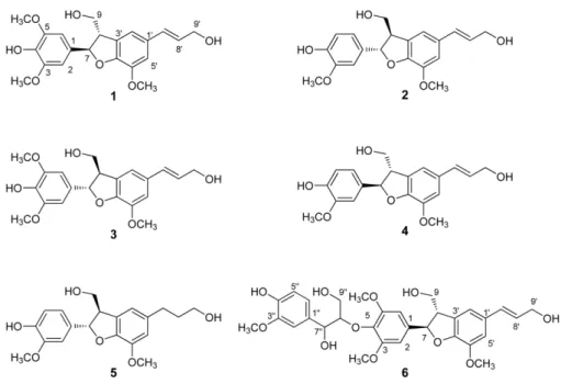
In our experimental system, dihydrobenzofuran neo- lignans (1 - 6) isolated from E. alatus (Fig. 1) showed the potent inhibitory effects on NO production induced by
Fig. 1. Structures of compounds 1 - 6 isolated from E.alatus leaves and twigs.Table 1. Inhibitory effects of compounds 1 - 6 isolated from E. alatus leaves and twigs on NO production induced by LPS in RAW264.7 cells
Concentration 10μM 20μM 50μM 100μM IC50
Nitrite (μM) μM
Control 65.1 ± 0.2
LPS 56.5 ± 0.3
1 33.2 ± 1.9* 20.2 ± 1.4* 18.8 ± 2.7* 15.4 ± 0.8** 11.7 ± 1.2
2 29.3 ± 2.3* 23.2 ± 1.6* 11.2 ± 1.2** 66.1 ± 0.1** 9.3 ± 1.4
3 32.9 ± 1.7* 26.4 ± 0.3* 17.3 ± 1.5** 14.9 ± 0.5** 12.8 ± 2.0
4 26.6 ± 2.0* 21.4 ± 1.5* 68.7 ± 0.2** 62.9 ± 0.1** 8.5 ± 0.8
5 31.7 ± 1.5* 24.2 ± 1.2* 12.7 ± 0.8** 66.9 ± 0.2** 9.8 ± 2.0
6 48.8 ± 2.5 31.9 ± 0.9* 29.8 ± 1.3* 28.9 ± 1.0* 21.3 ± 3.3
L-NAME 48.5 ± 2.3
L-NNA 60.3 ± 1.7
L-NMMA 32.9 ± 2.2
RAW 264.7 cells were pre-treated with each compound for 1 h before exposure to LPS for 24 h. The concentration of nitrite in culture medium was measured as described in the Methods. The values shown are mean± s.d. of data from three independent experiments.
Significant compared with LPS alone, *P < 0.001, **P < 0.0001. L-NAME (L-Nitro-Arginine Methyl Ester, the NOS inhibitor, L-NNA (NG-Nitro-L-arginine, a well known inhibitor of NOS) and L-NMMA (NG-methyl-L-arginine acetate, an inhibitor of iNOS-induced nitric oxide production) were used as positive controls.
LPS with IC
50values of 8.5 - 21.3 μM, which were more potent than positive control, L-NAME (Table 1). The inhibitory effects of compounds 2, 4 and 5 which had 1,3,4-trisubstituted ABX aromatic ring system on C-ring were more potent than those of compounds 1, 3 with a symmetrical 1,3,4,5-tetrasubstituted AB aromatic ring system, especially at the highest concentration, 100 μM.
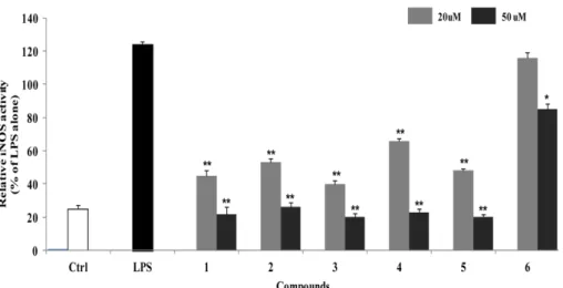
In addition, the presence of guaiacylglycerol moiety in compound 6 seemed to slightly decrease the inhibitory activity on NO production. There was no significant difference in the inhibitory effects of 1 - 5 on iNOS enzyme activity, however, the inhibitory activity of compound 6 was also appeared weak in consistent with the result of NO production (Fig. 2). In order to extend the understanding of anti-inflammatory effects of these neolignans, the effect of compound 4, which showed the most potent inhibitory activity, on the expression of inflammatory proteins, iNOS and COX-2, and the pro- duction of NO and PGE2 were evaluated in LPS- stimulated RAW264.7 cells. Treatment of RAW264.7 cells with LPS significantly increased the expression of iNOS and COX-2 proteins (Fig. 3 and 4). Pretreatment of compound 4 inhibited LPS-induced expression of these proteins in LPS-stimulated RAW264.7 cells. The expression levels of iNOS and COX-2 were significantly decreased up to control level at the concentration over 20 μM, and the production of their products, NO and PGE2 were also decreased.
In addition, compound 4 effectively inhibited LPS- induced production of pro-inflammatory cytokines, IL-1 β, IL-6 and TNF- α at the concentration over 20 μM (Fig. 5).
IL-6 is a cytokine released by LPS-activated monocytes
and plays a crucial role in immune response.
1It has been known that the overexpression of IL-6 is involved in
Fig. 2. Inhibitory effects of compounds 1 - 6 on iNOS enzyme activity in LPS-stimulated RAW264.7 cells. RAW264.7 cells were treated with compounds 1 - 6 for 1 h before exposure to LPS for 24 h. The fluorescent product was assessed as described in the Materials and Methods. The values shown are the mean± s.d. of data from three independent experiments. Results differ significantly from LPS alone,*P < 0.01, **P < 0.001.
Fig. 3. Inhibitory effects of compounds 4 on the expression of iNOS protein in LPS-stimulated RAW264.7 cells. RAW264.7 cells were treated with compound 4 for 1 h, and then exposed to LPS for 24 h. Cell lysates (40 ug protein) were prepared and subjected to Western blot analysis using iNOS-specific antibodies.
The relative protein levels were quantified by scanning densi- tometry and normalized to b-tubulin protein. NO production was measured by the Griess reaction and sodium nitrite was used as a standard. The values shown are the mean± s.d. of data from three independent experiments. Results differ significantly from LPS alone, *P < 0.01, **P < 0.001.
pathological conditions, such as, rheumatoid arthritis.
26In the present study, it was found that compound 4 significantly inhibited the production of IL-1 β and IL-6 induced by LPS in RAW264.7 macrophage cells without cytotoxicity. TNF- α is one of the important inflammatory cytokines required for the synergistic induction of NO synthesis in LPS-stimulated macrophage.TNF- α exhibits its pro-inflammatory activity by regulating several inter- cellular and vascular cell adhesion molecules, which results in the recruitment of leukocytes to sites of inflammation.
27The reduction of TNF- α by the pretreat- ment of compound 4 was also found at the concentration over 50 μM with statistical significance.
From above results, dihydrobenzofuran neolignans isolated from E. alatus leaves and twigs effectively inhibited NO production in LPS-stimulated RAW264.7 cell through attenuating the expressions of iNOS and COX-2, and the production of pro-inflammatory cytokines, IL-1 β, IL-6 and TNF-α. These neolignans are expected to
be potential candidates for development of therapeutics to prevent or treat inflammation-related diseases.
Acknowledgements
This research was supported by Basic Science Research Program through the National Research Foundation of Korea (NRF) funded by the Ministry of Science, ICT &
Fig. 4. Inhibitory effects of compounds 4 on the expression of COX-2 protein in LPS-stimulated RAW264.7 cells. RAW264.7 cells were treated with compound 4 for 1 h, and then exposed to LPS for 24 h. Cell lysates (40 ug protein) were prepared and subjected to Western blot analysis using COX-2-specific antibodies.
The relative protein levels were quantified by scanning densito- metry and normalized to b-tubulin protein. The concentrations of PGE2 in the culture medium were determined using ELISA system as described in Materials and Methods. The values shown are the mean± s.d. of data from three independent experiments.
Results differ significantly from LPS alone, *P < 0.01, **P < 0.001.
Fig. 5. Inhibitory effects of compounds 4 on the production of pro-inflammatory cytokines in LPS-stimulated RAW264.7 cells.
RAW264.7 cells were treated with compound 4 for 1 h, and then exposed to LPS for 24 h. The concentrations of IL-1β, IL-6 and TNF-α in the culture medium were determined using ELISA system as described in Materials and Methods. The values shown are the mean± s.d. of data from three independent experiments.
Results differ significantly from LPS alone, *P < 0.01, **P < 0.001.
Future Planning (NRF-2014R1A1A1008069) and partially supported by Gyeongnam National University of Science and Technology Grant.
References
(1) Kaplanski, G.; Marin, V.; Montero-Julian, F.; Mantovani, A.;
Farnarier, C. Trends Immunol. 2003, 24, 25-29.
(2) Adams, D. O.; Hamilton, T. A. Annu. Rev. Immunol. 1984, 2, 283- 318.
(3) Jeong, E. J.; Yang, H.; Kim, S. H.; Kang, S. Y.; Sung, S. H.; Kim, Y.
C. Food Chem. Toxicol. 2011, 49, 1394-1398.
(4) Jeong, E. J.; Cho, J. H.; Sung, S. H.; Kim, S. Y.; Kim, Y. C. Bioorg.
Med. Chem. Lett. 2011, 15, 2283-2286.
(5) Akihisa, T.; Yamamoto, K.; Tamura, T.; Iida, T.; Nambara, T.;
Chang, F. C. Chem. Pharm. Bull. 1992, 40, 789-791.
(6) De Fátima Silva, G. D.; Duarte, L. P.; Da Silva Paes, H. C.; De Sousa, J. R.; Nonato, M. C.; Portezani, P. J.; Mascarenhas, Y. P. J. Braz.
Chem. Soc. 1998, 9, 461-464.
(7) Liu, C. M.; Wang, H. X.; Wei, S. L.; Gao, K. J. Nat. Prod. 2008, 71, 789-792.
(8) Fang, J. M.; Lee, C. K.; Cheng, Y. S. Phytochemistry 1992, 31, 3659-3661.
(9) Yang, Y. P.; Cheng, M. J.; Teng, C. M.; Chang, Y. L.; Tsai, I. L.;
Chen, I. S. Phytochemistry 2002, 61, 567-572.
(10) Meng, J.; Jiang, T.; Bhatti, H. A.; Siddiqui, B. S.; Dixon, S.;
Kilburn, J. D. Org. Biomol. Chem. 2010, 8, 107-113.
(11) Lourith, N.; Katayama, T.; Suzuki, T. J. Wood Sci. 2005, 51, 370- 378.
(12) Matsuda, S.; Kadota, S.; Tai, T.; Kikuchi, T. Chem. Pharm. Bull.
1984, 32, 5066-5069.
(13) Dawson, V. L.; Brahmbhatt, H. P.; Mong, J. A.; Dawson, T. M.
Neuropharmacology 1994, 33, 1425-1430.
(14) Korhonen, R.; Lahti, A.; Kankaanranta, H.; Moilanen, E. Curr.
Drug Targets Inflamm. Allergy 2005, 4, 471-479.
(15) Yamashita, T.; Kawashima, S.; Ohashi, Y.; Ozaki, M.; Ueyama, T.;
Ishida, T.; Inoue, N.; Hirata, K.; Akita, H.; Yokoyama, M. Circulation 2000, 101, 931-937.
(16) Penglis, P. S.; Cleland, L. G.; Demasi, M.; Caughey, G. E.; James, M. J. J. Immunol. 2000, 165, 1605-1611.
(17) Nathan, C. FASEB J. 1992, 6, 3051-3064.
(18) Marletta, M. A. J. Biol. Chem. 1993, 268, 12231-12234.
(19) Duval, D. L.; Miller, D. R.; Collier, J., Billings, R. E. Mol.
Pharmacol. 1996, 50, 277-284.
(20) Son, H. J.; Lee, H. J.; Yun-Choi, H. S.; Ryu, J. H. Planta Med.
2000, 66, 469-471.
(21) Chen, T. H.; Kao, Y. C.; Chen, B. C.; Chen, C. H.; Chan, P.; Lee, H. M. Eur. J. Pharmacol. 2006, 541, 138-146.
(22) Hamasaki, Y.; Kobayashi, I.; Zaitu, M.; Tsuji, K.; Kita, M.;
Hayasaki, R.; Muro, E.; Yamamoto, S.; Matsuo, M.; Ichimaru, T.;
Miyazaki, S. Planta Med. 1999, 65, 222-226.
(23) Wang, J. P.; Raung, S. L.; Chen, C. C.; Kuo, J. S.; Teng, C. M.
Naunyn Schmiedebergs Arch. Pharmacol. 1993, 348, 663-669.
(24) Oh, J. H.; Kang, L. L.; Ban, J. O.; Kim, Y. H.; Kim, K. H.; Han, S.
B.; Hong, J. T. Chem. Biol. Interact. 2009, 180, 506-514.
(25) Choi, M. S.; Lee, S. H.; Cho, H. S.; Kim, Y.; Yun, Y. P.; Jung, H.
Y.; Jung, J. K.; Lee, B. C.; Pyo, H. B.; Hong, J. T. Eur. J. Pharmacol.
2007, 556, 181-189.
(26) Connell, L.; Mclnnes, I. B. Best Pract. Res. Clin. Rheumatol. 2006, 20, 865-878.
(27) Aggarwal, B. B.; Natarajan, K. Eur. Cytokine Netw. 1996, 7, 93- 124.
Received August 10, 2015 Revised September 16, 2015 Accepted September 16, 2015