217 http://dx.doi.org/10.4196/kjpp.2013.17.3.217
ABBREVIATIONS: AP-1, activator protein-1; COX-2, cyclooxygenase- 2; ERK, extracellular signal-related kinase; GAPDH, glyceraldehyde 3-phosphate dehydrogenase; IL-1β, interleukin-1β; IL-6, interleu- kin-6; iNOS, nitric oxide synthase; JNK, C-jun N-terminal kinase;
LPS, lipopolysacharide; TLR-4, Toll-like receptor-4; MAPK, mitogen- activated protein kinase; NF-κB, nuclear factor-kappa B; NO, nitric oxide; PARP, poly ADP-ribose polymerase.
Received March 5, 2013, Revised April 17, 2013, Accepted May 2, 2013
Corresponding to: Jin-Kyung Kim, Department of Biomedical Science, College of Natural Science, Catholic University of Daegu, 330 Geumrak-ri, Gyeoungsan 700-712, Korea. (Tel) 82-53-850-3774, (Fax) 82-53-850-3774, (E-mail) [email protected]
This is an Open Access article distributed under the terms of the Creative Commons Attribution Non-Commercial License (http://
creativecommons.org/licenses/by-nc/3.0) which permits unrestricted non-commercial use, distribution, and reproduction in any medium, provided the original work is properly cited.
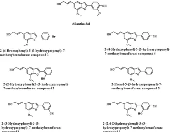
2-(4-Hydroxyphenyl)-5-(3-Hydroxypropenyl)-7-Methoxybenzofuran, a Novel Ailanthoidol Derivative, Exerts Anti-Inflammatory Effect through Downregulation of Mitogen-Activated Protein Kinase in Lipopolysaccharide-Treated RAW 264.7 Cells
Hyeon Jin Kim
1, Jong-Gab Jun
2, and Jin-Kyung Kim
11