The Frequency of Reexpansion Pulmonary Edema after Trocar and Hemostat Assisted Thoracostomy in Patients
with Spontaneous Pneumothorax
Kyoung Chul Cha,
1Hyun Kim,
1Ho Jin Ji,
2Woo Cheol Kwon,
3Hyung Jin Shin,
1Yong Sung Cha,
1Kang Hyun Lee,
1Sung Oh Hwang,
1Christopher C. Lee,
4and Adam J. Singer
4Departments of 1Emergency Medicine and 3Radiology, Wonju College of Medicine, Yonsei University, Wonju;
2Department of Emergency Medicine, Andong Hospital, Andong, Korea.
4Department of Emergency Medicine, Stony Brook University Hospital, Stony Brook, NY, USA.
Received: January 31, 2012 Revised: March 12, 2012 Accpetd: March 22, 2012
Corresponding author: Dr. Hyun Kim, Department of Emergency Medicine, Wonju College of Medicine, Yonsei University, 20 Ilsan-ro, Wonju 220-701, Korea.
Tel: 82-33-741-1614, Fax: 82-33-742-3030 E-mail: [email protected]
∙ The authors have no financial conflicts of interest.
© Copyright:
Yonsei University College of Medicine 2013 This is an Open Access article distributed under the terms of the Creative Commons Attribution Non- Commercial License (http://creativecommons.org/
licenses/by-nc/3.0) which permits unrestricted non- commercial use, distribution, and reproduction in any medium, provided the original work is properly cited.
Purpose: Several risk factors for development of reexpansion pulmonary edema (REPE) after drainage of pneumothoraces have been reported, but the association between the method of thoracostomy and the development of REPE is unknown.
The aim of this study was to compare the frequency of REPE after treatment of spontaneous pneumothorax with trocar or hemostat assisted closed thoracostomy.
Materials and Methods: We performed a prospective, observational study includ- ing 173 patients with spontaneous pneumothorax who visited the emergency de- partment from January 2007 to December 2008. In 2007, patients were treated with hemostat-assisted drainage, whereas patients in 2008 were treated with trocar-as- sisted drainage. The main outcome was the development of REPE, determined by computed tomography of the chest 8 hours after closed thoracostomy. Outcomes in both groups were compared using univariate and multivariate analyses. Results:
Ninety-two patients were included, 48 (42 males) of which underwent hemostat-as- sisted drainage and 44 (41 males) underwent trocar-assisted drainage. The groups were similar in mean age (24±10 vs. 26±14 respectively). The frequencies of REPE after hemostat- and trocar-assisted drainage were 63% (30 patients) and 86% (38 patients) respectively (p=0.009). In multivariate analysis, trocar-assisted drainage was the major contributing factor for developing REPE (odds ratio=5.7, 95% confi- dence interval, 1.5-21). Age, gender, size of pneumothorax, symptom duration and laboratory results were similar between the groups. Conclusion: Closed thoracosto- my using a trocar is associated with an increased risk of REPE compared with he- mostat-assisted drainage in patients with spontaneous pneumothorax.
Key Words: Pneumothorax, thoracostomy, pulmonary edema
INTRODUCTION
Since the first report by Carlson, et al.,1 reexpansion pulmonary edema (REPE) has been recognized as a potentially fatal yet uncommon complication occurring
ing closed thoracostomy, the end of the thoracostomy tube was connected to a single bottle system that was attached to a negative pressure (20 cm H2O using an Emerson post-op- erative suction pump model 55-JS, J. H. EMERSON Corp., Cambridge, MA, USA). The size of the pneumothorax was classified into one of the following four groups: small, medi- um, large and tension pneumothorax. A small pneumothorax was defined as a pneumothorax that was localized in the apex of the lung on chest radiography. A medium pneumo- thorax was defined as a pneumothorax that extended beyond one third of the width of a hemithorax. A large pneumotho- rax was defined as a pneumothorax leading to complete or nearly complete collapse of the lung parenchyma. A tension pneumothorax was defined as a pneumothorax associated with depression of diaphragm, or a shift of mediastinum and trachea away from the collapsed lung.5 PaO2, PCO2, SaO2, lactate, WBC count, troponin I and b-type natriuretic pep- tide were measured in all patients prior to tube thoracosto- my. Subsequent radiographs and chest computed tomogra- phy (CT) scans were obtained within 8 hours of closed thoracostomy to evaluate the need for bullectomy and verify the extent of reexpansion of collapsed lung and the presence or absence of pulmonary edema of the involved lung paren- chyma. The determination of the presence of REPE was made by two investigators (a board-certified emergency physician and a board-certified radiologist) who were blind to demographic and clinical information.
Estimation of pneumothorax size
The average interpleural distance (AID) was calculated in chest X-ray (CXR) as the arithmetic mean of the maximum apical interpleural distance (A) and two measurements of interpleural distances (B and C) at the midpoints of the up- per and lower halves of the lung.6
In erect PA radiographs, the percentage of pneumothorax volume was obtained from the following formula: Percentage pneumothorax in erect PA radiograph=4.2+[4.7×(A+B+C)].7
Due to severe dyspnea and pain, some patients were not able to maintain an erect position during chest radiography, and only supine radiographs were performed in these pa- tients. The above formula for calculating the size of pneu- mothorax could not be applicable with chest radiographs ob- tained in the supine position, in children (0-12 years old), or in patients with hydropneumothoraces.8 When supine radio- graphs were used, the percentage of pneumothorax volume was calculated using the following formula: percentage pneumothorax on supine AP radiograph=8.73+10.03×AID.9 after rapid expansion of the lung after closed thoracostomy
in patients with hemothorax, pneumothorax or pleural effu- sion.2 In previous studies, the prevalence of REPE has var- ied between 0.9% to 57%. Recognition of REPE has be- come more common with the development of improved diagnostic modalities such as computerized tomography.3-5 Management of patients with pneumothorax is most com- monly by closed thoracostomy. Closed thoracostomy may be performed by one of two methods. The first uses a trocar to insert a chest tube into the pleural cavity while the sec- ond uses a hemostat to enter the pleural cavity. The major potential advantages of the trocar-assisted method include its simplicity, rapid procedure time, and its ability to mini- mize associated tissue injury, pain, and subsequent scarring.
In contrast, the hemostat-assisted method is thought to be safer though slower than trocar-assisted thoracostomy.
A number of studies have explored potential risk factors for REPE after drainage of spontaneous pneumothorax, however, the association between REPE and the method of closed thoracostomy has not yet been reported. The objec- tive of the current study was to explore possible association between the method of closed thoracostomy and REPE af- ter drainage of spontaneous pneumothorax.
MATERIALS AND METHODS
Study design
A prospective observational study was conducted to deter- mine clinical characteristics and frequency of REPE, based on the method of closed thoracostomy used to treat spontane- ous pneumothorax. The study was approved by the Institu- tional Review Board and all patients gave informed consent.
Setting and subjects
The study was conducted at a suburban emergency depart- ment that is part of an academic medical center in Korea.
Patients presenting to the Wonju Christian Hospital with a spontaneous pneumothorax who were treated with either he- mostat- or trocar-assisted closed thoracostomy between Jan- uary 2007 and December 2008 were eligible for enrollment.
Measures and outcomes
Structured data collection was performed on study patients.
All patients underwent an erect posteroanterior (PA) or su- pine anteroposterior (AP) radiograph of the thorax to con- firm the presence of a spontaneous pneumothorax. Follow-
pressure (20 cmH2O using an Emerson post-operative suc- tion pump, model 55-JS, J. H. Emerson Corp., Cambridge, MA, USA).
Data analysis
Data were analyzed using SPSS 12.0 for Windows statistical software package (SPSS, Chicago, IL, USA). Continuous data are presented as means with standard deviations and compared with the independent sample t-test or Mann- Whitney U test if appropriate. Nominal data are presented as the percent frequency of occurrence and compared with a χ2 or Fischer’s exact test when appropriate. The association be- tween potential predictor variables and the occurrence of REPE was determined using logistic regression analysis.
The level of significance was preset at a p value of 0.05.
RESULTS
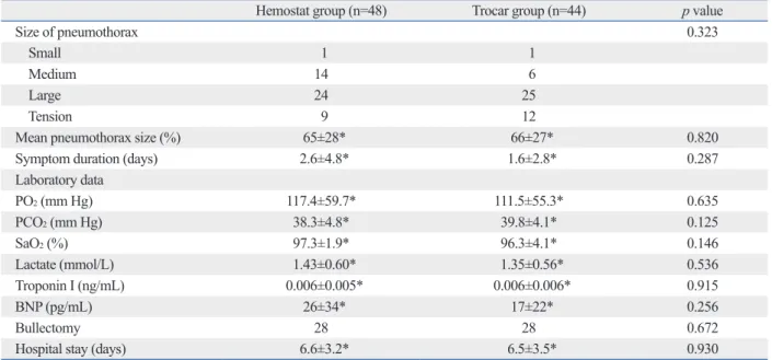
From January 2007 through December 2008, we prospective- ly evaluated 173 patients who were diagnosed with a sponta- neous pneumothorax. Eighty-one patients were excluded since they were treated by 100% oxygen inhalation alone, were transferred from another hospital after tube thoracosto- my, or had extensive adhesions on radiography (Fig. 2).
The study sample included 83 males with a mean age of 25±12 years. The mean time interval from symptom onset to ED visit was 2.2±4.1 days. Of all pneumothoraces, 20 were small, 20 were medium, 49 were large, and 21 were tension pneumothoraces.
Of all pneumothoraces, 41 (45%) involved the right lung and hemostat-assited thoracostomy was performed in 48 patients (52%). Based on subsequent CT imaging of the chest, REPE developed in 68 patients (74%) (Fig. 3) (Table 1). The frequency of REPE was higher in patients undergo- ing trocar-assisted thoracostomy (38 patients, 86%) than those undergoing hemostat-assisted thoracostomy (30 pa- tients, 63%; p=0.009). Age, gender, time interval from symptom onset to ED visit, location of pneumothorax, size of pneumothorax, time interval from thoracostomy to CXR evaluation, and time interval from thoracostomy to CT evaluation were not different between the two groups. There were also no between-group differences in PaO2, PCO2, SaO2, and lactate.
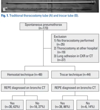
All of the study patients were admitted to the hospital and there were no subsequent major complications or deaths due to the development of REPE. Bullectomy was performed in Closed thoracostomy techniques (Fig. 1)
During the first year of the study (2007), all study patients underwent hemostat-assisted thoracostomy while patients presented during the second year (2008) underwent trocar- assisted thoracostomy.
The hemostat-assisted technique was performed using the following sequence:
1. A 2 cm-length skin incision was made at the rib level below the pleural puncture site.
2. Intercostal muscle dissection was performed using a straight hemostat.
3. Penetration of the parietal pleura was performed using a hemostat, and a thoracostomy tube (P.V.C Thoracic Cath- eter, 28 Fr., Sewoon Medical Co., Ltd., Cheonan, Korea) was inserted into the pleural cavity.
The trocar-assisted technique was performed using the following sequence:
1. A 1 cm-length skin incision was made at the rib level below pleural puncture site.
2. Penetration of parietal pleura was performed with a trocar tube (Trocar Catheter, 28 Fr., Mallinckrodt Medical Co., Ltd., Athlone, Ireland).
Following closed thoracostomy, the tube was connected to a single bottle system that was attached to a negative
Fig. 1. Traditional thoracostomy tube (A) and trocar tube (B).
Fig. 2. Study patient flow. CXR, chest X-ray; CT, computed tomography;
REPE, reexpansion pulmonary edema.
A B
(n=30, 63%)Yes
REPE diagnosed on broncho CT Hemostat technique (n=48)
Spontaneous pneumothorax (n=173)
(n=38, 86%)Yes
REPE diagnosed on broncho CT Trocar technique (n=44) Exclusion
1) No thoracostomy performed (n=35)
2) Thoracostomy at other hospital (n=19)
3) Lung adhesion in CXR or CT (n=27)
(n=18, 37%)No No
(n=6, 14%)
when the intercostal muscle and the parietal pleura are dis- sected. A second decompression phase occurs when the thoracic cavity is entered with a finger. The last phase of decompression occurs when the thoracostomy tube is in- serted into the pleural cavity using a hemostat. In contrast, the trocar technique is performed in one step with only a minimal skin incision and direct puncture and insertion of the thoracostomy tube through the parietal pleura. As a re- sult, intrathoracic pressure may decrease more rapidly with the trocar technique and thus could contribute to rapid reex- pansion of the collapsed lung, capillary injury and more frequent development of REPE.
Several investigators have hypothesized that REPE is the result of pulmonary vascular injury and an increase in cap- 28 patients undergoing hemostat-assisted thoracostomy
(58.3%) and in 28 patients undergoing trocar-assisted thora- costomy (63.4%). Mean length of hospital stay was 6.5±3.2 days after hemostat-assisted thoracostomy and 6.6±3.5 days after trocar-assisted thoracostomy (p=0.930) (Table 2).
We performed logistic regression analysis to determine the association between potential contributing factors and the de- velopment of REPE. The only factor significantly associated with REPE was trocar-assisted thoracostomy [odds ratio (OR) 5.7; 95% confidence interval (CI) 1.5 to 21.4; p=0.009].
The size of pneumothorax had little impact on the develop- ment of REPE (OR=1.1, 95% CI 1.0 to -1.1; p<0.001). Age, gender, and time interval from symptom onset to ED visit had no impact on the development of REPE (Table 3).
DISCUSSION
Our results demonstrate that while the frequency of REPE was increased when the trocar technique was used for closed thoracostomy in patients with spontaneous pneumothorax, it did not affect the ultimate outcome of the patients in this study.
The different rates of REPE between the two methods of closed thoracostomy might be due to differences in the rate of re-expansion of the collapsed lung. In an animal study, the investigators hypothesized that rapid decompression and re-expansion of the collapsed lung could result in capil- lary vascular injury and ipsilateral pulmonary edema while more gradual decompression might prevent pulmonary ede- ma.10,11 When the hemostat is used to decompress a pneu- mothorax, decompression of the collapsed lung may be more gradual. The first phase of decompression occurs
Table 1. Patient Characteristics at Clinical Presentation
Characteristics Values
Age (yrs) 25±12*
Male 83 (90%)
Symptom duration (days) 2.2±4.1*
Smoking 28 (30%)
Medical history
Asthma 2 (2%)
Pulmonary tuberculosis 3 (3%)
Prior pneumothorax 21 (23%)
Location of pneumothorax: right 41 (45%) Size of pneumothorax
Mean 65±27%*
Small 2 (2%)
Medium 20 (22%)
Large 49 (53%)
Tension 21 (23%)
REPE 68 (74%)
REPE, reexpansion pulmonary edema.
*Mean±standard deviation.
Fig. 3. Reexpansion pulmonary edema (REPE) after thoracostomy. No REPE (A) and REPE (B) after trocar technique. No REPE (C) and REPE (D) after hemo- stat technique.
A
C
B
D
ing to detect REPE.15 CT imaging of the chest was used in this study, because of its higher accuracy and sensitivity in diagnosing REPE. As a result, even small and asymptomat- ic cases of REPE were identified. In contrast, earlier studies have used CXR for the diagnosis of REPE, therefore, the incidence of REPE in our study was higher than previous studies, and it may be a more accurate reflection of true in- cidence of REPE after closed thoracostomy.
It is well known that most patients with REPE have no significant complications and do not require specific treat- ment modalities such as assisted ventilation or ICU care.2 All of the patients with REPE in our study also recovered after conservative management alone without any compli- cations. However, REPE-related mortality due to acute re- spiratory failure has been reported in several studies, and physicians should continue to be concerned with the clini- cal course of patients with REPE.8
The major limitation of our study is the fact that most of the clinical variables were limited to the ED and there was little information on the clinical course of pulmonary ede- ma after hospital admission. Therefore, we could not evalu- ate whether the development of REPE was related to dete- rioration of clinical symptoms such as dyspnea, tachypnea or palpitation during hospital stay.
Second, Emerson pump with a negative pressure of 20 cmH2O was applied to all enrolled patients. Rapid negative pressure application in long term lung collapse might be re- lated to increased incidence of REPE.3,11 Thus, it is likely illary permeability. Carlson, et al.1 earlier reported that pul-
monary capillary permeability increased when pulmonary circulation was normalized after hypoxic vascular injury during lung collapse. An increase in nitric oxide or xanthine oxidase after reexpansion of the collapsed lung was thought to result in an increase in pulmonary capillary permeabili- ty.12,13 The change in surface tension after long-standing col- lapse of lung has also been suggested to result in decreased surfactant production and pulmonary edema.14
Several risk factors for REPE have been proposed, in- cluding chronicity of collapse, larger size of pneumothorax, younger age, and underlying diseases of patients.4 To the best of our knowledge, however, no prior study has investi- gated the association between the method of thoracostomy and the development of REPE. Since we excluded patients with underlying lung adhesions, most of our cases had no underlying pulmonary diseases, and this may explain why we did not identify any such risk factor for REPE.
The detection rate of REPE was higher in our study than other earlier studies, possibly due to the application of neg- ative pressure after thoracostomy and the use of CT imag-
Table 2. Comparison of Patient Characteristics between Hemostat Group and Trocar Group
Hemostat group (n=48) Trocar group (n=44) p value
Size of pneumothorax 0.323
Small 1 1
Medium 14 6
Large 24 25
Tension 9 12
Mean pneumothorax size (%) 65±28* 66±27* 0.820
Symptom duration (days) 2.6±4.8* 1.6±2.8* 0.287
Laboratory data
PO2 (mm Hg) 117.4±59.7* 111.5±55.3* 0.635
PCO2 (mm Hg) 38.3±4.8* 39.8±4.1* 0.125
SaO2 (%) 97.3±1.9* 96.3±4.1* 0.146
Lactate (mmol/L) 1.43±0.60* 1.35±0.56* 0.536
Troponin I (ng/mL) 0.006±0.005* 0.006±0.006* 0.915
BNP (pg/mL) 26±34* 17±22* 0.256
Bullectomy 28 28 0.672
Hospital stay (days) 6.6±3.2* 6.5±3.5* 0.930
BNP, b-type natriuretic peptide.
*Mean±standard deviation.
Table 3. Results of Multivariate Analysis of Risk Factors for REPE
Risk factor Odds ratio 95% CI p value
Trocar technique 5.73 1.54-21.42 0.009 Size of pneumothorax 1.06 1.03-1.10 <0.001 Symptom duration 1.07 1.07-0.83 0.620 CI, confidence interval; REPE, reexpansion pulmonary edema.
4. Matsuura Y, Nomimura T, Murakami H, Matsushima T, Kake- hashi M, Kajihara H. Clinical analysis of reexpansion pulmonary edema. Chest 1991;100:1562-6.
5. Kim YK, Kim H, Lee CC, Choi HJ, Lee KH, Hwang SO, et al.
New classification and clinical characteristics of reexpansion pul- monary edema after treatment of spontaneous pneumothorax. Am J Emerg Med 2009;27:961-7.
6. Rhea JT, DeLuca SA, Greene RE. Determining the size of pneu- mothorax in the upright patient. Radiology 1982;144:733-6.
7. Collins CD, Lopez A, Mathie A, Wood V, Jackson JE, Roddie ME. Quantification of pneumothorax size on chest radiographs using interpleural distances: regression analysis based on volume measurements from helical CT. AJR Am J Roentgenol 1995;165:
1127-30.
8. Mahfood S, Hix WR, Aaron BL, Blaes P, Watson DC. Reexpan- sion pulmonary edema. Ann Thorac Surg 1988;45:340-5.
9. Choi BG, Park SH, Yun EH, Chae KO, Shinn KS. Pneumothorax size: correlation of supine anteroposterior with erect posteroanteri- or chest radiographs. Radiology 1998;209:567-9.
10. Miller WC, Toon R, Palat H, Lacroix J. Experimental pulmonary edema following re-expansion of pneumothorax. Am Rev Respir Dis 1973;108:654-6.
11. Pavlin J, Cheney FW Jr. Unilateral pulmonary edema in rabbits after reexpansion of collapsed lung. J Appl Physiol 1979;46:31-5.
12. Sivrikoz MC, Tunçözgür B, Cekmen M, Bakir K, Meram I, Koçer E, et al. The role of tissue reperfusion in the reexpansion injury of the lungs. Eur J Cardiothorac Surg 2002;22:721-7.
13. Saito S, Ogawa J, Minamiya Y. Pulmonary reexpansion causes xanthine oxidase-induced apoptosis in rat lung. Am J Physiol Lung Cell Mol Physiol 2005;289:L400-6.
14. Sautter RD, Dreher WH, MacIndoe JH, Myers WO, Magnin GE.
Fatal pulmonary edema and pneumonitis after reexpansion of chronic pneumothorax. Chest 1971;60:399-401.
15. Tan HC, Mak KH, Johan A, Wang YT, Poh SC. Cardiac output in- creases prior to development of pulmonary edema after re-expan- sion of spontaneous pneumothorax. Respir Med 2002;96:461-5.
16. Kernodle DS, DiRaimondo CR, Fulkerson WJ. Reexpansion pul- monary edema after pneumothorax. South Med J 1984;77:318-22.
17. Zehtabchi S, Rios CL. Management of emergency department pa- tients with primary spontaneous pneumothorax: needle aspiration or tube thoracostomy? Ann Emerg Med 2008;51:91-100, 100.e1.
18. Hassani B, Foote J, Borgundvaag B. Outpatient management of primary spontaneous pneumothorax in the emergency department of a community hospital using a small-bore catheter and a Heim- lich valve. Acad Emerg Med 2009;16:513-8.
19. Wakai A, O’Sullivan RG, McCabe G. Simple aspiration versus in- tercostal tube drainage for primary spontaneous pneumothorax in adults. Cochrane Database Syst Rev 2007:CD004479.
20. Hoi K, Turchin B, Kelly AM. How accurate is the Light index for estimating pneumothorax size? Australas Radiol 2007;51:196-8.
21. Cai W, Lee EY, Vij A, Mahmood SA, Yoshida H. MDCT for com- puterized volumetry of pneumothoraces in pediatric patients. Acad Radiol 2011;18:315-23.
that the use of Emerson pump with negative pressure may affect to the incidence of REPE. However, REPE can occur in patients without any negative pressure suction.16 Third, we used chest CT for diagnosing REPE instead of plain chest X-ray. It could have overestimated the incidence of REPE. However, chest CT is superior to chest X-ray for di- agnosing the REPE, we had used the most sensitive imag- ing tool.
Forth, we used single size thoracostomy tube in both groups regardless of pneumothorax size. Various thoracos- tomy techniques by pneumothorax size are introduced in recent studies.17-19 However, we used single size tube in or- der to exclude decompression pressure variation by diame- ter of pleural opening. Nevertheless, the opening of the pleura might be bigger in hemostat technique because the opening of the pleura was made manually prior to insertion of thoracostomy tube. Thus, decompression pressure could be different in two groups and it could affect the frequency of REPE. Fifth, patient allocation was not randomized. This may have led to the introduction of additional biases. Final- ly, while measuring the size of the pneumothorax with CT would have been more accurate than with CXR based for- mula,20,21 most patients had CT imaging after thoracostomy because of cost and patient convenience.
In conclusion, the incidence of REPE in ED patients with spontaneous pneumothorax was higher after trocar-assisted thoracostomy, however, ultimate clinical outcome was sim- ilar to hemostat-assisted thoracostomy. When using the tro- car technique for the treatment of spontaneous pneumotho- rax, intermittent clamping of thoracostomy tube to slow reexpansion of the collapsed lung should be considered.
REFERENCES
1. Carlson RI, Classen KL, Gollan F, Gobbel WG Jr, Sherman DE, Christensen RO. Pulmonary edema following the rapid reexpan- sion of a totally collapsed lung due to a pneumothorax: a clinical and experimental study. Surg Forum 1958;9:367-71.
2. Sohara Y. Reexpansion pulmonary edema. Ann Thorac Cardio- vasc Surg 2008;14:205-9.
3. Rozenman J, Yellin A, Simansky DA, Shiner RJ. Re-expansion pulmonary oedema following spontaneous pneumothorax. Respir Med 1996;90:235-8.