Korean Journal of Ophthalmology 22(1):66-69, 2008 DOI : 10.3341/kjo.2008.22.1.66
66
Bilateral Ophthalmic Artery Occlusion in Rhino-Orbito-Cerebral Mucormycosis
Yoo-Mi Song, MD
1, Sun Young Shin, MD
2
Department of Ophthalmology, Hanyang University College of Medicine
1, Seoul, Korea Department of Ophthalmology, The Catholic University College of Medicine
2, Seoul, Korea
Purpose: To report a case of bilateral ophthalmic artery occlusion in rhino-orbito-cerebral mucormycosis.
Methods: Reviewed clinical charts, photographs, and fluorescein angiography
Results: An 89‐year‐old man with poorly controlled diabetes developed sudden bilateral ptosis, complete ophthalmoplegia of the right eye, and superior rectus palsy of the left eye. Brain and orbit magnetic resonance imaging showed midbrain infarction and mild diffuse sinusitis. On the 2nd day of hospitalization, sudden visual loss and light reflex loss developed. There were retinal whitening, absence of retinal arterial filling, and a total lack of choroidal perfusion on fluorescein angiography of the right eye. The left eye showed a cherry red spot in the retina and the absence of retinal arterial filling and partial choroidal perfusion on fluorescein angiography. On rhinologic examination, mucormyosis was noticed. Despite treatment, visual acuity and light reflex did not recover and he died 4 days after admission.
Conclusions: Bilateral ophthalmic artery occlusion can occur in rhino‐orbital‐cerebral mucormycosis.
Korean Journal of Ophthalmology 22(1):66-69, 2008
Key Words: Bilateral ophthalmic artery occlusion, Mucormycosis
Received: May 30, 2006 Accepted: September 10, 2007
Reprint requests to Sun Young Shin, MD. Department of Ophthal-
mology, Gangnam St. Mary’s Hospital, College of Medicine, The
Catholic University of Korea. 505 Banpo-dong, Seocho-gu, Seoul
137-040, Korea. Tel: 82-2-590-1523, Fax: 82-2-533-6718, E-mail:
[email protected]
* Presented at the 95th Meeting of the Korean Ophthalmological
Society, Pusan, Korea, April 14, 2006.
Rhino-orbito-cerebral mucormycosis (ROCM) is a fatal fulminant opportunistic infection leading to death in some cases, which mostly occurs among patients with a weakened immune system. This case involved bilateral ophthalmic artery occlusion with third nuclear, as well as fourth and sixth cranial nerve palsies of the right eye, which is a rare initial presentation of mucormycosis.
Case Report
An 89-year-old man presented with sudden onset of bilateral ptosis on November 2, 2005. He had a one week history of xerostomia. Three days prior to hospitalization, the patient was diagnosed with diabetes mellitus and had a history of surgery for gastric cancer, which occurred four years ago.
His fasting glucose level and postprandial (2 hr-after-meal) glucose level were 196 mg/dL and 300 mg/dL, respectively.
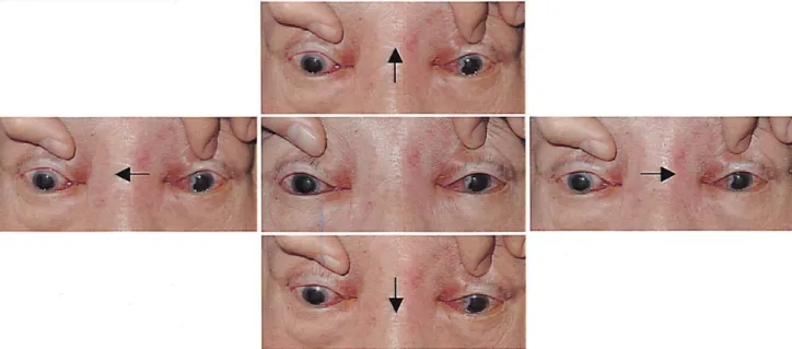
His visual acuity was 20/100 in both eyes. The right pupil was mid-dilated and had no light reflex. Complete ophthlamoplegia of the right eye and superior rectus palsy of the left eye were observed. Bilateral ptosis, mild proptosis, and periorbital puffiness were also observed. His anterior segment examination was notable for moderate nuclear sclerotic cataracts and mild chemosis, while fundoscopic examination revealed no specific abnormal finding.
Brain and orbit magnetic resonance imaging (MRI) revealed no specific abnormal findings in the cavernous sinus and orbit, except bilateral midbrain infarction and mild diffuse sinusitis (Fig. 1). Based on the brain MRI finding, the bilateral ptosis and superior rectus palsy of the left eye were regarded to be caused by 3
rd nuclear palsy. The results of CSF analysis showed that white blood cell count and protein were elevated and intravenous antibiotics were given for a central nervous system infection.
On November 3, 2005, the visual acuity of both eyes
decreased to perception of hand motion and bilateral
complete ophthlamoplegia developed (Fig. 2). Anterior
segment examination revealed no specific change compared
with the previous finding. Fundoscopic examination revealed
that the optic disc was pale and the posterior pole was
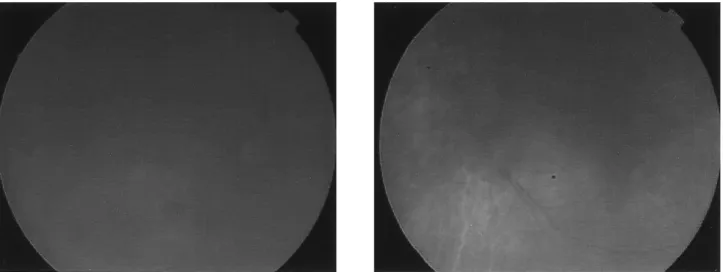
generally edematous in both eyes. A cherry-red spot was
found in the left eye (Fig. 3). Fluorescein angiography (FAG)
demonstrated that retinal vessels were not imaged, even 10
minutes after injecting the dye.