http://dx.doi.org/10.5468/ogs.2015.58.5.368 pISSN 2287-8572 · eISSN 2287-8580
Introduction
Carcinoma of the uterine cervix is the second most common cancer among women in developing countries and the fourth most common cancer in women worldwide [1]. The incidence and mortality of cervical cancer have been greatly reduced due to screening programs and vaccination [1]. However, cer- vical cancer is still one of the major causes of cancer deaths in women worldwide. Among different histological subtypes of cervical cancer, squamous cell carcinoma is the most common, accounting for about 80% of cervical cancer cases.
Squamous cell carcinoma antigen (SCC-Ag) is the most com- monly used serum tumor marker for squamous cell cervical
cancer [2,3]. Most studies reported that elevated pretreatment SCC-Ag levels correlate with extent of disease such as tumor
The prognostic value of squamous cell carcinoma antigen for predicting tumor recurrence in cervical squamous cell carcinoma patients
Hyun Kyung Ryu, Ji Sun Baek, Woo Dae Kang, Seok Mo Kim
Department of Obstetrics and Gynecology, Chonnam National University Medical School, Gwangju, Korea
Objective
The aim of this study was to evaluate the prognostic value of squamous cell carcinoma antigen (SCC-Ag) and the optimal cut-off value for predicting recurrence in cervical squamous cell carcinoma patients with complete remission after primary treatment.
Methods
We reviewed the records of 783 cervical squamous cell cancer patients who underwent primary therapy and showed complete remission at our institution between January 2000 and April 2014. A receiver operating characteristic curve was used to determine the optimal SCC-Ag threshold to predict recurrence. Cox regression model for disease free survival was used to assess differences in outcome.
Results
The median follow-up period was 41.2 months, and 154 patients (19.7%) had recurrent disease. The median pretreatment and posttreatment SCC-Ag level was 2.6 ng/mL (range, 0.1 to 532.0 ng/mL) and 0.7 ng/mL (range, 0.0 to 46.8 ng/mL), respectively. Both pretreatment and posttreatment SCC-Ag levels were higher in the recurrence group (P=0.017 and P=0.039). Optimal cut-off value of pretreatment and posttreatment SCC-Ag for predicting recurrence was 1.86 ng/mL (area under the curve, 0.663; P=0.000), and 0.9 ng/mL (area under the curve, 0.581; P=0.002), respectively. In the multivariate Cox regression model, pretreatment SCC-Ag >1.86 ng/mL (odds ratio, 2.11; 95% confidence interval, 1.38 to 3.22; P=0.001) and posttreatment SCC-Ag >0.9 ng/mL (odds ratio, 1.64; 95% confidence interval, 1.18 to 2.28;
P=0.003) were significantly associated with poor disease free survival.
Conclusion
Patients with pretreatment SCC-Ag >1.86 ng/mL or posttreatment SCC-Ag >0.9 ng/mL should be considered at high risk for cancer recurrence after complete remission, and therefore, closer surveillance is needed.
Keywords: Cervical squamous cell cancer; Cut-off value; Disease-free survival; Squamous cell carcinoma-related antigen
Received: 2015.3.17. Revised: 2015.4.26. Accepted: 2015.4.29.
Corresponding author: Seok Mo Kim
Department of Obstetrics and Gynecology, Chonnam National University Medical School, 160 Baekseo-ro, Dong-gu, Gwangju 61467, Korea
Tel: +82-61-379-7687 Fax: +82-62-227-1637 E-mail: [email protected]
Articles published in Obstet Gynecol Sci are open-access, distributed under the terms of the Creative Commons Attribution Non-Commercial License (http://creativecommons.
org/licenses/by-nc/3.0/) which permits unrestricted non-commercial use, distribution, and reproduction in any medium, provided the original work is properly cited.
Copyright © 2015 Korean Society of Obstetrics and Gynecology
diameter, depth of cervical stromal invasion, lymphovascular space invasion, parametrial involvement, and lymph node metastasis [4-10]. Monitoring of SCC-Ag during radiotherapy and/or chemotherapy reflects both the tumor response to the treatment and the clinical outcome of patients [11-13]. SCC-Ag has also been recognized as a sensitive indicator of recurrence [14,15].
The clinical relevance of pretreatment SCC-Ag level is still debated [16]. Some studies reported that it has no prognos- tic value [2,17,18], and some other studies reported that it is associated with disease free survival or overall survival [4- 7,10,19]. Several studies reported variable cut off values of pretreatment SCC-Ag level for predicting cervical cancer recur- rence, but most of the studies included limited patient groups considering the cancer stage or primary treatment modalities.
The purpose of this study is to investigate the prognostic value and the optimal cut off value of pretreatment and post- treatment SCC-Ag level for predicting cancer recurrence in cervical squamous cell carcinoma patients who have achieved complete remission after primary treatment, regardless of the cancer stage and primary treatment modality.
Materials and methods
We retrospectively reviewed the records of total 783 patients with squamous cell carcinoma of the uterine cervix who had achieved complete remission after primary treatment at the Department of Obstetrics and Gynecology of Chonnam Na- tional University Hospital between January 2000 and April 2014. Patients were considered eligible for the study if they fulfilled the following criteria: histologically confirmed squa- mous cell carcinoma by punch biopsy, loop electrosurgical excision procedure, or hysterectomy specimen; patients in whom SCC-Ag levels were checked before and after primary treatment; and patients who had achieved complete response after primary treatment. Patients with underlying disease that can influence the SCC-Ag level, such as chronic liver disease or renal disease, benign lesions of the lung, or skin disease were excluded.
Primary treatment was selected considering the clinical stage of disease, age, and underlying disease in the patient. In pa- tients with early stage cervical cancer (IA to IIA), surgery-based treatment was performed. Patients with locally advanced cervical cancer (IIB to IV) underwent radiotherapy (RT) only or
concurrent chemotherapy with radiation therapy (CCRT). Ad- juvant radiotherapy or CCRT after surgery was performed in patients who had one or more intermediate- risk factors (large tumor size, deep cervical stromal invasion, and lymph-vascular space involvement) or high- risk factors (positive lymph node involvement, microscopic parametrial invasion, and positive resection margins with the tumor). Cisplatin-based chemo- therapy was administered in all cases that received CCRT.
Serum SCC-Ag level was checked at the time of diagnosis of cervical cnacer (pretreatment SCC-Ag level) and at the first visit after the completion of treatment (posttreatment SCC-Ag lev- el). Clinical stage was determined according to the guidelines of the International Federation of Gynecology and Obstetrics (FIGO). Tumor size determined as the largest diameter of the primary tumor was evaluated by computed tomography (CT), magnetic resonance imaging (MRI), or hysterectomy specimen.
Status of lymph node involvement was confirmed by either pathological verification of the surgical specimen or it was de- fined as nodal size >1 cm on CT or MRI [20,21]. Biochemical response was defined as the change in SCC-Ag levels before and after primary treatment. Positive biochemical response was defined when the SCC-Ag level was reduced, and biochemical failure was defined when there were two consecutive increases in the SCC-Ag level above 1 ng/mL than immediately before or elevation above 1.5 ng/mL. The rest of the responses were defined as negative biochemical responses.
Follow-up after primary treatment was performed approxi- mately every 3 months for the first 2 years, every 6 months for the next 3 years, and every year thereafter. During the routine follow-up, imaging studies including CT or MRI were per- formed each year. Imaging and biopsy were performed when tumor recurrence was suspected based on the clinical finding or imaging study. Local recurrence was diagnosed by biopsy, and distant metastasis was diagnosed by imaging studies.
Disease free survival (DFS) was defined as the time elapsed between the initiation of primary treatment and first detection of cancer recurrence or the date of the last visit for patients with no evidence of disease. The study protocol was evaluated and approved by the institutional review board at Chonnam National University Hospital.
Statistical analysis was performed using IBM SPSS ver. 21.0 (IBM Corp., Armonk, NY, USA). Statistical comparison was carried out using Student’s t-test or Pearson chi-square test.
To determine the sensitivity, specificity, and the optimal cut-
off values of the parameters, receiver operating characteristic
(ROC) curve analysis was performed. Variables showing a sig- nificant association with DFS were included in the multivariate analysis based on the Cox proportional-hazard model. Distant DFS curves were plotted using the Kaplan-Meier method. All reported P-values are two-sided, and P-values <0.05 were considered statistically significant.
Results
Seven hundred and eighty-three patients with squamous cell carcinoma of the uterine cervix who had achieved complete response after primary treatment were included. The median age of the patients was 58 years (range, 27 to 98 years) and the median follow-up period was 41.2 months (range, 1.0 to
165.9 months). The median pretreatment and posttreatment SCC-Ag level was 2.6 ng/mL (range, 0.1 to 532 ng/mL) and 0.7 ng/mL (range, 0 to 46.8 ng/mL), respectively. Five-year disease free survival and 5-year survival rate were 76.1% and 98.5%, respectively.
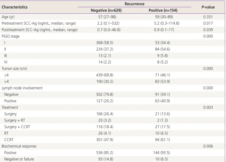
During the follow-up period, 154 patients (19.7%) expe- rienced cervical cancer recurrence. Median SCC-Ag level at recurrence was 2.0 ng/mL (range, 0.2 to 117.0 ng/mL). The patient characteristics according to the presence of recur- rent disease are shown in Table 1. Both pretreatment and posttreatment SCC-Ag levels were higher in the recurrence group (P=0.017 and P=0.039 respectively). The percentages of advanced FIGO stage, tumor >4 cm in diameter, and posi- tive lymph node involvement were also significantly higher in the recurrence group. Negative biochemical response or
Table 1. Patient characteristics according to cancer recurrence
Characteristics Recurrence
P-value Negative (n=629) Positive (n=154)
Age (yr) 57 (27–98) 59 (30–89) 0.331
Pretreatment SCC-Ag (ng/mL, median, range) 2.2 (0.1–532) 5.2 (0.3–114.8) 0.017
Posttreatment SCC-Ag (ng/mL, median, range) 0.7 (0.0–46.8) 0.9 (0.1–17) 0.039
FIGO stage 0.000
I 368 (58.5) 53 (34.4)
II 234 (37.2) 84 (54.6)
III 13 (2.1) 9 (5.8)
IV 14 (2.2) 8 (5.2)
Tumor size (cm) 0.000
≤4 439 (69.8) 71 (46.1)
>4 190 (30.2) 83 (53.9)
Lymph node involvement 0.000
Negative 502 (79.8) 91 (59.1)
Positive 127 (20.2) 63 (40.9)
Treatment 0.003
Surgery 166 (26.4) 21 (13.6)
Surgery + RT 20 (3.2) 2 (1.3)
Surgery + CCRT 116 (18.4) 27 (17.5)
RT 26 (4.1) 10 (6.5)
CCRT 301 (47.9) 94 (61.1)
Biochemical response 0.006
Positive 536 (85.2) 144 (93.5)
Negative or failure 93 (14.8) 10 (6.5)
Values are presented as median (range) or number (%).
SCC-Ag, squamous cell carcinoma antigen; FIGO, International Federation of Gynecology and Obstetrics; RT, radiotherapy; CCRT, concurrent chemotherapy and radiotherapy.
failure was more frequently observed in the recurrence group.
The distribution of treatment modalities was different among the two groups. The rate of primary surgery was significantly lower, and on the contrary, the rate of CCRT utilization was significantly higher in the recurrence group.
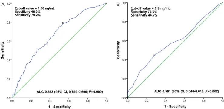
ROC curve analysis was used to obtain the optimal cut- off point values for pretreatment and posttreatment SCC-Ag levels for predicting cancer recurrence (Fig. 1). In this analysis, both pretreatment and posttreatment SCC-Ag levels had a statistically significant influence on predicting cancer recur- rence (area under the curve=0.663 and 0.581 respectively,
P=0.000 and 0.002 respectively). After studying the ROC anal-ysis results and considering the best sensitivity and specificity, the optimal cut-off value of pretreatment and posttreatment SCC-Ag level was 1.86 ng/mL (sensitivity 79.2%, specificity 46.0%) and 0.9 ng/mL (sensitivity 44.2%, specificity 72.0%), respectively.
Univariate and multivariate analyses for DFS are shown in Ta- ble 2. In the multivariate Cox regression model, pretreatment SCC-Ag >1.86 ng/mL (odds ratio [OR], 2.11; 95% confidence interval [CI], 1.38 to 3.22; P=0.001) and posttreatment SCC- Ag >0.9 ng/mL (OR, 1.64; 95% CI, 1.18 to 2.28; P=0.003) were identified as independent risk factors associated with poor disease free survival. FIGO stage III to IV (OR, 1.74; 95%
CI, 1.38 to 3.22; P=0.035), tumor size >4 cm (OR, 1.57; 95%
CI, 1.09 to 2.26; P=0.015), and positive lymph node involve- ment (OR, 1.45; 95% CI, 1.01 to 2.07; P=0.043) were also associated with poor disease free survival.
Distant DFS curves according to pretreatment and posttreat- ment SCC-Ag levels, tumor size, and lymph node involve- ment are shown in Fig. 2. Women with pretreatment SCC- Ag >1.86 ng/mL (P=0.000), posttreatment SCC-Ag >0.9 ng/
mL (P=0.000), tumor size >4 cm in diameter (P=0.000), and positive lymph node involvement (P=0.000) had a significantly poor DFS (Fig. 2).
Discussion
The risk factors for recurrent disease in early stage cervical cancer include large tumor size, positive lymph node, parame- trial involvement, and lymph-vascular space invasion [22]. Our finding that tumor size, lymph node involvement, and FIGO stage are associated with tumor recurrence in the multivariate analysis is in concurrence with this fact.
SCC-Ag is one of the major tumor markers, which is cur- rently widely employed as a tumor marker in squamous cell carcinoma of the uterine cervix. Previous literature reported
Fig. 1. Receiver operating characteristic curve for pretreatment squamous cell carcinoma antigen (SCC-Ag) level (A), and posttreatment SCC-Ag level (B) for predicting recurrence. Optimal cut-off value of pretreatment and posttreatment SCC-Ag for predicting tumor recurrence was 1.86 and 0.9 ng/mL, respectively.
AUC, area under the curve; CI, confidence interval.
A B
that the pretreatment level of SCC-Ag is associated with FIGO stage, tumor size, and parametrial involvement [4-10], and that continued or increasing levels of SCC-Ag reflect persistent or recurrent disease [23,24]. However, the clinical relevance of pretreatment SCC-Ag level is still debated [16]. In the study by Gaarenstroom et al. [10], SCC-Ag was significantly related to progression free survival and overall survival in the univariate analysis, but not in the multivariate analysis. Similarly, Yuan et al. [6] reported that elevated preoperative serum SCC-Ag level was a poor prognostic factor for survival in the univariate anal- ysis but not in the multivariate analysis. In studies by Hong et al. [19] and Duk et al. [5], elevated pretreatment serum SCC- Ag level was an independent prognostic variable for survival.
Elevated serum SCC-Ag level at the time of diagnosis was as- sociated with a three times higher risk of recurrence [25]. Simi- larly, Strauss et al. [7] found that elevated pre-operative serum SCC-Ag level above 3 ng/mL was an independent poor prog- nostic factor for recurrence free survival and overall survival.
Our findings support the prognostic value of pretreatment SCC-Ag level in predicting recurrence. Pretreatment SCC-Ag
>1.86 ng/mL was an independent risk factor of cancer recur- rence in the multivariate analysis and disease progression in the survival analysis. However, the cut off values of elevated SCC- Ag level vary from study to study. Strauss et al. [7] reported a SCC-Ag cut off level of 3 ng/mL in patients with stages IA2 to IIB squamous cell carcinoma of the cervix who underwent radi- cal hysterectomy. In another study of early stage squamous cell carcinoma of the cervix, a cut off value of 1.9 ng/mL was used to predict postoperative radiotherapy [9]. Davelaar et al. [26]
reported that a cut off level of 1.1 ng/mL was associated with poor prognosis in early stage squamous cell carcinoma of the cervix. Gadducci et al. [27] reported that pretreatment serum SCC-Ag <7 ng/mL was associated with longer progression free survival and overall survival in the univariate analysis in patients with stage IB to IIIB disease who underwent neo-adjuvant chemotherapy followed by radical surgery. The differences in cut off values of pretreatment SCC-Ag may be contributable to differences in the cancer stages of patients included in each study. Since an increased pretreatment SCC-Ag level is known to correlate with advanced FIGO stage, the median level of
Table 2. Univariate and multivariate analyses for disease free survivalVariable Median DFS
(mo)
Univariate Multivariate analysis
P-value HR (95% CI) P-value
Pretreatment SCC-Ag (ng/mL) 0.000 0.001
≤1.86 148.5 1
>1.86 132.0 2.11 (1.38–3.22)
Posttreatment SCC-Ag (ng/mL) 0.000 0.003
≤0.9 147.5 1
>0.9 108.0 1.64 (1.18–2.28)
Biochemical response 0.001 0.821
Positive 147.2 1
Negative or failure 108.0 0.82 (0.43–1.95)
FIGO stage 0.000 0.035
stage I–II 147.2 1
stage III–IV 108.0 1.74 (1.38–3.22)
Tumor size (cm) 0.000 0.015
≤4 132.0 1
>4 146.3 1.57 (1.09–2.26)
Lymph node involvement 0.000 0.043
Negative 147.0 1
Positive 132.0 1.45 (1.01–2.07)
DFS, disease free survival; HR, hazard ratio; CI, confidence interval; SCC-Ag, squamous cell carcinoma antigen; FIGO, International Federation of Gynecology and Obstetrics.