Original Article
Clinical significance of tumor volume and lymph node involvement assessed by MRI in stage IIB cervical cancer patients treated with concurrent chemoradiation therapy
Dae Woo Lee1, Young Tae Kim1, Jae Hoon Kim1, Sunghoon Kim1, Sang Wun Kim1, Eun Ji Nam1, Jae Wook Kim2
Division of Gynecologic Oncology, Department of Obstetrics and Gynecology,
1Yonsei University College of Medicine, 2Kwandong University College of Medicine, Seoul, Korea
Objective: The purpose of this study was to evaluate the prognostic significance of tumor volume assessed by pretreatment MRI in stage IIB cervical cancer patients with concurrent chemoradiation therapy.
Methods: A retrospective chart review was performed on seventy five patients with cervical cancer who were treated with concurrent weekly cisplatin (40 mg/m2) and radiotherapy between January 2000 and April 2007. Potential prognostic factors were age, chemotherapy numbers, histology, tumor diameter and volume, lymph node (LN) involvement and pretreatment squamous cell carcinoma antigen (SCC-Ag) levels.
Results: The median follow-up time was 55 months (range, 8 to 104 months). The median tumor size and volume (range) were 4.5 cm (2 to 10) and 33.1 mL (4.2 to 392.7), respectively. Pelvic LN enlargement rate was 58.7%.
Para-aortic LN enlargement rate was 14.7%. Using multivariate analysis, a tumor volume (>33 mL, p=0.025), pelvic LN enlargement (p=0.044) revealed a significantly unfavorable outcome on overall survival. PFS was influenced by tumor histology (p<0.001), pelvic LN enlargement (p=0.015) and pretreatment SCC-Ag levels (p=0.018). We found that 22 (29.3%) patients had recurrences and 14 (18.7%) patients died of disease. The 5-year overall survival rate was 80.6% (standard error, 4.9%) and 5-year PFS rate was 71.3% (standard error, 5.3%).
Conclusion: Tumor volume and pelvic LN involvement showed possibility to predict overall survival in patient with stage ⅡB cervical cancer. Optimal tumor volume and pelvic LN assessment by pretreatment MRI might be helpful to predict treatment outcome.
Key Words: Cervical neoplasms, Chemoradiation therapy, MRI, Tumor volume
Received October 11, 2009, Revised January 8, 2010, Accepted January 24, 2010
Correspondence to Young Tae Kim
Department of Obstetrics and Gynecology, Yonsei University College of Medicine, 250 Seongsanno, Seodaemun-gu, Seoul 120-752, Korea Tel: 82-2-2228-2230, Fax: 82-2-313-8357
E-mail: [email protected]
INTRODUCTION
Cervical cancer is the only gynecological cancer staged clin- ically according to the International Federation of Gynecology and Obstetrics (FIGO) classification system. However, clin- ical staging has limitations in evaluation of several parameters including parametrial invasion, lymph node (LN) metastasis, pelvic wall invasion.1 Clinical evaluation of tumor size in cer- vical cancer remains inaccurate when compared with surgical staging. Additionally the FIGO clinical staging system has limited accuracy with staging errors increasing for more ad- vanced disease. Since National Cancer Institute (NCI) issued
clinical announcement that noted the improved survival with concurrent chemoradiation therapy (CCRT) compared to ra- diation alone among women with locally advanced cervical cancer in 1999, cisplatin-based combined chemotherarpy dur- ing external beam irradiation has been a standard treatment.2 It is known that increasing tumor size and volume affect over- all survival and tumor recurrence.3,4 Pretreatment squamous cell carcinoma antigen (SCC-Ag) levels correlated with extent of disease, the response to treatment, and can be used to pre- dict the tumor recurrence.5,6 In operable patients, accurate evaluations of tumor size, extension to surrounding tissue or LN metastasis are possible by pathologic report. However, prognostic factors assessment in inoperable patients who are planned for CCRT, should be evaluated by clinical examina- tion and imaging studies.
Among imaging study modalities, magnetic resonance imag- ing (MRI) has been widely used to evaluate the size and volume of primary tumor, parametrial invasion and LN enlargement.
Although computed tomography (CT) and MRI have a com- parable accuracy in staging, MRI is regarded as the most reliable
tool for the treatment planning of cervical cancer due to superior soft tissue contrast and multiplanar capability.7,8 Even though clinical stage is important prognostic factors, stage does not necessarily correlate with tumor size, volume, and LN involvement.9 And treatment outcome may vary according to tumor size, volume or other prognostic factors in patients with same stage IIB cervical cancer.
The aim of this study was to evaluate the prognostic sig- nificance of tumor size, volume and LN enlargement assessed by pretreatment MRI in presence of other prognostic factor such as age, histology, pretreatment SCC-Ag levels.
MATERIALS AND METHODS 1. Patient population
A retrospective chart review of patient with stage IIB cervical cancer who received an MRI scan before curative aimed CCRT was performed. Between January 2000 and April 2007, sev- enty five patients were treated with CCRT at the Yonsei University College of Medicine were diagnosed between. The cut-off date for follow-up was June 2008.
The staging was based on FIGO classification system. The pro- cedure for clinical staging included a medical history, physical examination, routine laboratory tests, chest radiography, intra- venous pyelography, cystoscopy, sigmoidoscopy and MRI scan.
LN diameter greater than 1 cm in minimum diameter were con- sidered positive node. SCC-Ag levels were measured before the start of CCRT and 1 month after completing treatment.
Potential prognostic factors were age, numbers of chemo- therapy cycle, tumor histology, tumor diameter and volume, LN involvement and pretreatment SCC-Ag levels.
2. Treatment policy
Radiotherapy was delivered with a combination of external irradiation and high-dose rate intracavitary radiation by a re- mote afterloading system using iridium192 sources (Gamma- Med II). External whole-pelvis irradiation was performed with a dose of 1.8 Gy per fraction 5 times per week to a midline dose of 27.0 to 36.0 Gy. This was followed by high-dose rate intracavitary radiation with 6 insertions (twice per week) with a fractional dose of 5.0 Gy to a total dose of 30.0 Gy at point A. After high-dose rate intracavitary radiation, patients received a second course of external irradiation with central shielding up to a total external dose of 45.0 to 50.4 Gy. In case of para-aortic LN enlargement on MRI, extended-field radia- tion was administered. During radiotherapy, cisplatin was given intravenously once a week at a dose of 40 mg/m2 of body surface area (BSA) with the total dose not exceeding 70 mg per week.
3. MRI imaging and tumor volume measurement
MRI was performed using a 1.5 Tesla scanner (INTERA ACHIEVA 1.5T; Philips Medical Systems, Best, Nederland) with a SENSE-body coil. Routine spin echo transverse, coronal,
and sagittal plane T2 weighted images (TR/TE; 3632-4182/90 ms, number of excitation (NEX); 2, section thickness; 5 mm, intersection gap; 2 mm, field of view; 24 cm, matrix; 512×256) and transverse spin-echo T1 weighted images (TR/TE; 678.2/11 ms, NEX; 3, section thickness; 5 mm, intersection gap; 2 mm, field of view (FOV); 24 cm, matrix; 256×256) of the pelvis were acquired. Gradient echo precontrast fat suppressed sagittal T1 weighted images (THRIVE : TR/TE 3.1×1.9 ms; NEX; 1, echo- train length; 48, flip angle; 10o, section thickness; 4 mm, inter- section gap; 2 mm, field of view; 37 cm, matrix; 336×307) were obtained. A gadolinium chelate (Dotarem; Guerbet, Aulnay- sous-Bois, France) was administered intravenously at a dose of 0.2 mL per kilogram of body weight by hand injection. When contrast agent was seen at the pelvic aorta on the bolus tracking scan, 6 sequential sets of images were scanned during non-breath hold with a time interval of 20 seconds with parameters identi- cal to the precontrast images. Contrast enhanced T1-weighted axial images (THRIVE: TR/TE=4.5/2.2 ms; NEX; 1, echo-train length; 60, flip angle; 15o, section thickness; 4 mm, intersection gap; 2 mm, field of view; 40 cm, matrix; 320×224) were obtained. The diameter-based calculation was computed by measuring the largest tumor diameter in each orthogonal plane on MRI scan. The longitudinal diameter (d1) along the long axis of the endometrial cavity on the sagittal images and the ante- roposterior diameter (d2, orthogonal to the longitudinal diam- eter) were measured on the sagittal images. The largest lateral diameter (d3) was measured on the axial images. Diameter-based measurements were computed as an ellipsoid (V=d1×d2×d3
×π/6) to calculate diameter-based volume (V).
4. Statistical analysis
Overall survival (OS) was assessed from the date of CCRT to death from any cause or the date of last contact. Progression free survival (PFS) was measured from the treatment start to either progression/relapse or to the date of last contact for pa- tients who are alive and progression free. Survival curves were measured by the Kaplan-Meier method. Differences in survival were compared using the log-rank statistical test. Prognostic factor analyses for OS and PFS were performed using Cox re- gression method. Hazard ratio is given with 95% confidence in- tervals (95% CI). SPSS 12.0 (SPSS Inc., Chicago, IL, USA) was used for the statistical analysis. A p-value <0.05 was consid- ered to be statistically significant.
RESULTS 1. Patient characteristics
Seventy five patients were reviewed for this study. The pa- tients and tumor-associated characteristics are summarized in Table 1. Fifty four patients (72.0%) received 6 cycles che- motherapy, but nine patients (12.0%) did not complete plan- ned chemotherapy. Forty four patients (58.7%) showed pelvic LN enlargement on MRI scan. Eleven patients (14.7%) ex- hibited both pelvic and para-aortic LN enlargement. The me-
Table 1. Patient characteristics (N=75)
Variables No. of patients (%)
Age, yr
Follow-up, mo Histology
No. of chemotherapy cycle
Lymph node involvement
Tumor diameter, cm Tumor volume, mL
Pre-treatment SCC-Ag, ng/mL (excluded adenocarcinoma) Post-treatment SCC-Ag, ng/mL (excluded adenocarcinoma)
Median (range) ≤39 40-49 50-59 60-69 ≥70 Median (range) Squamous cell carcinoma Adenocarcinoma Median (range) 3-5
6 7-10 Pelvic Para-aortic Median (range) Median (range) Mean±SD Median (range) Mean±SD Median (range)
51 (33-71) 7 (9.3) 30 (40.0) 19 (24.0) 18 (25.3) 1 (1.3) 55 (8-104)
67 (89.3)
8 (19.7) 6 (3-10) 9 (12.0) 54 (72.0) 12 (16.0) 44 (58.7) 11 (14.7) 4.5 (2-10) 33.1 (4.2-392.7)
7.9 ± 10.4 4.6 (0.1-61.2)
1.1±2.2 0.5 (13.7) SCC-Ag: squamous cell carcinoma antigen.
Table 3. Univariate and multivariate analysis of prognostic factor for PFS (Cox proportional hazard)
Variables Univariate analysis Multivariate analysis
p-value Hazard ratio (95% CI) p-value Hazard ratio (95% CI) Tumor histology (SCC, adenocarcinoma)
Tumor diameter (<4.5 cm, ≥4.5 cm) Tumor volume (<33 mL, ≥33 mL)
Pretreatment SCC-Ag* (<4.5 ng/mL, ≥4.5 ng/mL) Pelvic lymph node (negative, positive)
Para-aortic lymph node (negative, positive) Age (<50, ≥50 yr)
0.019 0.087 0.050 0.013 0.010 0.375 0.479
3.33 (1.22-9.12) 2.09 (0.89-4.83) 2.35 (1.00-5.51) 3.34 (1.28-8.66) 4.15 (1.40-12.32)
1.64 (0.55-4.85) 0.74 (0.32-1.71)
0.000
0.507 0.018 0.015
18.38 (3.61-93.43)
1.38 (0.53-3.57) 3.77 (1.25-11.31) 6.78 (1.45-31.06)
PFS: progression free survival, SCC-Ag: squamous cell carcinoma antigen.
*Excluded adenocarcinoma.
Table 4. Five-yr overall survival (OS) for patient subgroup Variables No. of patients 5-yr OS (%) %SE Age, yr
<50 ≥50 Histology
Squamous cell carcinoma Adenocarcinoma Tumor diameter, cm <4.5
≥4.5
Tumor volume, mL <33
≥33
No. of chemotherapy cycle <6
≥6
Pelvic lymph node Negative Positive
Para-aortic lymph node Negative
Positive
Pretreatment SCC-Ag, ng/mL*
<4.5 ≥4.5
37 38
67 8
45 30
47 28
9 66
31 44
64 11
31 36
76.3 85.1
81.6 75.0
88.1 64.6
94.0 49.3
66.8 79.5
94.7 69.5
82.5 71.6
89.3 70.7
7.5 6.2
5.1 15.3
5.1 10.2
3.9 12.3
20.8 5.4
5.1 7.5
5.1 14.0
5.2 8.5 SCC-Ag: squamous cell carcinoma antigen.
*Excluded adenocarcinoma.
Table 2. Site of recurrence
Site No. of patients (%)
Pelvic cavity Pelvic sidewall Distant metastasis
Pelvic cavity+Pelvic sidewall Pelvic cavity+Distant metastasis Pelvic sidewall+Distant metastasis
4 (5.3) 2 (2.7) 8 (10.7)
1 (1.3) 6 (8.0) 1 (1.3)
dian tumor diameter and volume were 4.5 cm (range, 2 to 10 cm) and 33.1 mL (range, 4.2 to 392.7 mL), respectively.
2. Progression free survival
Twenty two patients (29.3%) had recurred. Eight patients
had developed distant metastasis and 7 patients developed both local and distant metastasis (Table 2). The 5-year PFS rate was 71.3% (standard error, 5.3%). In univariate analysis, tumor histology, tumor volume, pretreatment SCC-Ag levels and pelvic LN involvement were statistically significant prog- nostic factor on PFS. By multivariate analysis of PFS according to these factors, tumor histology (squamous cell carcinoma), SCC-Ag levels (≥4.5 ng/mL) and pelvic LN involvement showed statistically significant factors on PFS (Table 3).
3. Overall survival
Among seventy five patients, 14 (18.7%) have died. The
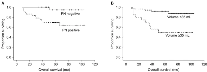
Fig. 1. Overall survival curves for (A) pelvic LN involvement (left, p=0.044) and (B) tumor volume (right, p=0.025). PN: pelvic lymph node.
Table 5. Univariate and multivariate analysis of prognostic factor for overall survival (Cox proportional hazard)
Variables Univariate analysis Multivariate analysis
p-value Hazard ratio (95% CI) p-value Hazard ratio (95% CI) Tumor histology (SCC, adenocarcinoma)
Tumor diameter (<4.5 cm, ≥4.5 cm) Tumor volume (<33 mL, ≥33 mL)
Pretreatment SCC-Ag* (<4.5 ng/mL, ≥4.5 ng/mL) Pelvic lymph node (negative, positive)
Para-aortic lymph node (negative, positive) Age (<50, ≥50 yr)
0.330 0.026 0.001 0.044 0.019 0.199 0.503
2.22 (0.47-9.71) 3.33 (1.15-9.61) 6.25 (2.09-18.75) 3.30 (1.03-10.59) 11.47 (1.50-87.81) 2.33 (0.64-8.42) 0.70 (0.24-2.01)
0.314 0.025 0.426 0.044
0.38 (0.060-2.49) 8.31 (1.30-53.15) 1.64 (0.48-5.58) 9.80 (1.07-90.09)
SCC-Ag: squamous cell carcinoma antigen.
*Excluded adenocarcinoma.
5-year OS rate was 80.6% (standard error, 4.9%). Estimated (Kaplan-Meier) five-year survival rate according to patient subgroups were summarized in Table 4. The 5-year survival rate of patients with tumor volume of <33 mL was 94.0%
compared with 49.3% in patients with tumor volume of ≥33 mL. In univariate analysis, large tumor diameter and volume, pelvic LN involvement and elevated pretreatment SCC-Ag levels were statistically significant relation to worse survival (Table 5). There was no significant relation between patient age, tumor histology or para-aortic LN involvement and OS.
Using multivariate analysis, tumor volume of >33 mL (p=0.025) and positive pelvic LN involvement (p=0.044) re- vealed a significantly unfavorable outcome on overall survival. The hazard ratio for tumor volume of ≥ 33 mL was 8.31 (95% CI, 1.30 to 54.15) and for positive pelvic LN in- volvement was 9.80 (95% CI, 1.07 to 90.09). Overall survival curves for tumor volume and pelvic LN involvement were shown in Fig. 1.
DISCUSSION
Tumor size and tumor infiltration into surrounding normal
tissue can be accurately estimated in surgically treated patients. However it is usually not available in patients treated with CCRT. Previous studies have shown that MRI indicates the closest correlation to actual tumor volume that is cur- rently performable in the clinical setting.10 In the evaluation of cervical cancer mass, MRI has several benefits with compar- ison to CT. The soft tissue contrast and multiplanar capability of MRI provides clear delineation of tumor. Tumor location including local invasion and diameter in T2 weighted MRI cor- related well with that in surgical specimen.11,12 Therefore, more accurate tumor size measurement and assessment of in- vasion to surrounding normal tissue, the depth of stromal in- vasion and LN evaluation has been possible.
In this study, we evaluated the relationship of prognostic fac- tors (tumor diameter, volume, LN involvement, age, histol- ogy and pretreatment SCC-Ag) to treatment outcome in 75 stage IIB cervical cancer patients treated with CCRT. Our re- sults indicated that the tumor volume and pelvic LN involve- ment were significantly associated with OS. However, the da- ta showed that tumor diameter, pretreatment SCC-Ag levels and para-aortic LN involvement had a poor relationship with OS. Burghardt et al.4 reported that OS was more closely re-
lated to tumor volume than to clinical or pathologic stage in early cervical cancer patients treated by radical hysterectomy and lymphadenectomy. In their series, 5-year survival rates of 91% for patients with tumor volume less than 2.5 mL, 79% for patients with tumor volume 2.5 to 10 mL, 70% for those with tumor volume 10 to 50 mL, and 48% for those with tumor vol- ume more than 50 mL. In advanced FIGO stage I and II tumors treated with radical radiotherapy, the treatment outcome was dependent on clinically determined tumor size rather than stage.9 Our results revealed that the tumor diameter of ≥4.5 cm correlated with worse OS by univariate analysis, but there was no correlation with OS by multivariate analysis. In gen- eral, tumor mass of stage IIB cervical cancer is larger than ear- ly cervical cancer. We suppose that tumor volume is the ap- propriate parameter to predict treatment outcome than tumor diameter in large tumor mass.
FIGO criteria currently used to stage cervical cancer do not account for LN involvement, but the LN metastasis is major factor to determining prognosis and proper treatment.
Increasing tumor size and increasing depth of stromal in- vasion correlated most strongly with LN metastasis and a shorter disease-free survival in previous studies of patients with FIGO stage 1B.3,13 The accuracy and sensitivity rates with MRI for detecting LN metastasis from cervical cancer are be- tween 76 to 100% and 36 to 71%, respectively.14,15 However, the limitation of MRI is that it is not possible to differentiate metastatic LN from non-metastatic LN hyperplasia of similar size and shape. In our study, pelvic LN involvement correlated significantly with OS and PFS by multivariate analysis.
Para-aortic LNs does not correlate with treatment outcome.
The incidence of pelvic LN metastasis in stage II cervical can- cer undergoing radical hysterectomy was approximately 35 to 45.8%. Our data revealed that pelvic LN metastasis and para-aortic LN metastasis were 58.7% and 14.7%, respectively.
Also, patients with positive pelvic LNs have a 5-year survival rate of 69.5% whereas patients with positive para-aortic LNs were higher than patients with positive pelvic LNs. Para-aort- ic metastases were found in 4.5 to 7.2% of patients in this stage.16-18 Higher 5-year survival rates of >90% are reported among patients treated surgically with no evidence of LN metastasis, compared to patients with positive pelvic node (50 to 60%, 5-year survival) or para-aortic nodes (20 to 30%, 5-year survival).19 Although it is accepted commonly that pa- tients with para-aortic LN metastases have lower OS and higher recurrence, the incidence of pelvic or para-aortic LN in- volvement and 5-year survival rates were relatively higher than previous studies. We can assume that a portion of LN in- volvement on MRI might be actually LN hyperplasia.
In addition, actual parametrial involvement was pathologi- cally confirmed in approximately 21 to 55% in the clinical stage IIB cervical cancer.16,20,21 This suggests that almost one half of the patients are overstaged due to difficulty in dis- tinguishing parametrial extension from inflammatory change, endometriosis, adhesion, and irregular shape of large cervical
tumor.20,21 The 5-year survival rate of stage stage IIB cervical cancer patients with radical hysterectomy ranged between 55 to 77%.22 In comparison with our 5-year survival rates, a part of our patients might be overstaged. Exact evaluations of LN status in the staging of cervical cancer are essential to direct treatment and reduce morbidity. More recently, positron emission tomography is more sensitive than MRI or CT for de- tecting LN metastases in patients with cervical cancer.23,24 Therefore, other diagnostic modalities such as positron emis- sion tomography or sentinel node biopsy may have a potential role for the exact assessment of LN metastases.
For imaging-based tumor size assessment, simple diameter measurement of three orthogonal diameters or three-dimen- sional (3D) volumetry with region-of-interest (ROI) quanti- tative image analysis are commonly used. The measurement of orthogonal tumor diameters is the simplest, fastest, and most practical methods extensively used in the radiology department. Simple diameter-based tumor measurement ap- pears to be equivalent to contour tracing/ROI analysis 3D tu- mor volumetry for tumor size assessment for predicting outcome.25,26 Diameter-based measurement can be easily computed as an ellipsoid-shaped tumor volume, but does not consider irregularities in tumor border and shape, which can be accounted for by 3D ROI volumetry. Thus it may be less ac- curate because cervical cancers have irregular contour that de- viate from the idealized ellipsoid volume.
Tumor histology (adenocarcinoma) and pretreatment SCC-Ag levels of ≥4.5 ng/mL revealed poor prognosis in PFS, but no correlation with OS. Generally, adenocarcinoma has a poor prognosis and relatively resistant response to radiation therapy.27 SCC-Ag levels correlate with the extent of the dis- ease, the response to treatment, survival and recurrence.28 There were only 9 patients with adenocarcinoma histology in our study. The results may not have been large enough to be able to make a clear declaration. Thus, we suggest that a pro- spective study involving a large cohort of patients will be re- quired to confirm this possibility.
We conclude that in patient with stage IIB cervical cancer, tumor volume and pelvic LN involvement provided possibility to predict OS. Therefore, optimal tumor volume and pelvic LN assessment by pretreatment MRI might be helpful to predict treatment outcome.
CONFLICT OF INTEREST
No potential conflict of interest relevant to this article was reported.
ACKNOWLEDGEMENTS
This study was supported by the Brain Korea (BK) 21 Project for Medical Sciences, Yonsei University and a grant from the Korean Health 21 R&D Project, Ministry of Health & Welfare and Family, Republic of Korea (0412-CR01-0704-0001).
REFERENCES
1. Hricak H, Yu KK. Radiology in invasive cervical cancer. AJR Am J Roentgenol 1996; 167: 1101-8.
2. National Cancer Institute. NCI issues clinical announcement on cervical cancer: chemotherapy plus radiation improves survival.
Bethesda: National Cancer Institute; 1999.
3. Eifel PJ, Morris M, Wharton JT, Oswald MJ. The influence of tumor size and morphology on the outcome of patients with FIGO stage IB squamous cell carcinoma of the uterine cervix.
Int J Radiat Oncol Biol Phys 1994; 29: 9-16.
4. Burghardt E, Baltzer J, Tulusan AH, Haas J. Results of surgical treatment of 1028 cervical cancers studied with volumetry.
Cancer 1992; 70: 648-55.
5. Micke O, Bruns F, Schafer U, Prott FJ, Willich N. The impact of squamous cell carcinoma (SCC) antigen in patients with ad- vanced cancer of uterine cervix treated with (chemo-)radio- therapy. Anticancer Res 2005; 25: 1663-6.
6. Ogino I, Nakayama H, Okamoto N, Kitamura T, Inoue T. The role of pretreatment squamous cell carcinoma antigen level in locally advanced squamous cell carcinoma of the uterine cervix treated by radiotherapy. Int J Gynecol Cancer 2006; 16: 1094-100.
7. Scheidler J, Heuck AF. Imaging of cancer of the cervix. Radiol Clin North Am 2002; 40: 577-90.
8. Amendola MA, Hricak H, Mitchell DG, Snyder B, Chi DS, Long HJ 3rd, et al. Utilization of diagnostic studies in the pretreat- ment evaluation of invasive cervical cancer in the United States: results of intergroup protocol ACRIN 6651/GOG 183. J Clin Oncol 2005; 23: 7454-9.
9. Thoms WW Jr, Eifel PJ, Smith TL, Morris M, Delclos L, Wharton JT, et al. Bulky endocervical carcinoma: a 23-year experience. Int J Radiat Oncol Biol Phys 1992; 23: 491-9.
10. Ebner F, Tamussino K, Kressel HY. Magnetic resonance imag- ing in cervical carcinoma: diagnosis, staging, and follow-up.
Magn Reson Q 1994; 10: 22-42.
11. Narayan K, McKenzie A, Fisher R, Susil B, Jobling T, Bernshaw D. Estimation of tumor volume in cervical cancer by magnetic resonance imaging. Am J Clin Oncol 2003; 26: e163-8.
12. Oellinger JJ, Blohmer JU, Michniewicz K, Siewert C, Wust P, Gutberlet M, et al. Pre-operative staging of cervical cancer:
comparison of magnetic resonance imaging (MRI) and com- puted tomography (CT) with histologic results. Zentralbl Gynakol 2000; 122: 82-91.
13. Zaino RJ, Ward S, Delgado G, Bundy B, Gore H, Fetter G, et al.
Histopathologic predictors of the behavior of surgically treated stage IB squamous cell carcinoma of the cervix: a Gynecologic Oncology Group study. Cancer 1992; 69: 1750-8.
14. Choi SH, Kim SH, Choi HJ, Park BK, Lee HJ. Preoperative mag- netic resonance imaging staging of uterine cervical carcinoma:
results of prospective study. J Comput Assist Tomogr 2004; 28:
620-7.
15. Yu KK, Hricak H, Subak LL, Zaloudek CJ, Powell CB.
Preoperative staging of cervical carcinoma: phased array coil fast spin-echo versus body coil spin-echo T2-weighted MR
imaging. AJR Am J Roentgenol 1998; 171: 707-11.
16. Kawagoe T, Kashimura M, Matsuura Y, Sugihara K, Toki N, Aoki T. Clinical significance of tumor size in stage IB and II car- cinoma of the uterine cervix. Int J Gynecol Cancer 1999; 9:
421-6.
17. Sakuragi N, Satoh C, Takeda N, Hareyama H, Takeda M, Yamamoto R, et al. Incidence and distribution pattern of pelvic and paraaortic lymph node metastasis in patients with Stages IB, IIA, and IIB cervical carcinoma treated with radical hysterectomy. Cancer 1999; 85: 1547-54.
18. Takeda N, Sakuragi N, Takeda M, Okamoto K, Kuwabara M, Negishi H, et al. Multivariate analysis of histopathologic prog- nostic factors for invasive cervical cancer treated with radical hysterectomy and systematic retroperitoneal lymphadenectomy.
Acta Obstet Gynecol Scand 2002; 81: 1144-51.
19. Tinga DJ, Timmer PR, Bouma J, Aalders JG. Prognostic sig- nificance of single versus multiple lymph node metastases in cervical carcinoma stage IB. Gynecol Oncol 1990; 39: 175-80.
20. Inoue T, Morita K. The prognostic significance of number of positive nodes in cervical carcinoma stages IB, IIA, and IIB.
Cancer 1990; 65: 1923-7.
21. Kamura T, Tsukamoto N, Tsuruchi N, Kaku T, Saito T, To N, et al. Histopathologic prognostic factors in stage IIb cervical carci- noma treated with radical hysterectomy and pelvic-node dis- section: an analysis with mathematical statistics. Int J Gynecol Cancer 1993; 3: 219-25.
22. Suprasert P, Srisomboon J, Kasamatsu T. Radical hysterectomy for stage IIB cervical cancer: a review. Int J Gynecol Cancer 2005; 15: 995-1001.
23. Rose PG, Adler LP, Rodriguez M, Faulhaber PF, Abdul-Karim FW, Miraldi F. Positron emission tomography for evaluating para-aortic nodal metastasis in locally advanced cervical cancer before surgical staging: a surgicopathologic study. J Clin Oncol 1999; 17: 41-5.
24. Narayan K, Hicks RJ, Jobling T, Bernshaw D, McKenzie AF. A comparison of MRI and PET scanning in surgically staged lo- co-regionally advanced cervical cancer: potential impact on treatment. Int J Gynecol Cancer 2001; 11: 263-71.
25. Mayr NA, Taoka T, Yuh WT, Denning LM, Zhen WK, Paulino AC, et al. Method and timing of tumor volume measurement for outcome prediction in cervical cancer using magnetic reso- nance imaging. Int J Radiat Oncol Biol Phys 2002; 52: 14-22.
26. Hatano K, Sekiya Y, Araki H, Sakai M, Togawa T, Narita Y, et al.
Evaluation of the therapeutic effect of radiotherapy on cervical cancer using magnetic resonance imaging. Int J Radiat Oncol Biol Phys 1999; 45: 639-44.
27. Lea JS, Sheets EE, Wenham RM, Duska LR, Coleman RL, Miller DS, et al. Stage IIB-IVB cervical adenocarcinoma: prog- nostic factors and survival. Gynecol Oncol 2002; 84: 115-9.
28. Molina R, Filella X, Lejarcegui JA, Pahisa J, Torne A, Rovirosa A, et al. Prospective evaluation of squamous cell carcinoma and carcinoembryonic antigen as prognostic factors in patients with cervical cancer. Tumour Biol 2003; 24: 156-64.