Prognosis of hepatic epithelioid
hemangioendothelioma after living donor liver transplantation
Byeong-Gon Na
1, Shin Hwang
1, Chul-Soo Ahn
1, Ki-Hun Kim
1, Deok-Bog Moon
1, Tae-Yong Ha
1, Gi-Won Song
1, Dong-Hwan Jung
1, Gil-Chun Park
1, Young-In Yoon
1, Woo-Hyoung Kang
1, Hwui-Dong Cho
1, Sang Hoon Kim
1, Seung-Mo Hong
2, Sung-Gyu Lee
11Department of Surgery, Asan Medical Center, University of Ulsan College of Medicine, Seoul, Korea
2Department of Pathology, Asan Medical Center, University of Ulsan College of Medicine, Seoul, Korea
Background: Epithelioid hemangioendothelioma (EHE) is a rare borderline vascular tu- mor. Due to its rarity and protean behavior, the optimal treatment of hepatic EHE has not yet been standardized. This single-center study describes outcomes in patients with he- patic EHE who underwent living donor liver transplantation (LDLT).
Methods: The medical records of patients who underwent LDLT for hepatic EHE from 2007 to 2016 were reviewed.
Results: During 10-year period, four patients, one man and three women, of mean age 41.3±11.1 years, underwent LDLT for hepatic EHE. Based on imaging modalities, these pa- tients were preoperatively diagnosed with EHE or hepatocellular carcinoma, with percu- taneous liver biopsy confirming that all four had hepatic EHE. The tumors were multiple and scattered over entire liver, precluding liver resection. Blood tumor markers were not elevated, except that CA19-9 and des-γ-carboxy prothrombin was slightly elevated in one patient. Mean model for end-stage liver disease score was 10.8±5.7. All patients under- went LDLT using modified right liver grafts, with graft-recipient weight ratio of 1.11±0.19, and all recovered uneventfully after LDLT. One patient died due to tumor recurrence at 9 months, whereas the other three have done well without tumor recurrence, resulting in 5-year disease-free and overall patient survival rates of 75% each. The patient with tumor recurrence was classified as a high-risk patient based on the original and modified he- patic EHE-LT scoring systems.
Conclusions: LDLT can be an effective treatment for patients with unresectable hepat- ic EHEs that are confined within the liver and absence of macrovascular invasion and lymph node metastasis.
Keywords: Borderline tumor; Recurrence; Vascular tumor; Metastasis; Immunohistochemical stain
Received October 12, 2020 Revised November 5, 2020 Accepted November 6, 2020 Corresponding author: Shin Hwang Department of Surgery, Asan Medical Center, University of Ulsan College of Medicine, 88 Olympic-ro 43-gil, Songpa- gu, Seoul 05505, Korea
Tel: +82-2-3010-3930 Fax: +82-2-3010-6701 E-mail: [email protected]
© The Korean Society for Transplantation This is an Open Access article distributed under the terms of the Creative Commons Attribution Non-Commercial License (http://creativecommons.org/licenses/
by-nc/4.0/) which permits unrestricted non-commercial use, distribution, and reproduction in any medium, provided the original work is properly cited.
pISSN 2671-8790
eISSN 2671-8804
INTRODUCTION
Epithelioid hemangioendothelioma (EHE) is a rare tumor composed of cords of epithelioid cells on a background of myxohyaline stroma. The 2002 World Health Organization classification described EHE as lesions with metastatic potential [1,2]. Hepatic EHE is a rare borderline vascular tumor, with an aggressiveness graded between hemangi- oma and hepatic hemangiosarcoma [3,4]. Because many patients with hepatic EHE are asymptomatic, these lesions are frequently detected incidentally [4-7]. Due to their rarity and protean behavior, the optimal treatment of hepatic EHE has not yet been standardized [8]. Partial hepatec- tomy has been recommended for patients with unilobar hepatic EHE, although aggressive recurrences have been reported after hepatectomies [9,10]. Liver transplantation (LT) is indicated for patients with advanced liver involve- ment, with approximately 200 such patients being report- ed in literature, mostly from the United States, Europe, and Canada [11-13]. To our knowledge, only three patients in Korea have undergone LT cases for hepatic EHE [14,15].
This study describes the clinicopathological features and prognosis of patients with hepatic EHE who underwent living donor liver transplantation (LDLT) at a single center over a 10-year period.
METHODS
The study protocol was approved by of the Institutional Re- view Board at Asan Medical Center (IRB No. 2019-1347), which waived the requirement for informed consent due to the retrospective nature of this study. This study was performed in accordance with the ethical guidelines of the
World Medical Association Declaration of Helsinki 2013.
Patients
The LT database at our institution was searched to identify patients who had been diagnosed with hepatic EHE and underwent LDLT over a 10-year period from January 2007 to December 2016. During this study period, 3,467 patients had undergone adult LT at our center. The medical records of these patients were retrospectively reviewed, with all patients followed up until July 2020.
Preoperative Evaluation, Surgical Procedures, and Postoperative Follow-up
Routine preoperative evaluation of primary liver tumors has been described elsewhere [16]. The protocol for ABO-incompatible LDLT includes desensitization with rituximab and plasmapheresis [17]. The LDLT recipients were followed up every month during the first year, every 2 months for the next 4 years, and every 3 months there- after. Patients with recurrent liver tumors were treated as described [18].
Immunohistochemical Staining
Formalin-fixed paraffin-embedded tissue samples were im- munohistochemically stained for CD34 (1:500, QBEND10;
Immunotec Inc., Monrovia, CA, USA), CD31 (1:800, JC70;
Cell Marque, Rocklin, CA, USA) and coagulator factor VIII-related antigen (FVIII:Ag) (1:2000; DAKO, Glostrup, Denmark) using a Benchmark autostainer (Ventana Med- ical System, Tucson, AZ, USA). The diagnosis of hepatic EHE was based on the histological features and immuno- histochemical profiles described in the 2010 WHO classifi- cation of liver tumors [19].
Hepatic EHE-LT Score for Assessing Risk of Posttransplant Tumor Recurrence
Hepatic EHE-LT scores were calculated using the fol- lowing formula: 5×(pathological macrovascular inva- sion)+3×(waiting time ≤120 days)+2×(pathological inva- sion hilar lymph node). Hepatic EHE-LT scores of 0–2, 3–5, and 6–10 were regarded as indicating low, intermediate, and high risk, respectively, for posttransplant tumor recur- rence [20].
Calculation of Model for End-Stage Liver Disease Score The model for end-stage liver disease (MELD) score is calculated using the following formula: “9.57×log
e(creati- nine, mg/dL)+3.78×log
e(total bilirubin, mg/dL)+11.2×log
eHIGHLIGHTS
• Four cases of hepatic epithelioid hemangioendothelio- ma who underwent living donor liver transplantation are presented in this study.
• Living donor liver transplantation can be an effective
treatment for patients with unresectable hepatic epithe-
lioid hemangioendotheliomas that are confined within
the liver and absence of macrovascular invasion and
lymph node metastasis.
(INR)+6.43” [21].
Statistical Analysis
Numerical data are presented as mean and standard devi- ation. Survival rates were estimated using the Kaplan-Mei- er method. Statistical analyses were performed using IBM SPSS ver. 22.0 (IBM Corp., Armonk, NY, USA).
RESULTS
Patient Demographics and Preoperative Diagnosis
During the 10-year study period, four patients (0.11% of the 3,647 patients who underwent adult LT) underwent LDLT for hepatic EHE. The clinicopathological features of these four patients are described in Table 1. The four patients in- cluded one man and three women, of mean age 41.3±11.1 years. One (25%) was positive for hepatitis B virus infec- tion, but none were positive for hepatitis C virus infection or had alcoholic liver disease. Three patients (75%) were experiencing jaundice or abdominal pain at the time of their initial visit to the outpatient clinic, which led to further examination.
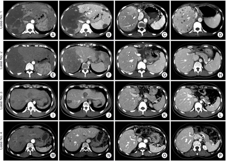
Imaging modalities indicated suspected preoperative diagnoses of hepatic EHE or hepatocellular carcinoma (Fig. 1). Preoperative liver biopsy resulted in a pathologic diagnosis of hepatic EHE in all four patients. Analysis of mean blood concentrations of preoperative tumor markers showed a mean alpha-fetoprotein concentration of 2.2±1.4 ng/mL (reference, 7.5 ng/mL), a mean des-γ-carboxy pro- thrombin (DCP) concentration of 31.0±16.8 mAU/mL (refer- ence, 40 mAU/mL), and a mean carbohydrate antigen 19-9 (CA19-9) concentration of 22.7±13.2 ng/mL (reference, 37 ng/mL). Only one patient presented with slightly elevated lev- els of CA19-9 and DCP (Table 1).
Peritransplant Clinical Courses
All four patients were indicated for LT because they had unresectable multiple tumors scattered throughout the entire liver (Fig. 1). Their mean MELD score was 10.8±5.7.
Because none had any likelihood of undergoing deceased donor liver transplantation (DDLT), all four underwent LDLT using modified right liver grafts. Three donors (75%) were siblings of the recipient, and one (25%) was a daughter. The mean graft-recipient weight ratio was 1.11±0.19 (Table 2).
All patients recovered uneventfully from LDLT operation
without major surgical complications (Fig. 1).
Table 1.Clinical pr ofiles of the recipients Case No. Age (yr) Sex Clinical presentation HB V infection Preoper ative imaging diagnosis Preoper ative biopsy
MELD score
AFP (ng/mL) DCP (mA U/ mL)
CA19-9 (ng/ mL)
Original HEHE-L
T score
Modified HEHE-L
T score Tumor recurrence Diseas e- free s ur viv al period (mo)
Recurrence treatment
Sur viv al status Over all pat ient su rviv al pe riod (mo) 1 41 F Jaundice No R/O EHE EHE-confirmed 19 4.1 56 40.1 10 2 Yes 2
Lung Mx+C
Tx Dead 9 2 39 F Abdominal pain No R/O EHE EHE-confirmed 10 1.3 26 9.8 8 1 No 105 No Alive 105 3 29 M Abdominal pain No R/O EHE EHE-confirmed 7 1.1 21 16 3 0 No 50 No Alive 50 4 56 F
Routine health screening
Yes R/O HCC, R/O EHE EHE-confirmed 7 2.2 21 25 3 0 No 47 No Alive 47 HB V, he pat itis B viru s; M ELD, m ode l f or end-st age live r dise ase ; AF P, alpha-f et opr ot ein; DCP , de s-γ -carbo xy pr ot hr om bin; CA 19-9, carbohydr at e ant ige n 19-9; HEHE, he pat ic epit he lioid hemangioendothelioma; L T, liver tr ansplantation; R/O , rule out; EHE, epithelioid hemangioendothelioma; Mx, metastasect omy; C Tx, chemother apy; HCC, hepat ocellular car cinoma.
Explant Pathology
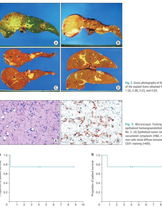
The pathological features of the explanted livers are sum- marized in Table 2. All four patients had multiple tumors scattered over the entire liver (Fig. 2). Two patients (case No. 1 and 2) had portal vein tumor thrombi, and one (case No. 1) had lymph node metastases. Microscopic examina- tion showed low cellular epithelioid or spindle tumor cells on a background of fibromyxoid stroma, with the epitheli- oid tumor cells containing vacuolated cytoplasm (Fig. 3A).
Immunohistochemical staining showed that the tumor cells in all four patients were diffusely immune-positive for
CD31, CD34, and FVIII:Ag (Fig. 3B). Each one patient had hepatic EHE-LT scores of 7 and 5, whereas two had hepat- ic EHE-LT scores of 0 (Table 1).
Posttransplant Outcomes
One patient, who had a large-sized tumor, 15 cm in di- ameter, and 13 small liver masses with portal vein tumor thrombosis and lymph node metastasis, was positive for lung metastases 2 months after LT. Pulmonary metasta- sectomy and systemic chemotherapy were performed, but this patient died due to tumor progression 9 months
CaseNo.1CaseNo.2CaseNo.3CaseNo.4
A B C D
E F G H
I J K L
M N O P
Fig. 1. Pretransplant and posttransplant computed tomography (CT) findings. Case No. 1 (A-D): pretransplant (A) arterial-phase and (B) portal-phase CT
images show a large tumor and small masses, along with involvement of the right portal vein branches. (C) CT scan taken at 1 month after transplanta-
tion shows the usual posttransplant findings. (D) CT scan taken at 7 months after transplantation shows multiple liver metastases. Case No. 2 (E-H): pre-
transplant (E) arterial-phase and (F) portal-phase CT images show a large tumor and small masses. CT scans taken at 1 month (G) and 7 years (H) after
transplantation show the usual posttransplant findings. Case No. 3 (I-L): pretransplant (I) arterial-phase and (J) portal-phase CT images show multiple
small tumors. CT scans taken at 1 month (K) and 4 years (L) after transplantation show the usual posttransplant findings. Case No. 4 (M-P): pretrans-
plant (M) arterial-phase and (N) portal-phase CT images show multiple small tumors. CT scans taken at 1 month (O) and 3 years (P) after transplantation
show the usual posttransplant findings.
after transplantation (Fig. 1). The other three patients have done well without tumor recurrence. The 1-, 3-, and 5-year disease-free and overall patient survival rates were all 75%
(Fig. 4).
DISCUSSION
Treatments for hepatic EHE include hepatic resection, LT, chemotherapy, radiotherapy, hormone therapy, radiofre- quency ablation, and surveillance alone. The 5-year patient survival rates have been reported to be 75% in 22 patients who underwent hepatic resection, 20% in 60 patients treat- ed with chemotherapy/radiotherapy, and 4.5% in 70 pa- tients who underwent surveillance alone [5]. Another study found that the 5-year patient survival rates were 86% in 11 patients who underwent hepatic resection and 73% in 11 LT recipients [22]. The 3-year patient survival rates have been reported to be 74.1% in 17 patients who underwent hepatic resection patients and 81.6% in 12 patients who underwent transarterial chemoembolization (TACE) [6]. Al- though these studies reported similar survival rates in pa- tients who underwent hepatic resection, LT and TACE, the indications for each treatment modality were different. He- patic resection is indicated for resectable intrahepatic le- sions, whereas LT and TACE are indicated for unresectable lesions. Extrahepatic involvement, including lymph node and distant metastasis, is a contraindication for surgical treatment. The roles of nonsurgical therapies, including systemic/regional chemotherapy, radiotherapy, hormone therapy, and immunotherapy, have been investigated in only a few small case series [23-25].
The Europe Liver Transplant Registry (ELTR) reported a 5-year survival rate of 83% in 59 LT recipients with he- patic EHE, whereas the United Network for Organ Sharing (UNOS) registry reported a 5-year survival rate of 64%
in 110 LT recipients, respectively [11,12]. LT is regarded as primary or salvage therapy for patients with multiple unresectable tumors not responsive to other nonsurgical treatments. However, DDLT in patients with hepatic EHE is limited by various factors, including donor shortage, high medical costs, the need for lifelong immunosuppressant therapy, patient willingness, and risk of tumor recurrence.
The indications of LT for hepatic EHE have not been well-defined. A prognostic score based on analysis of the ELTR-ELITA (European Liver Intestine Transplant Associa- tion) registry suggested that macrovascular invasion, short
Table 2.Pr ofiles of living donors and tr ansplantation Case No. Donor age (yr) Donor sex Donor relationship ABO- incompatibility Gr aft type
Gr aft volume (mL)
GRWR (%) CIT (min) WIT (min) TIT (min)
Maximal tumor size (cm)
Tumor number Lym ph node metas tas is Tumor location CD31 stain CD34 stain Fact or VIII antigen stain 1 44 F Sibling No MRL 460 1.15 95 48 143 15 14 Yes Whole liver Positive Positive Positive 2 40 F Sibling No MRL 640 1.36 99 38 137 13 8 No Whole liver Positive Positive ND 3 26 M Sibling No MRL 723 0.94 114 36 150 3.8 7 No Whole liver ND Positive ND 4 30 F Daughter Yes MRL 503 1.01 60 25 85 2 15 No Whole liver Positive ND ND GRWR, gr aft-recipient weight r atio; CIT , cold ischemic time; WIT , warm ischemic time; TIT , t otal ischemic time; MRL, modified right liver; ND, not done.
Fig. 2. Gross photographs of the cut surface
of the explant livers obtained from case No.
1 (A), 2 (B), 3 (C), and 4 (D).
A B
C D
A B
Fig. 3. Microscopic findings of hepatic
epithelioid hemangioendothelioma in case No. 2. (A) Epithelioid tumor cells containing vacuolated cytoplasm (H&E, ×400). (B) Tu- mor cells show diffuse immune-positivity for CD31 staining (×400).
Fig. 4. Kaplan-Meier analysis of disease-free (A) and overall (B) patient survival.
1 2 3 4 5 6 7 8 9
1.0
0.8
0.6
0.4
0.2
10
Proportionofpatientsurvival
Posttransplant year 0
A B
1 2 3 4 5 6 7 8 9
1.0
0.8
0.6
0.4
0.2
10
Proportionofpatientsurvival
Posttransplant year 0