185 https://e-kcj.org
An 89-year-old woman underwent transfemoral transcatheter aortic valve replacement with a 26-mm Evolut PRO (Medtronic, Minneapolis, MN, USA) bioprosthesis because of severe aortic stenosis.
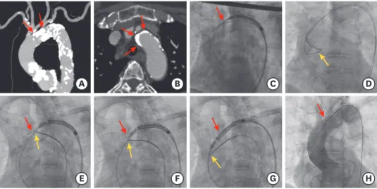
As shown in Figure 1, the delivery catheter failed to pass through the severely calcified aortic arch, because of a dense calcified mass in the outer curvature of the aortic arch (Supplementary Video 1). Eventually, a snare-assisted delivery using a 35-mm loop Amplatz Goose Neck Snare kit (ev3 Endovascular, Inc., Plymouth, MN, USA) was attempted. We tightened the snare to grasp and retain the stiff wire and the nose cone of the delivery catheter. Further, we advanced the delivery catheter while simultaneously pulling the snare catheter downward. Finally, the delivery catheter was passed and the valve implantation was successful without major aortic complications (Supplementary Videos 2 and 3).
Korean Circ J. 2021 Feb;51(2):185-186 https://doi.org/10.4070/kcj.2020.0384 pISSN 1738-5520·eISSN 1738-5555
Images in
Cardiovascular Medicine
Received: Aug 28, 2020 Revised: Oct 9, 2020 Accepted: Nov 25, 2020 Correspondence to Umihiko Kaneko, MD
Department of Cardiovascular Medicine Cardiovascular Medicine, Sapporo Cardiovascular Clinic, Sapporo Heart Center, North 49, East 16, 8-1 Higashi Ward, Sapporo, Hokkaido 007-0849, Japan.
E-mail: [email protected] Copyright © 2021. The Korean Society of Cardiology
This is an Open Access article distributed under the terms of the Creative Commons Attribution Non-Commercial License (https://
creativecommons.org/licenses/by-nc/4.0) which permits unrestricted noncommercial use, distribution, and reproduction in any medium, provided the original work is properly cited.
ORCID iDs Umihiko Kaneko
https://orcid.org/0000-0003-2392-5084 Daisuke Hachinohe
https://orcid.org/0000-0003-4828-1836 Tsutomu Fujita
https://orcid.org/0000-0002-3651-6231 Funding
The authors received no financial support for the research, authorship, and/or publication of this article.
Conflict of Interest
The authors have no financial conflicts of interest.
Umihiko Kaneko , MD, Daisuke Hachinohe , MD, Ken Kobayashi, MD, and Tsutomu Fujita , MD
Department of Cardiovascular Medicine, Sapporo Cardio Vascular Clinic, Sapporo Heart Center, Sapporo, Japan
Snare-Assisted Valve Delivery to
Overcome a Severely Calcified Aortic Arch during Transcatheter Aortic Valve Replacement
A B C D
E F G H
Figure 1. Snare-assisted delivery of a self-expanding valve.
(A, B) Baseline computed tomography reveals a severely calcified aortic arch with dense calcified plaques
protruding into the lumen. (C) The protruding calcification hampers the passage of a delivery catheter. (D) First,
we insert a standard wire inside the snare introduced from the contralateral femoral access. Second, the standard
wire inserted in the left ventricle is exchanged with the stiff wire. (E-G) Next, the snare is tightened and the stiff wire
and nose cone of the delivery catheter are retained. Simultaneous pulling of the snare results in successful passage
of the delivery catheter. (H) Final angiography shows an optimal valve position without aortic complications. Red
arrows indicate the dense calcified plaque of the aortic arch, and yellow arrows indicate the snare position.
Author Contributions
Supervision: Fujita T; Validation: Hachinohe D, Kobayashi K; Writing - original draft: Kaneko U.
Severely calcified aortic arches are a major limitation in the implantation of currently available self-expanding valves due to the rigidity and non-steerable features of these valves, with the risk of delivery failure or fatal complications including aortic dissection or rupture.
1)The inflexible and bulky delivery catheter tends to follow the outer curvature of the aortic arch, and massive calcification can obstruct the device passage. The snare technique can direct the wire and delivery catheter away from the aortic wall, leading to successful crossing.
In an extremely calcified aortic valve and a horizontal aorta that blocks the passage of a bioprosthetic valve, a snare catheter can achieve coaxiality with the aortic orifice and successful crossing of the aortic valve.
2)SUPPLEMENTARY MATERIALS
Supplementary Video 1
The delivery catheter fails to pass through the severely calcified aortic arch.
Click here to view
Supplementary Video 2
Simultaneous pulling of the snare results in successful passage of the delivery catheter.
Click here to view
Supplementary Video 3
Final aortography confirms no major aortic complications.
Click here to view
REFERENCES
1. Szlapka M, Michel E, Ricciardi MJ, Malaisrie SC. Valve-in-valve-prosthesis embolization and aortic dissection: single procedure, double complication. Eur J Cardiothorac Surg 2019;56:204-5.
PUBMED | CROSSREF
2. Kaneko U, Hachinohe D, Kobayashi K, et al. Evolut self-expanding transcatheter aortic valve replacement in patients with extremely horizontal aorta (aortic root angle ≥ 70°). Int Heart J 2020;61:1059-69.
PUBMED | CROSSREF