저작자표시-비영리-변경금지 2.0 대한민국 이용자는 아래의 조건을 따르는 경우에 한하여 자유롭게 l 이 저작물을 복제, 배포, 전송, 전시, 공연 및 방송할 수 있습니다. 다음과 같은 조건을 따라야 합니다: l 귀하는, 이 저작물의 재이용이나 배포의 경우, 이 저작물에 적용된 이용허락조건 을 명확하게 나타내어야 합니다. l 저작권자로부터 별도의 허가를 받으면 이러한 조건들은 적용되지 않습니다. 저작권법에 따른 이용자의 권리는 위의 내용에 의하여 영향을 받지 않습니다. 이것은 이용허락규약(Legal Code)을 이해하기 쉽게 요약한 것입니다. Disclaimer 저작자표시. 귀하는 원저작자를 표시하여야 합니다. 비영리. 귀하는 이 저작물을 영리 목적으로 이용할 수 없습니다. 변경금지. 귀하는 이 저작물을 개작, 변형 또는 가공할 수 없습니다.
A Doctoral Dissertation
A study on the regulation of neuronal
cell death through mitochondrial
calcium and membrane potential
Jin-Ji Wu
Department of Medicine
Graduated School
Jeju National University
미토콘드리아
칼슘과 막전압을 통한
신경세포
손상사멸의 조절에 관한 연구
지도교수
: 은 수 용
오
금 희
이
논문을 의학 박사학위 논문으로 제출함
2013년 8월
오금희의
의학 박사학위 논문을 인준함
심사위원장
위
원
위
원
위
원
위
원
제주대학교
대학원
2013년 8
A study on the regulation of neuronal cell death through
mitochondrial calcium and membrane potential
Jin-Ji Wu
(Supervised by professor Su-Yong Eun)
A thesis submitted in partial fulfillment of the requirement for the
degree of doctor of philosophy in medicine
August, 2013
This thesis has been examined and approved.
Date
Department of Medicine
Graduated School
Jeju National University
1
ABSTRACT
It has been demonstrated that even a small mitochondrial depolarization is sufficient to prevent neuronal cell death by suppressing mitochondrial calcium overload since mitochondrial membrane potential (∆Ψm) contributes to determining a driving force for calcium to enter the mitochondria. Therefore, mitochondrial depolarization has been recently evaluated as a novel mechanism of neuroprotection via inhibiting neurotoxic mitochondrial calcium overload during neuronal insults. The active compounds from the peel of citrus fruits are known to exert significant biological activities including anti-inflammatory and anti-oxidant properties. A growing body of evidence has recently demonstrated that these compounds recover damaged cognitive function in the models of neurodegenerative disease. However, the specific mechanism of neuroprotective effects has not to be clearly elucidated yet. Therefore, we investigated the neuroprotective mechanism of citrus peel extracts (CPE) against oxidative neurotoxicity implicated in neurodegenerative diseases. The results showed that neuronal viability was significantly increased by CPE in H2O2-treated neuronal HT-22 cells, which were used as an in vitro model of oxidative neurotoxicity. The CPE demonstrated a robust scavenging activity of intracellular reactive oxygen species (ROS). CPE treatment significantly blocked H2O2-induced Ca2+ overload in both the cytosol and the mitochondria as indicated by Fluo-4 and Rhod-2 respectively, and inhibited neuronal apoptosis cascades including caspase 3. Additionally, we determined using TMRM and JC-1 fluorometric probes for mitochondrial membrane potential (∆Ψm) that the CPE and CPE compounds such as nobiletin are capable of inducing a mild mitochondrial depolarization which has been recently evaluated as a novel mechanism of neuroprotection via inhibiting mitochondrial Ca2+ overload during neuronal insults. Our findings suggest a dual neuroprotective mechanism of CPE via not only anti-oxidant activity but also ∆Ψm regulation.
2
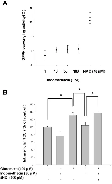
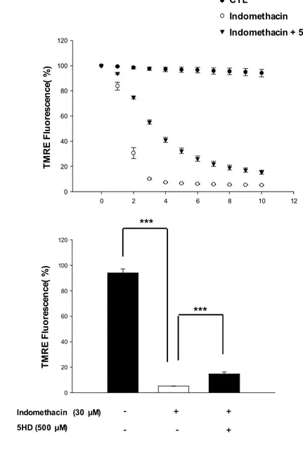
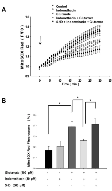
In the next study, we investigated the exact neuroprotective mechanism of mitochondrial membrane depolarization in neuronal insults-induced cell death, separated from anti-oxidant mechanism. For this experimental aim, we tested the neuroprotective mechanism of indomethacin, non-steroid anti-inflammatory drugs (NSAIDs), which does not have antioxidant activity but evokes mitochondrial membrane depolarization. The results demonstrated that neuronal viability was significantly increased by indomethacin treatment in glutamate-exposed primary cortical neurons as a glutamate-induced neurotoxicity model. This neuroprotective effect was abolished by 5-hydorxydecanoate (5HD), a ATP-sensitive K+ channels (mitoKATP) channel blocker. This blockade of mitoKATP channels by 5HD treatment significantly inhibited indomethacin-induced mitochondrial depolarization and also abolished indomethacin-induced inhibitory effect on mitochondrial calcium overload. It also suppressed mitochondrial dysfunction-associated parameters such as ROS generation and mitochondrial permeability transition pore (mPTP) open.
Taken together, these results suggest that the active compounds of CPE and indomethacin may be considered as promising neuroprotective agents via inducing a mitochondrial depolarization. In addition, we propose here that mitochondrial ion channels and transporters such as mitoKATP channels could be evaluated as the novel therapeutic targets for neuroprotection against neuronal cell death implicated in one of the critical causes of brain ischemia and neurodegenerative diseases.
Keywords: mitochondrial calcium, mitochondrial membrane potential, calcium, mitochondria, Indomethacin, Citrus
3
CONTENTS
ABSTRACT ...1
CONTENTS...3
LIST OF FIGURES AND TABLE...4
PART Ⅰ...6
PART Ⅱ...8
REFERENCE...67
4
LIST OF FIGURES AND TABLE
Fig.1. Neuroprotective effects of CPE against H2O2-induced oxidative neurotoxicity ...22
Fig.2. Effect of CPE on procaspase-3 and PARP against H2O2-induced oxidative
neurotoxicity...23
Fig.3. Anti-oxidant activities of CPE... 24
Fig.4. CPE evokes a mild mitochondrial depolarization, as indicated in real-time measurement of ∆Ψm...27
Fig.5. Differential effects of single compounds of CPE on JC-1 Fred/Fgreen ratio indicative of ∆Ψm...32
Fig.6. Inhibitory effects of CPE on H2O2-induced cytosol calcium overload... 34
Fig.7. Inhibitory effects of CPE on H2O2-induced mitochondrial calcium overload...36
Fig.8. Neuronal purity in primary cortical neuron cultures...50
5
Fig.10. Anti-oxidant activities of indomethacin...52
Fig.11. Effect of indomethacin on the mitochondrial membrane potential, cytosol calcium and mitochondrial calcium...54
Fig.12. Inhibitory effect of 5HD on indomethacin-induced mitochondrial depolarization...58
Fig.13. Effects of indomethacin and 5HD on glutamate-induced mitochondrial superoxide generation...59
Fig.14. Effects of indomethacin and 5HD on glutamate-induced mitochondrial mitochondrial calcium overload...62
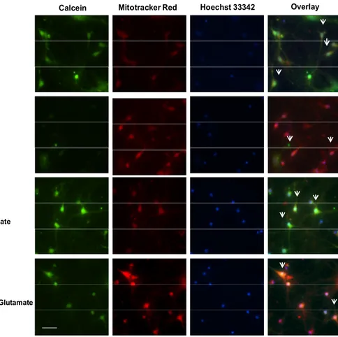
Fig.15. Effects of indomethacin and 5HD on glutamate-induced mPTP opening...64
6
PART Ⅰ
1. INTRODUCTION... 11
2. MATERIALS AND METHODS... 13
2.1. Materials
2.2. Preparation of the extract and compound 2.3. Cell culture
2.4. Real-time measurement of mitochondrial membrane potential (∆Ψm) 2.5. Flow cytometric and imaging analyses of ∆Ψm
2.6. Flow cytometric analyses of cytosol and mitochondrial Ca2+ 2.7. Mitochondrial Ca2+ Imaging
2.8. Measurement of cell viability 2.9. Morphological analysis of apoptosis 2.10. Western blotting analysis
2.11. Measurement of intracellular reactive oxygen species (ROS) 2.12. DPPH free radical scavenging assay
2.13. Measurement of superoxide dismutase (SOD) activities 2.14. Measurement of catalase activities
2.15. Statistics analysis
3. RESULTS ...20
7
3.2. Antioxidant activities of CPE
3.3. Mild-depolarizing effects of CPE on ∆Ψm
3.4. Differential effects of single compounds of CPE on ∆Ψm
3.5. Inhibitory effects of CPE on H2O2-induced overload of Ca2+ in both the cytosol and the mitochondria
8
PART
Ⅱ
1. INTRODUCTION... 41
2. MATERIALS AND METHODS...43
2.1. Materials 2.2. Cell culture
2.3. Immunofluorescence microscopy and labeling of mitochondria 2.4. Measurement of cell viability
2.5. Real-time measurement of ∆Ψm
2.6. Dural recording of cytosolic and mitochondrial calcium 2.7. DPPH radical scavenging assay
2.8. Intracellular reactive oxygen species (ROS) measurement 2.9. Real-time measurement of mitochondrial superoxide generation 2.10. Mitochondrial permeability transition pore (mPTP) assay 2.11. Statistics analysis
3. RESULTS ...48
3.1. Purity of primary cortical neuron
3.2. Mitochondrial KATP channel blockade abolishes neuroprotective effect of indomethacin on glutamate-induced neurotoxicity in primary cortical neuron 3.3. Mitochondrial KATP channel blockade abolishes neuroprotective effect of
9
3.4. Effect of indomethacin on the mitochondrial membrane potential, cytosol calcium and mitochondrial calcium
3.5. Mitochondrial KATP channel blockade abolishes neuroprotective effect of indomethacin on mitochondrial membrane potential
3.6. Mitochondrial KATP channel blockade abolishes neuroprotective effect of indomethacin on glutamate-induced mitochondiral superoxide generation
3.7. Mitochondrial KATP channel blockade abolishes neuroprotective effect of indomethacin on glutamate-induced mitochondrial calcium overload
3.8. Mitochondrial KATP channel blockade abolishes neuroprotective effect of indomethacin on glutamate-induced mPTP opening
10
PART Ⅰ
Dual neuroprotective mechanisms of
Citrus sunki Peel Extract against
neuronal oxidative stress
11
1. Introduction
The electron transport chains maintain an electrochemical gradient of approximately -180 mV across the inner mitochondrial membrane(Kroemer et al., 2007)(Hung et al., 2010). Mitochondrial membrane potential (∆Ψm) contributes to determining a driving force for Ca2+ to enter the mitochondria via Ca2+-permeable channels such as the mitochondrial Ca2+ uniporter(Kirichok et al., 2004). Therefore, the entry of Ca2+ into the mitochondria is highly dependent on ∆Ψm. It has been demonstrated that even a small mitochondrial depolarization is sufficient to prevent mitochondrial Ca2+ overload(Nunez et al., 2006)(Valero et al., 2008) and the subsequent apoptosis(Garcia-Martinez et al., 2010). This partial depolarization of mitochondrial membrane should be fundamentally distinguished from ∆Ψm dissipation, which means a full-blown mitochondrial depolarization at the almost final stage in mitochondria-dependent apoptotic pathways.
The neuroprotective effects of the minocycline(Garcia-Martinez et al., 2010), NSAIDs (Sanz-Blasco et al., 2008), and KB-R7943 (Storozhevykh et al., 2010) against excitotoxic insults have recently been proposed to be attributable to their intrinsic ability to induce mild mitochondrial depolarization in resting neurons. Owing to a growing body of evidences, ∆Ψm is evaluated as a promising target to modulate mitochondrial Ca2+ overload which might be the triggering point of neuronal cell death. In this context, the compounds that are able to reduce the driving force for Ca2+ entry into the mitochondria by inducing a mild mitochondrial depolarization, may be considered as promising neuroprotective agents.
Natural extracts from fruits of the genus Citrus have been reported to harbor an abundance of flavonoids and to exert significant biological activities such as anti-inflammatory effects, anti-atherogenic effects and anti-cancer activity(Galati et al., 1994) (Lee et al., 2001)(Ko et al., 2010). The majority of these flavonoids are localized in Citrus fruit peel, rather than in the flesh of the fruit. In particular, Citrus peel is a rich source of
12
flavonol glycosides (rutin, etc.), flavanone glycosides (hesperidin and naringin, etc.) and polymethoxylated flavones (nobiletin, tangeretin and sinensetin, etc.) which are quitely rare in other plants(Choi et al., 2007).
Recently, the neuroprotective activities of active compounds from Citrus peel extract have been demonstrated in several studies. Nobiletin, a polymethoxylated flavone isolated from Citrus fruits, has been shown to enhance damaged cognitive function in several animal models such as ischemia, learning and memory impairment, olfactory bulbectomy-induced , and transgenic Alzheimer’s disease animal models (Onozuka et al., 2008). We reported in the previous study that nobiletin suppresses excess microglia activation, which has been implicated in both neuroinflammation and neurodegeneration (Clapham, 2007). However, the cellular and molecular mechanisms underlying these neuroprotective effects of Citrus peel have not to be clearly elucidated yet.
In this study, the neuroprotective mechanism of ethanolic peel extract (CPE) of Citrus
sunki Hort. ex Tanaka was investigated against H2O2-inducedoxidative neurotoxicity model.
Finally, we suggest a possible dual neuroprotective mechanisms of CPE in the present study. One of the dual mechanisms is associated with anti-oxidant activities of CPE via scavenging reactive oxygen species (ROS). And the other is associated with the partial blockade of mitochondrial Ca2+ uptake owing to the mild depolarization of the mitochondrial membrane.
13
2. Materials and methods
2.1. Materials
Dulbecco’s Modified Eagle Medium (DMEM), fetal bovine serum (FBS), penicillin/streptomycin, 5,5’,6,6’-tetrachloro-1,1’,3,3’-tetraethylbenzimidazolocarbocyanineiodide (JC-1), tetramethylrhodamine methyl ester (TMRM), fluo-4 acetoxymethyl ester (Fluo-4 AM) and rhod-2 acetoxymethyl ester (Rhod-2 AM) were purchased from Invitrogen (Carlsbad, CA, USA). Anti-procaspase 3 and anti-poly (ADP-ribose) polymerase (PARP) were purchased from Santa Cruz Biotechnology (Santa Cruz, CA, USA), anti-b-actin from Cell Signaling Technology (Danvers, MA, USA) and horseradish peroxidase (HRP)-conjugated secondary antibodies from Vector Laboratories (Burlingame, MA, USA). All other reagents were purchased from Sigma-Aldrich (St Louis, MO, USA), unless indicated otherwise.
2.2. Preparation of the extract and compounds
CPE was prepared from the peel of Citrus sunki Hort. ex Tanaka, as described previously (Choi et al., 2007). Briefly, the peels from mature fruits of C. sunki were obtained from Seogwipo-si on Jeju island, South Korea in September, 2008. The powered peels (10 g) were extracted twice for 72 h with 200 ml of ethanol-water (7:3, v/v) at room temperature. The extract was filtered, lyophilized and stored at -20 °C until use. High performance liquid chromatography (HPLC) analyses using a Waters 2695 Alliance HPLC system (Waters Corp., Milford, MA) showed that CPE contained abundant flavonoids, such as rutin (5.32 mg/g), hesperidin (14.15 mg/g), nobiletin(8.67 mg/g), tangeretin (14.52 mg/g), sinensetin (0.91mg/g). Rutin, hesperidin, nobiletin and tangeretin were purchased from Sigma-Aldrich
14
(St Louis, MO, USA) to explore single compounds responsible for CPE effects on ∆Ψm.
2.3. Cell culture
HT-22 neurons as an immortalized hippocampal neuronal cell line(Breyer et al., 2007) (a generous gift from Dr. B.H. Lee, Gachon University of Medicine and Science, South Korea) were cultured in DMEM containing 10% FBS, and 1% penicillin/streptomycin, and maintained at 37°C in a humidified atmosphere of 5% CO2.
2.4. Real-time measurement of mitochondrial membrane potential (∆Ψm)
TMRM is a cell-permeant, cationic fluorescent probe that is sequestered by mitochondria in proportion to ∆Ψm(Scaduto and Grotyohann, 1999). The cells on poly-L-lysine-coated cover glasses were loaded with 100 nM TMRM for 30 min, washed for three times and mounted on a recording chamber of an epifluorescence inverted microscope Olympus IХ71 (Olympus, Japan). The cells were superfused with normal Tyroid solution by fast flow system using tubing. Normal Tyrode solution contained (mM): 140 NaCl, 0.5 MgCl2, 1.8 CaCl2, 5.4 KCl, 10 Hepes, 5 glucose. CPE application was accomplished by changing perfusion medium containing CPE. Digitized fluorescence images were acquired at 30 s intervals with a cooled-charged device (CCD) camera (Cascade, Roper Scientific, USA), and analyzed in a personal computer using Metafluor software (Universal Imaging, Sunnyvale, CA, USA).
2.5. Flow cytometric and imaging analyses of ∆Ψm
∆Ψm was also determined by the membrane potential-sensitive ratiometric dye JC-1 in accordance with the manufacturer’s recommended protocols(Salvioli et al., 1997). After CPE
15
treatment, the cells were loaded with JC-1 (5 μM) for 30 min, washed 3 times and immediately analyzed with a flow cytometer (Becton Dickinson, USA) or imaged on a confocal laser scanning microscope (FV500, Olympus, Japan). Carbonyl cyanide m-chlorophenylhydrazone (CCCP, 10 μM) was used as a mitochondrial uncoupler to induce total mitochondrial membrane depolarization which mimics apoptotic total ∆Ψm dissipation shown at the almost final stage in mitochondria-dependent apoptotic pathway.
JC-1 is a cell-permeant, cationic fluorescent probe which exhibits ∆Ψm-dependent accumulation in the mitochondria, the membrane potential of which is extremely hyperpolarized to approximately -180 mV. The more depolarized the mitochondrial membrane becomes, the fewer JC-1 molecules are accumulated in the mitochondria. Therefore, mitochondrial membrane depolarization decreases the ratio of JC-1 aggregates to JC-1 monomers. This is indicated by a decrease in the ratio of red to green fluorescence intensity (Fred/Fgreen ratio) since red fluorescence emissions (~590 nm) are obtained from JC-1 aggregates and green fluorescence emissions (~525 nm) are generated by JC-JC-1 monomers.
2.6. Flow cytometric analyses of cytosol and mitochondrial Ca2+
For the measurement of cytosolic and mitochondrial Ca2+ levels, cell-permeable Fluo-4 AM and Rhod-2 AM are used as the selective Ca2+ indicators respectively. Fluorescence intensities of them increase upon Ca2+ binding (>100-fold). As cationic Rhod-2 AM shows potential-driven uptake into the mitochondria, it has been used as a selective indicator for mitochondrial Ca2+. After CPE treatment, the cells were loaded with 2 mM Fluo-4 AM and 2 mM Rhod-2 AM respectively for 30 min, washed 3 times. The fluorescence intensities were immediately analyzed with a flow cytometer (Becton Dickinson, USA).
16 2.7. Mitochondrial Ca2+ Imaging
For Rhod-2 confocal microscopic imaging, the cells were seeded on poly-L-lysine coated cover glasses. After CPE treatment, the cells were loaded with Rhod-2 AM (5 μM) for 2 h at 4°C, washed 3 times and transferred to 37°C for an additional 2 h. After fixing and mounting, Rhod-2 fluorescence was imaged on a confocal laser scanning microscope (FV500, Olympus, Japan) using a cooled charge-coupled device (CCD) camera controlled by Flow View 4.2 software (Olympus, Japan).
2.8. Measurement of cell viability
MTT [3-(4,5-dimethylthiazol-2-yl)-2,5-diphenyl tetrazolium bromide] was used to evaluate the effects of CPE on cell viability, as described previously(Cui et al., 2010). In brief, after CPE treatment, 200 ml MTT (2 mg/ml) was added to each well in 24-well plates and incubated for an additional 2 hours. The liquid in each well was then aspirated and 500 ml dimethyl sulfoxide (DMSO) was added, and then absorbance was read with a microplate reader (Sunrise, Tecan, Austria).
2.9. Morphological analysis of apoptosis
The degree of apoptosis was determined by nuclear staining with Hoechst 33342, a cell-permeant DNA-specific fluorescent dye. Cells were placed in 24-well plates, treated, and incubated for 10 min with Hoechst 33342 (5 μg/ml). Nuclear chromatin condensation was then observed under a IX-71 fluorescent microscope (Olympus, Japan) equipped with a CoolSNAP-Pro color digital camera (Media Cybernetics, Silver Spring, MD, USA).
17 2.10. Western blotting analysis
Cells were washed once with phosphate-buffered saline (PBS; pH 7.4) and lysed with modified RIPA buffer (10 mM Tris-HCl; pH 7.4, 150 mM NaCl, 1 mM EGTA, 0.1% SDS, 1 mM NaF, 1 mM Na3VO4, 1 mM PMSF, 1 mg/ml aprotinin, and 1 mg/ml leupeptin). Protein (50 μg) was separated by sodium dodecyl sulfate-polyacrylamide gel electrophoresis (SDS-PAGE) and then electro-transferred onto a polyvinylidene fluoride (PVDF) membrane (BIO-RAD, CA, USA) using Towbin transfer buffer (192 mM glycine, 25 mM Tris, and 20% methanol; pH 8.3). The blots were incubated with 5% skim milk in TTBS (25 mM Tris, 150 mM NaCl, PH 7.4, containing 0.1% Tween 20) for 2 h at room temperature to block nonspecific binding. Subsequently, the membranes were incubated overnight at 4℃ with anti-procaspase 3 (1:1000), anti-PARP (1:1000) and anti-b-actin antibody (1:5000). The blots were washed 3 times with TTBS and incubated with the appropriate horseradish peroxidase (HRP)-conjugated secondary antibodies (1:5000) for 1 h at room temperature. After several washes, the blots were developed using enhanced chemiluminescence detection reagents (Intron Biotechnology, South Korea) according to the manufacturer’s instructions. Optical densities of the band were quantified with an Image J analyzer (http://rsb.info.nih.gov/ij/) and normalized with those of b-actin.
2.11. Measurement of intracellular reactive oxygen species (ROS)
The cells were seeded on 96-well tissue culture plates at 2ⅹ104 cells/well and pretreated for 30 min with CPE followed by 1 mM H2O2 for an additional 30 min. After the addition of 50 mM of DCF-DA (2′,7′-dichlorodihydrofluorescein diacetate), fluorometric analysis was conducted at an excitation/emission wavelength of 485 nm/535 nm using a microplate reader (Spectra Fluor, Tecan, Austria).
18 2.12. DPPH free radical scavenging assay
Various concentrations of CPE (10 ml) were added to 190 ml (0.15 mM in ethanol) of DPPH (1,1-diphenyl-2-picrylhydrazyl radical) and vigorously mixed. The mixture was incubated for 1 h in darkness at room temperature, and the absorbance was read at 517 nm using a microplate reader (Sunrise, Tecan, Austria). The percentage level of DPPH scavenging was calculated according to the following formula: % Radical Scavenging=[(A-As)/A]ⅹ100, in which A is the absorbance of DPPH and As is its absorbance after citrus peel extract treatment.
2.13. Measurement of superoxide dismutase (SOD) activities
The cells were scraped and suspended in 10 mM phosphate buffer (pH 7.5) after treatment and then lysed via sonication. The lysates were incubated on ice for an additional 10 min in 1% Triton X-100 and centrifuged for 30 min at 5,000ⅹg at 4°C. The protein content of the supernatant was determined by the Bradford method. Total SOD activity was evaluated by measuring the inhibition rate of epinephrine auto-oxidation(Misra and Fridovich, 1972). Fifty micrograms of protein were added to 50 mM phosphate buffer (pH 10.2) containing 1mM epinephrine. Epinephrine rapidly undergoes auto-oxidation to generate adrenochrome, a pink-colored product, which was analyzed at 480 nm with a UV-visible spectrophotometer (Shimadzu, Germany) in kinetic mode. The SOD activities were expressed in units/ mg protein.
2.14. Measurement of catalase activities
19
superoxide dismutase (SOD) activities). Fifty micrograms of protein were added to 50 mM
phosphate buffer (pH 7.0) containing 100 mM H2O2 and subsequently incubated for 2 min at 37°C, after which the absorbance was monitored for 2 min at 240 nm. The change in absorbance was proportional to the breakdown of H2O2. Catalase activities were expressed in terms of units/ mg protein.
2.15. Statistics analysis
Data are expressed as mean value ± standard error of the mean (SEM) of three or four samples in one independent experiment. Statistical analyses were conducted using Student’s
t-test. The differences between groups were regarded as statistically significant when p <
20
3. Results
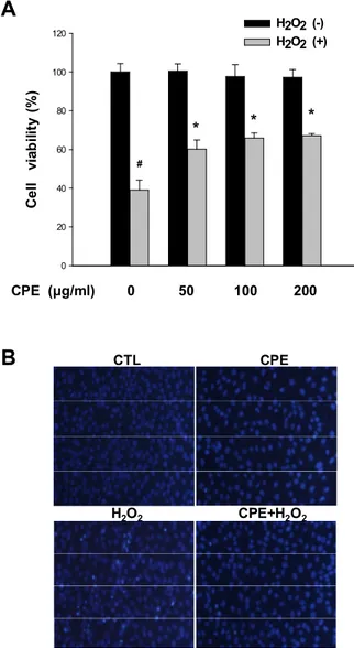
3.1. Neuroprotective effects of CPE against H2O2-induced oxidative neurotoxicity
The neuroprotective effects of CPE against H2O2-induced oxidative neurotoxicity were evaluated via MTT assays (Fig. 1-A). HT-22 neurons were treated with H2O2 (1mM) for 12 h in the presence or absence of CPE. Pretreatment with various concentrations (50, 100 and 200 μg/ml) of CPE was carried out for 1 h prior to H2O2 stimulation. The CPE treatments markedly increased cell viability against H2O2-induced oxidative neurotoxicity in a dose- dependent manner, by 34.8±3.8%, 43.9±7.9% and 46±10.0% respectively, relative to the group treated with H2O2 alone. The CPE alone evidenced no cytotoxicity at concentrations below 200 μg/ml. The cell viability was slightly decreased in case of 400 mg/ml of CPE alone in lactate dehydrogenase (LDH) release assays or MTT assays (data not shown).
The degree of apoptosis was determined by nuclear staining with Hoechst 33342, a cell-permeant DNA-specific fluorescent dye (Fig. 1-B). The H2O2-treated cells demonstrated significant chromatin condensation, which is indicative of apoptosis. However, CPE (200 mg/ml) also markedly reduced H2O2-induced chromatin condensation, which is consistent with the cell viability data shown in Fig. 1-A.
Generally, ROS induces mitochondrial dysfunction and the subsequent opening of the mitochondrial permeability transition pore (mPTP). Mitochondrial cytochrome C is then released into cytoplasm through the mPTP, which evokes the activation of caspase 3. This protease, in turn, degrades several proteins including PARP, an important DNA repair enzyme. Regarding these apoptotic cascades, the results of Western blotting analyses demonstrated H2O2-induced reductions of procaspase-3 (Fig. 2-A) and poly PARP (Fig. 2-B), which are associated with caspase 3 activation and PARP cleavage. They were almost fully recovered by 64.9±5.9% and 52.2±11.3%, respectively in the CPE (200 mg/ml)-treated cells.
21
These results demonstrate that CPE exerts neuroprotective effects against H2O2-induced oxidative neurotoxicity.
3.2. Antioxidant activities of CPE
We investigated the antioxidant activities of CPE against H2O2-treated HT-22 neurons (Fig. 3). Various concentrations (50, 100, 200 and 400 mg/ml) of CPE were incubated for 2 h with DPPH in cell-free system. The amounts of DPPH radicals were spectrophotometrically determined, which indicate radical scavenging activities. The results of DPPH assays showed that CPE (50, 100, 200 and 400 μg/ml) have marked ROS-scavenging activity in cell-free system in a dose-dependent manner (Fig. 3-A). The fluorescence spectrometric data from DCF-DA assays revealed that CPE treatment (200 mg/ml, 1 h) significantly attenuated the intracellular ROS levels by 44.6±1.6% (Fig 3-B) in H2O2 (1 mM, 30 min)-stimulated neurons. Additionally, we evaluated the enzyme activities of catalases and SOD, which are important antioxidant intracellular enzymes. CPE treatment (200 mg/ml, 1 hr) protected HT-22 neurons from H2O2 (1 mM, 30 min)-induced deterioration of these enzyme activities by 81.9±4.5% and 98.5±27.7%, respectively (Fig 3-C and D).
22 0 20 40 60 80 100 120 H2O2 (-) H2O2 (+) C e ll vi ab ili ty ( % ) CPE (μg/ml) 0 50 100 200 # * * *
A
CTL CPEB
H2O2 CPE+H2O2Fig. 1. Neuroprotective effects of CPE against H2O2-induced oxidative neurotoxicity.
A. HT-22 neurons were treated with H2O2 (1 mM) for 12 h in the presence or absence of CPE (50, 100 and 200 mg/ml). CPE was pretreated for 1 hr before H2O2 stimulation. The neuroprotective effects of the CPE were evaluated using MTT cell viability assays. B. Chromatin condensation as an apoptotic nuclear appearance was observed under a fluorescent microscope after Hoechst 33342 staining. #, p<0.05 as compared to the untreated controls and *, p<0.05 as compared to the group treated with H2O2 alone. Scale bar, 100 mm.
23 0 20 40 60 80 100 120 CTL CPE H2O2 CPE+H2O2 P ro -c as p as e-3 ( % ) #
*
Procaspase-3 β-actinA
0 20 40 60 80 100 120 P A R P (% ) #*
B
PARP β-actin CTL CPE H2O2 CPE+H2O2Fig.2. Effect of CPE on procaspase-3 and PARP against H2O2-induced oxidative
neurotoxicity
The effects of CPE on procaspase-3 (A) and PARP (B) expression levels were determined via Western blotting analyses. Groups were compared with % of untreated control group. #,
p<0.05 as compared to the untreated controls and *, p<0.05 as compared to the group treated
24 0 200 400 600 800 1000 1200 CTL CPE H2O2 CPE+H2O2 # ** In tr a c e ll u la r R O S ( % ) B 0 10 20 30 40 50 60 70 C a ta la s e a c ti vi ty (U /m g ) # * CTL CPE H2O2 CPE+H2O2 C 0 10 20 30 40 50 S O D a c ti v it y ( U /m g ) # * CTL CPE H2O2 CPE+H2O2 D 0 10 20 30 40 CPE (μg/ml) 50 100 200 400 ** D P P H ra d ic a l s c a ve n g in g a c ti vi ty (% ) A ** ** **
Fig.3. Anti-oxidant activities of CPE.
A. Various concentrations (50, 100, 200 and 400 mg/ml) of CPE were incubated for 2 h with DPPH in cell-free system. The amounts of DPPH radicals were spectrophotometrically determined, which indicate radical scavenging activities. B. HT-22 neurons were treated with H2O2 (1 mM) for 30 min in the presence or absence of CPE (200 mg/ml). CPE was pretreated for 1 hr before H2O2 stimulation. Then intracellular ROS levels were detected using a spectrofluorometer using DCF-DA. C-D. The effects of CPE on enzyme activities of catalase (C) and SOD (D) were measured with a spectrophotometer. #, p<0.05 as compared to the untreated control group and *, p<0.05; **, p<0.01 as compared to the group treated with H2O2 alone.
25 3.3. Mild-depolarizing effects of CPE on ∆Ψm
We evaluated the effects of CPE on ∆Ψm in resting neurons using real-time fluorescence recording analyses. TMRM is a cell-permeant, cationic fluorescent probe that is sequestered by mitochondria in proportion to ∆Ψm (Scaduto and Grotyohann, 1999). The cells on poly-L-lysine-coated cover glasses were loaded with 100 nM TMRM for 30 min, washed for three times and mounted on a recording chamber of an epifluorescence inverted microscope Olympus IХ71 (Olympus, Japan). TMRM fluorescence values from individual cells were normalized to the value before drug treatment and traces shown in Fig. 4-A show average recordings of TMRM fluorescence intensities.
Treatment of TMRM-loaded cells with CPE induced a dose (100, 200 and 400 mg/ml)-dependent decrease in TMRM fluorescence intensities, indicating mild mitochondrial depolarization (Fig. 4). The mitochondrial uncoupler CCCP (10 mM) markedly decreased TMRM fluorescence intensity, which means total ∆Ψm dissipation. At 10 min-time point after drug treatment, the normalized TMRM fluorescent intensities were 71.7%, 67.6%, 62.2% , 41.3 %, 10.9% in control, CPE (100, 200 and 400 mg/ml) and CCCP (10 mM)-treated groups, respectively (Fig. 4-B). These data indicate that mitochondria-depolarizing activity of CPE was significantly smaller than that of the uncoupler CCCP. This CPE-induced partial depolarization of mitochondrial membrane should be fundamentally distinguished from ∆Ψm dissipation, which means a full-blown mitochondrial depolarization at the almost final stage in mitochondria-dependent apoptotic pathways. This mild mitochondrial depolarization has been recently evaluated as a novel mechanism of neuroprotection via inhibiting mitochondrial Ca2+ overload during neuronal insults (Garcia-Martinez et al., 2010; Sanz-Blasco et al., 2008; Storozhevykh et al., 2010).
JC-1 is another cationic fluorescent probe which exhibits ∆Ψm-dependent accumulation in the mitochondria and has different optical properties from TMRM (See ‘Materials and
26
methods’). Therefore, we tried to investigate CPE effects on ∆Ψm with JC-1 in addition to TMRM. As mitochondrial membrane depolarization is also indicated by a reduction in the JC-1 aggregate forms (red) to JC-1 monomers (green) ratio of JC-1 fluorescence intensities, we evaluated the effects of CPE on the JC-1 ratio using flow cytometric analyses (Fig. 5-B) and confocal microscopy (Fig. 5-A), in addition to real-time fluorescence measurement with TMRM (Fig. 4). HT-22 neurons were treated with CPE (200 mg/ml, 1 h), subsequently loaded for 30 min with JC-1 fluorescent indicator (2 mM) and analyzed with a flow cytometer. This flow cytometric data with JC-1 (Fig. 5) indicate that CPE treatment resulted in a partial depolarization of the mitochondrial membrane, consistent with ∆Ψm data using TMRM (Fig. 4). The confocal microscopic images for ∆Ψm visually demonstrated the reduction of red to green ratio of JC-1 fluorescence intensities after treatments of CPE (Fig. 5-A), consistent with quantitative flow cytometric data with JC-1 (Fig. 5-B).
27
A
CTL CPE 100 μg/ml CPE 200 μg/ml CPE 400 μg/ml CCCP 10 μM min Δ Ψm T M R M F lu o re sc en ce ( % ) 0 20 40 60 80B
** *** Δ Ψm T M R M F lu o re sc en ce ( % ) *** CPE (μg/ml) - 100 200 400 -CCCP (μM) - - - - 1028
Fig.4. CPE evokes a mild mitochondrial depolarization, as indicated in real-time measurement of ∆Ψm.
A. Real-time measurement of ∆Ψm was conducted with MetaFluor software using a cationic fluorescent probe TMRM that is sequestered by mitochondria in proportion to ∆Ψm. A decrease in TMRM fluorescence reflects mitochondrial depolarization. The cells on poly-L-lysine-coated cover glasses were loaded with 100 nM TMRM for 30 min, washed for three times and mounted on a recording chamber of an epifluorescence inverted microscope Olympus IХ71. Various doses (100, 200 and 400 mg/ml) of CPE were superfused on TMRM-loaded HT-22 neurons in recording chamber to evaluate CPE effects on ∆Ψm. TMRM fluorescence values from individual cells were normalized to the value before drug treatment and averaged (n=12 cells). Traces show average recordings of TMRM fluorescence intensities. B. Quantification of TMRM fluorescence. At 10 min-time point after drug treatment, the normalized TMRM fluorescent intensities were compared among controls, CPE (100, 200 and 400 mg/ml) and CCCP (10 mM)-treated groups. **, p<0.01; ***,
29
3.4. Differential effects of single compounds of CPE on ∆Ψm
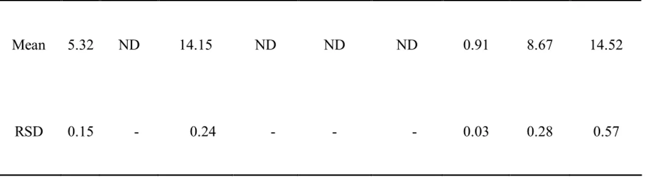
The major flavonoids of CPE (Table. 1) were investigated to explore single compounds responsible for CPE effects on ∆Ψm. Since HPLC analyses showed that CPE contained abundant flavonoids, such as rutin (5.32 mg/g), hesperidin (14.15 mg/g), nobiletin(8.67 mg/g), tangeretin (14.52 mg/g), sinensetin (0.91 mg/g).
The normalized ratios of red (JC-1 aggregate) to green (JC-1 monomers) fluorescence intensities (Fred/Fgreen ratio) of JC-1 were 56.6±4.5%, 73.8±0.6%, 67.9±0.6%, 78.8±1.7%, 98.2±1.9% and 95.7±6.1%, respectively in CCCP (10 mM, 30 min), CPE (200 mg/ml, 1 hr), nobiletin (50 mM, 1 h), tangeretin (50 mM, 1 h), rutin (50 mM, 1 h), and hesperidin (50 mM, 1 h)-treated groups compared to control group (Fig. 2-A). This flow cytometric data indicate that only polymethoxylated flavones such as nobiletin and tangeretin were capable of inducing a mild mitochondrial depolarization, while rutin as flavonol glycosides and hesperidin as flavanone glycosides did not affect on ∆Ψm. These results suggest that single compounds responsible for CPE effects on ∆Ψm are polymethoxylated flavones such as nobiletin and tangeretin.
30
Table 1. Flavonoids content (mg/g) contained in CPE.
Flavonoid Rutin Naringin Hesperedin Quercetin Naringenin Hesperetin Sinensetin Nobiletin Tangeretin
Mean 5.32 ND 14.15 ND ND ND 0.91 8.67 14.52
RSD 0.15 - 0.24 - - - 0.03 0.28 0.57
RSD, relative standard deviation (%) ND, not detected
The numbers are the means calculated from three experiments
31 0 20 40 60 80 100 120 *** *** *** *** JC -1 Fred / F g re en ( % o f c o n tr o l) B A JC-1 monomers Green JC-1 aggregates Red Overlay Control CCCP CPE C
32
Fig 5. Differential effects of single compounds of CPE on JC-1 Fred/Fgreen ratio indicative
of ∆Ψm.
A. The major flavonoids of CPE were investigated to explore single compounds responsible for CPE effects on ∆Ψm. HT-22 neurons were treated with CCCP (10 mM, 30 min), CPE (200 mg/ml, 1 hr), nobiletin (50 mM, 1 h), tangeretin (50 mM, 1 h), rutin (50 mM, 1 h), and hesperidin (50 mM, 1 h), subsequently loaded for 30 min with JC-1 (2 mM). The red (JC-1 aggregates) to green (JC-1 monomers) fluorescence ratio of JC-1 were measured using flow cytometry analyses to evaluate the effects on ∆Ψm and normalized compared to values in untreated control group. A decrease in JC-1 Fred/Fgreen ratio indicates mitochondrial depolarization. ***, p<0.001 as compared to controls. B. The representative confocal microscopic images for ∆Ψm were shown by the JC-1 dual emission of red and green fluorescences. The upper panels for JC-1 green fluorescent emission, the middle panels for JC-1 red fluorescent emission, and the lower panels for the overlay of green and red ones. Scale bar, 100 mm. C. Chemical structures of major flavonoids contained in CPE.
33
3.5. Inhibitory effects of CPE on H2O2-induced overload of Ca2+ in both the cytosol and
the mitochondria
Ca2+ deregulation of intracellular Ca2+ homeostasis is known to be involved in pathological processes such as oxidative neurotoxicity (Feissner et al., 2009). Therefore, we investigated the regulatory effect of CPE (10, 100, 200 and 400 μg/ml) on Ca2+ deregulation in H2O2 (1 mM, 30 min)-induced oxidative neurotoxicity. Flow cytometric analyses were accomplished using Fluo-4 AM and Rhod-2 AM fluorescent indicators for cytosolic and mitochondrial Ca2+,respectively. H
2O2 treatment remarkably increased both cytosolic and mitochondrial Ca2+ levels ([Ca2+]
i and [Ca2+]m) and the H2O2–induced increases in Ca2+ levels were significantly attenuated by CPE treatment (200 mg/ml) in both the cytosol and the mitochondria, by 26.6±0.7% and 27.0±1.7%, respectively (Fig. 5-A). These inhibitory effects of CPE (10, 100, 200 and 400 μg/ml) on both cytosolic and mitochondrial Ca2+ levels were dose-dependent, showing a bell-shaped dose-response relation. The maximal inhibitions were shown in 200 μg/ml of CPE treatment (Fig. 5-B). Fig. 5-C provides representative confocal microscopic images demonstrating the inhibitory effect of CPE on H2O2-induced mitochondrial overload of Ca2+, indicated by rhod-2 fluorescence.
34 0 50 100 150 200 250 300 F lu o -4 f lu o re sc en ce (% ) CTL CPE H2O2 CPE+H2O2 ** # B CTL CPE H2O2 CPE+H2O2 A
Fig.6. Inhibitory effects of CPE on cytosol Ca2+ overload.
A and B After pretreatment of CPE for 1 h, HT-22 neurons were treated with H2O2 (1 mM, 30 min) in the presence or absence of CPE (200 mg/ml), then loaded for 30 min with Fluo-4 AM (5 mM) for the measurement of cytosolic Ca2+ level. The fluorescent intensities were immediately evaluated via flow cytometric analyses. The inhibitory effects of CPE on H2O2 -induced overload of Ca2+ in cytosol. Groups were compared with % of untreated control group. #, p<0.05 as compared to the untreated controls and *, p<0.05; **, p<0.01, as compared to the group treated with H2O2 alone.
35 0 20 40 60 80 100 120 140 160 180 200 *** # R h o d -2 F lu o re sc en ce (% o f co n tr ol )
A
Control CPE H2O2 CPE+H2O2
10 100 200 400 0 5 10 15 20 25 30 35 *** * *** *
B
CPE (μg/ml) 10 100 200 400 In h ib it io n ( % ) Control CPEC
H2O2 CPE+H2O236
Fig.7. Inhibitory effects of CPE on H2O2-induced mitochondrial calcium overload.
A. After pretreatment of CPE for 1 h, HT-22 neurons were treated with H2O2 (1mM, 30 min) in the presence or absence of CPE (200 mg/ml), then loaded for 30 min with Rhod-2 AM (5 mM) for the measurement of mitochondrial Ca2+ levels. The fluorescent intensities were immediately evaluated via flow cytometric analyses. B. Dose (10, 100, 200 and 400 μg/ml)-response relations of the inhibitory effects of CPE on H2O2-induced overload of Ca2+ in mitochondria. C. The representative confocal microscopic images demonstrating the inhibitory effect of CPE on H2O2-induced mitochondrial overload of Ca2+, indicated by rhod-2 fluorescence. Nuclear counterstaining was accomplished with DAPI. Groups were compared by % of untreated control group. #, p<0.05 as compared to the untreated controls and *, p<0.05; ***, p<0.001, as compared to the group treated with H2O2 alone. Scale bar, 100 mm.
37
4. Discussion
In this study, we investigated the neuroprotective mechanism of CPE against oxidative neurotoxicity. Treatment with CPE markedly increased neuronal viability in an H2O2 -induced oxidative neurotoxicity model using HT-22 cells (Fig. 3-A and B). Additionally, CPE treatment inhibited neuronal apoptosis cascades including caspase 3 and PARP (Fig.3-C and D). CPE treatment demonstrated the robust anti-oxidant activity of ROS scavenging (Fig. 4), and significantly blocked H2O2–induced mitochondria Ca2+ overload in both the cytosol and the mitochondria (Fig. 5) and induced a mild depolarization of the mitochondrial membrane (Fig. 1 and 2). Taken together, the results of the present study suggest dual mechanisms underlying the neuroprotective effects of CPE: these dual mechanisms involves both anti-oxidant activity and partial blockade of mitochondrial Ca2+ overload due to the mild depolarization of the mitochondrial membrane.
The flow cytometric data indicate that only polymethoxylated flavones (PMFs) such as nobiletin and tangeretin were capable of inducing a mild mitochondrial depolarization, while rutin as flavonol glycosides and hesperidin as flavanone glycosides did not affect ∆Ψm. These results suggest that single compounds responsible for CPE effects on ∆Ψm are PMFs such as nobiletin and tangeretin. PMFs, are flavones bearing two or more methoxy groups on their basic benzo-c-pyrone (15-carbon, C6–C3–C6) skeleton with a carbonyl group at the C4 position (Li et al., 2009). PMFs were shown to the most potentially inhibit NO release in lipopolysaccharide(LPS)-stimulated RAW 264.7 murine macrophage cell line out of Citrus peel flavonoids, suggesting stronger anti-inflammatory activity than any other Citrus peel flavonoids (Choi et al., 2007). PMFs are the most commonly investigated due to their outstanding biological activities out of Citrus flavonoids. The possible targets by which PMFs regulate ∆Ψm might be mitochondrial channels/transporters, or possibly the components of electron transport chains. The underlying mechanism how PMFs induces
38
mild mitochondrial depolarization remains unclear, and should be elucidated in future studies. The balance of ROS levels between generation and scavenging is crucial to cellular functions and homeostasis. However, excessively high levels of ROS activate the apoptotic cascades through mitochondrial dysfunction. ROS-induced mitochondria dysfunction involves ATP depletion, ∆Ψm dissipation, MPTP opening, and cytosolic cytochrome C release. These cascades activate caspase 9 and caspase 3 in turn, and then the activated caspase 3 degrades important proteins, including the DNA repair-related enzyme PARP. These apoptotic processes are referred to as the intrinsic pathway or the mitochondria-dependent pathway.
Hydrogen peroxide (H2O2) is a freely diffusible form of ROS through the cell membrane and is produced by many intracellular reactions, and is implicated in apoptosis in various cells. H2O2 treatment is the most commonly used experimental model of oxidative cytotoxicity. H2O2 treatment induces excess generation of ROS such as the superoxide (O2-·) and hydroxyl radical (·OH), which subsequently result in mitochondrial dysfunction and ATP depletion(Ishimura et al., 2008). Intracellular ATP depletion induces the dysfunction of ATP-dependent ionic pumps. Dysfunction of Na+-K+ ATPases evokes cell membrane depolarization and increases [Ca2+]
i due to activation of voltage-gated Ca2+ channels and a reversed operation of the Na+- Ca2+ exchanger (Wang et al., 2003). In addition, sarco-endoplasmic reticulum Ca2+ ATPases (SERCA), and plasma membrane Ca2+ ATPases (PMCA) are susceptible to ATP depletion, all of which are involved in the increase of [Ca2+]
i (Clapham, 1995). It is known that the cytosolic Ca2+ overload ultimately results in the uptake of Ca2+ into the mitochondria (Rizzuto et al., 2009). Therefore, H
2O2 treatment increases both [Ca2+]
i and [Ca2+]m, as shown in Fig. 5.
The physiological level of mitochondrial Ca2+ regulates intracellular Ca2+ signals as well as the rate of ATP synthesis through the tricarboxylic acid (TCA) cycle-associated metabolic enzymes (i.e., pyruvate, alpha-ketoglutarate and isocitrate dehydrogenases)(Jouaville et al., 1999; Lasorsa et al., 2003). However, excess mitochondrial Ca2+ overload is crucial to
39
trigger neuronal apoptosis by opening the mPTP, evoking cytochrome C release, activating caspase cascades, and ultimately inducing cell death (Pinton et al., 2008).
In this study, we demonstrated that CPE treatment significantly reduced H2O2-induced increases in the levels of cytosolic and mitochondrial Ca2+ (Fig. 5). The inhibitory effects of CPE on cytosolic Ca2+ levels are attributed principally to their ROS scavenging activity, because excess intracellular ROS-induced mitochondrial dysfunction increases cytosolic Ca2+ levels. It is known that the cytosolic Ca2+ overload ultimately results in the uptake of Ca2+ into the mitochondria. However, the inhibitory effect of CPE on mitochondrial Ca2+ overload is difficult to explain with single mechanism because inhibiting of H2O2-induced cytosol Ca2+ overload finally reduces mitochondrial Ca2+ overload.
Taken together, we suggest that CPE may exert neuroprotective activities against oxidative neurotoxicity implicated in neurodegenerative diseases. The neuroprotective mechanism is associated with the intrinsic ability of CPE to induce mild mitochondrial depolarization as well as with its anti-oxidant activities. These findings might raise the recent issue of a novel role of mild mitochondrial depolarization in neuroprotection.
40
PART
Ⅱ
A novel neuroprotective mechanism of
indomethacin against excitotoxicity
via mitochondrial K
ATP
in the primary
41
1.
Introduction
Indomethacin is a NSAID commonly used to reduce fever, pain and swelling. It was a potent, nonselective inhibitor of the cyclooxygenase enzymes (COX-1 and COX-2) and is a powerful anti-inflammatory agent and also exhibits anticancer activity as suggested by a report demonstrating that indomethacin significantly increased the lifespan of terminally ill patients suffering from a range of cancers (Lundholm et al., 1994). The neuroprotective effects of the minocycline, NSAIDs, and KB-R7943 (Storozhevykh et al., 2010) against excitotoxic insults have recently been proposed to be attributable to their intrinsic ability to induce mitochondrial depolarization in resting neurons. ∆Ψm is evaluated as a potent target to modulate mitochondrial Ca2+ overload which might be the triggering point of neuronal cell death.
Steady-state Ca2+ level within mitochondria of living cells have many physiological functions. 1) Activation of dehydrogenases in TCA cycle, respiratory chain and ATP synthesis. 2) Buffering local cytosolic Ca2+ rises. 3) Inducing mPTP opening and cell death (Pizzo et al., 2012). But in many neuronal insults, mitochondria calciumoverload induces the collapse of ∆Ψm, opening of the mPTP and release of pro-apoptotic factors to trigger cell death. The exact molecular composition of mPTP is unknown, but possible regulation components, such as adenine nucleotide transporter (ANT), voltage-dependent anion channel (VDAC), and cyclophilin D were shown to be directly or indirectly involved in mPTP opening(Crompton, 1999).
Owing to a growing body of evidences, mitochondrial potassium channels, especially mitKATP, are believed to be involved in cytoprotection of injured cardiac and neuronal tissues (Liu et al., 1999) (Garlid et al., 2003). KATP is a potassium channel which is inactivated by ATP. The molecular identity of the KATP channel is still unclear. It is believed that KATP channels are composed of a pore-forming subunit (Kir6.2 subunit) and a mitochondrial sulfonylurea receptor (mitoSUR) (Choma et al., 2009). It is located in various parts of the
42
cell, including the plasma membrane (sKATP) and inner mitochondrial membrane (mitoKATP). Function of KATP channel opening will depend upon location: activation of sKATP leads to hyperpolarization of cells while opening of mitoKATP causes depolarization of mitochondria. The mitoKATP channel was first identified using the patch clamp technique in rat liver mitochondria (Inoue et al., 1991). Previously, the properties of brain mitochondrial mitoKATP channels were also determined using isolated mitochondria (Bajgar et al., 2001). It was demonstrated that a physiological role of the mitoKATP channels is to buffer potential perturbations of matrix volume and the intermembrane space so that ATP production and transport are at optimal levels for cellular needs (Busija et al., 2004).
In the present study, we investigated the neuroprotective effect of indomethacin, a non-steroidal anti-inflammatory drug on glutamate-induced excitotoxicity model. And we also examined if 5HD, a specific mitKATP channel blocker, abolishes the protection by indomethacin in cultured cortical neuron.
43
2. Materials and methods
2.1. Materials
Minimum Essential Medium (MEM), Neurobasal Medium, fetal bovine serum (FBS), penicillin/streptomycin, B-27 Serum-Free Supplement were purchased from Gibco BRL(Grand Island, NY, USA). Tetramethylrhodamine ethyl ester (TMRE), Fura-2 acetoxymethyl ester (Fura-2 AM), rhod-2 acetoxymethyl ester (Rhod-2 AM), Imag-It live Mitochondrial Transition Pore Assay Kit and MitoSOX Redwere purchased from Invitrogen (Carlsbad, CA, USA). We purchased anti-cytochrome C from Santa Cruz Biotechnology (Santa Cruz, CA, USA) and Alexa 488 a rabit IgG were purchased from Invitrogen (Invitrogen Barcelona, Spain). All other reagents were purchased from Sigma-Aldrich (St Louis, MO, USA), unless indicated otherwise.
2.2. Cell culture
All animal experiments were approved by the Institutional Review Board (IRB) of animal, Jeju University College of Medicine, and the procedure was carried out in accordance with the guidelines of the IRB. Primary cortical neuronal culture was obtained from 20-day-old embryonic Sprague-Dawley rats. The cortex was dissected and placed in ice-cold Ca2+ free Normal Tyrode solution. Then the cortex was removed to Plating Media (MEM supplemented with 10% FBS, 0.45% Glucose, 1 mM sodium pyruvate and penicillin/streptomycin) and dissociated by glass pasteur pipettes. Then cortical neuron cells were plated onto poly-L-lysine coated glass coverslips and maintain at 37°C in a humidified atmosphere of 5% CO2. After six hours later, aspirate plating medium and add neurobasal medium to each well and change the media twice for week. In some experiment, at 4th day,
44
cultures were treated with 1 μM arabinofuranosyl cytidine (Ara-C) to prevent glial proliferation. Neurons at 10-13 DIV were used for the experiments.
2.3. Immunofluorescence microscopy and labeling of mitochondria
Cells were treated under the various experimental conditions and then fixed in 4% paraformaldehyde for 30 min and permeabilized in PBS containing 0.5% Triton X-100 in PBS for 10 min twice at room temperature. Following blocking incubation in 5% normal goat serum in PBS for 1 h. Neurons were incubated at room temperature for 2 h with anti-Tuj-1 or anti-NeuN (1:200) in 5% NGS in PBS. Then the coverglass were exposed to green-fluorescent Alexa Fluor 488 anti-rabbit IgG (1:200) for 1 h at room temperature. After washing and mount with Prolong antifade kit solution with DAPI. In some experiment, 0.2 μM Mito Tracker Red was been used to labeling of mitochondria. The fluorescence images were captured by an epifluorescence inverted microscope Olympus IХ71 (Olympus, Japan) and MetaMorph (Molecular Devices, CA, USA) software.
2.4. Measurement of cell viability
MTT [3-(4,5-dimethylthiazol-2-yl)-2,5-diphenyl tetrazolium bromide] was used to examine the effect on cell viability, as previously described (Cui et al., 2010). After treatment, MTT was added to the cultured medium and incubated for 2 h at 37°C. Then, medium was removed, and DMSO was added to dissolve formazan. After gently shaking, absorbance was subsequently read at 540 nm using a microplate reader (Model 550, Bio-Rad, USA).
45 2.5. Real-time measurement of ∆Ψm
The cells on poly-L-lysine-coated cover glasses were loaded with 25 nM TMRM for 10 min, washed for three times and mounted on a recording chamber of an epifluorescence inverted microscope Olympus IХ71 (Olympus, Japan). Digitized fluorescence images were acquired at 30 s intervals with a cooled-charged device (CCD) camera (Cascade, Roper Scientific, USA), and analyzed in a personal computer using Metafluor software (Molecular Devices, CA, USA). During recording, the cells were superfused with Normal Tyroid solution by fast flow system using tubing. Normal Tyrode solution contained (mM): 145 NaCl, 1.3 MgCl2, 2 CaCl2, 5 KCl, 10 Hepes, 10 glucose. Drug application was accomplished by changing perfusion medium.
2.6. Dual recording for Cytosolic and mitochondrial Ca2+
For simultaneous fluorescence imaging of cytosolic Ca2+, neurons were loaded for 45 min in the cell culture medium at 37°C with Fura-2 AM (5 μM) and Pluronic F127 (0.1%) . For mitochondrial calcium measurement, Rhod-2 AM (2 μM) was used and incubated the cells at 4°C for 45 min. After washing, the cell was transfer to the recording chamber for acquirement. Fluorescence was excited with the high speed filter changer of 340 nm, 380 nm, 548/20 nm wavelengths using a rapidly tunable (<1.2msec wavelength change) with Lamber DG4 (Sutter Instrument, Novato, CA) and D510/80m emitter filter (Chroma Technology). Digitized fluorescence images were acquired at 6 s intervals with a cooled-charged device (CCD) camera (Cascade, Roper Scientific, USA), and analyzed in a personal computer using Metafluor software (Molecular Devices, CA, USA). The changes in cytosolic and mitochondrial calcium were normalized to the baseline fluorescence at the beginning.
46 2.7. DPPH radical scavenging assay
Various concentrations of CPE (10 ml) were added to 190 ml (0.15 mM in ethanol) of DPPH (1,1-diphenyl-2-picrylhydrazyl radical) and vigorously mixed. The mixture was incubated for 1 h in darkness at room temperature, and the absorbance was read at 517 nm using a microplate reader (Sunrise, Tecan, Austria). The percentage level of DPPH scavenging was calculated according to the following formula: % Radical Scavenging=[(A-As)/A]ⅹ100, in which A is the absorbance of DPPH and As is its absorbance after citrus peel extract treatment.
2.8. Intracellular reactive oxygen species (ROS) measurement
The cells were seeded on 96-well tissue culture plates at 2ⅹ104 cells/well and pretreated for 30 min with CPE followed by 1 mM H2O2 for an additional 30 min. After the addition of 50 mM of DCF-DA (2′,7′-dichlorodihydrofluorescein diacetate), fluorometric analysis was conducted at an excitation/emission wavelength of 485 nm/535 nm using a microplate reader (Spectra Fluor, Tecan, Austria).
2.9. Real-time measurement of mitochondrial superoxide generation
The cells on poly-L-lysine-coated cover glasses were loaded with 5 μM MitoSOX Red for 10 min, washed and mounted on a recording chamber of an epifluorescence inverted microscope Olympus IХ71 (Olympus, Japan). Digitized fluorescence images were acquired at 30 s intervals with a cooled-charged device (CCD) camera (Cascade, Roper Scientific, USA), and analyzed in a personal computer using Metafluor software (Molecular Devices, CA, USA).
47
2.10. Mitochondrial permeability transition pore (mPTP) assay
mPTP opening was assessed directly by the calcein/cobalt method according to the manufacturer's protocol. Calcein AM a nonfluorescent esterase substrate, can passively diffuse into the cells and accumulates in cytosolic compartment, including mitochondria. After loading, intracellular esterase cleaves the acetoxymethyl esters to liberate fluorescent dye calcein, which does not cross the mitochondrial or plasma membranes. To quench fluorescence of cytosolic calcein, CoCl2 was added to the media to maintain only mitochondrial calcein fluorescence. Cells were co-loaded with calcein-AM 1 μM and CoCl2 1 mM for 30 min at 37°C. The cells then subjected to inverted microscope Olympus IХ71 (Olympus, Japan) and MetaMorph (Molecular Devices, CA, USA) software.
2.11. Statistics analysis
Data are expressed as mean value ± standard error of the mean (SEM). Statistical analyses were conducted using Student’s t-test. The differences between groups were regarded as statistically significant when p < 0.05.
48
3. Results
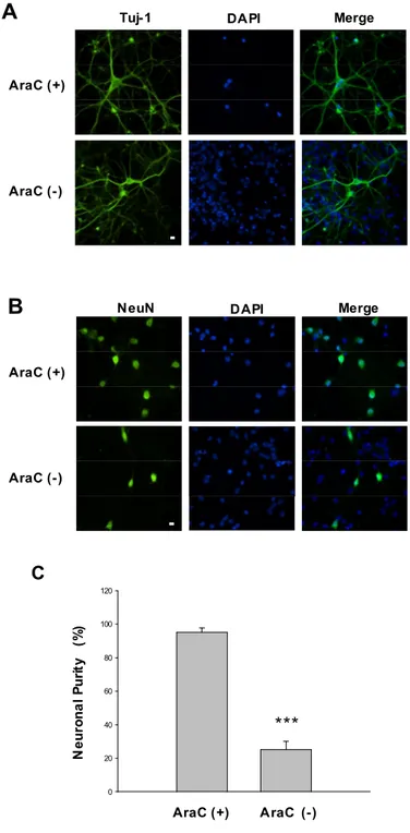
3.1. Purity of primary cortical neuron
Cells, at 10-15 DIV were used in our study. In real time recording, neurons were easily distinguishable from glia in morphology. Under microscopy, neurons had smooth rounded soma and distinct processes and lay just above the focal plane of the glial layer. To get a pure neuron in some experiments except real time recording, cultures were treated with 1 μM arabinofuranosyl cytidine (Ara-C) to prevent glial proliferation. Therefore we investigated the purity of neurons in our culture system using immunofluorescence microscopy using anti-Tu-j1 (Fig. 8-A) and anti-NeuN (Fig. 8-B) which was specific neuronal microtubules marker and neuronal nuclear marker. Neurons were counted in the presence or absence of Ara-C and their purities of neuron were 95.2±2.5 % and 25.1±5.0 % respectively (Fig. 8-C).
3.2. Mitochondrial KATP channel blockade abolishes neuroprotective effect of
indomethacin on glutamate-induced neurotoxicity in primary cortical neuron
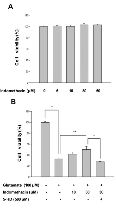
To induce a calcium dependent excitotoxic cell death, we exposed cultured cortical neurons to glutamate (100 mM) and glycine (10 mM) for 30 min. Treatments were terminated by washing cells three times before incubating with regular medium. Cell viability was analyzed 24 h later via MTT assays. Neurons were treated with glutamate(100 mM) for 30 min in the presence or absence of indomethacin. Pretreatment with various concentrations (10, 30) of indomethacin were carried out for 5 min prior to glutamate expose. Indomethacin treatments markedly increased cell viability against glutamate-induced neurotoxicity to 41.6±4.0%, and 50.0±5.6% respectively in a dose- dependent manner, relative to the group
49
treated with glutamate alone was 32.8±1.7% (Fig. 9-B). Indomethacin alone evidenced no cytotoxicity at concentrations below 50 μM (Fig. 9-A). Treatment of 5HD (500 μM), a specific mitoKATP channels blocker partially abolished these protective effects of indomethacin. These data indicate that mitoKATP may be regulate neuroprotective effect of indomethacin.
3.3. Mitochondrial KATP channel blockade abolishes neuroprotective effect of
indomethacin on glutamate-induced ROS generation
In the next study, we investigated the antioxidant activities of indomethacin. Various concentrations (1, 10, 50 and 100 mM) of indomethacin were incubated for 2 h with DPPH in cell-free system. The amounts of DPPH radicals were spectrophotometrically determined, indicating radical scavenging activities. The results of DPPH assays showed that indomethacine did not have a ROS-scavenging activity in cell-free system (Fig. 10-A). While the fluorescence spectrometric data from DCF-DA assays revealed that indomethacin treatment (30 mM) significantly attenuated the intracellular ROS levels (Fig 10-B) in glutamate (100 mM)-stimulated neurons. Therefore, these results indicate that neuroprotective effect of indomethacin is not caused by antioxidant activity of indomethacin.
50 C 0 20 40 60 80 100 120 N e u ro n a l P u ri ty (% ) *** AraC (+) AraC (-) AraC (+) AraC (-)
Tuj-1 DAPI Merge
A
NeuN DAPI Merge
AraC (+)
AraC (-)
B
Fig. 8. Neuronal purity in primary cortical neuron cultures. Cultures were treated with 1
μM arabinofuranosyl cytidine (Ara-C) to prevent glial proliferation at 4th days. A-B. Cultured primary cortical neurons were marked with Tuj-1 and NeuN by immunofluorescence. C. Quantification of Ara-C positive cell and negative cell. Blue colors show nuclei stained with DAPI and green colors show the fluorescence of Tuj-1 and NeuN. Scale bar, 10 mm.