교신저자: 박수영, 700-721 대구광역시 중구 삼덕2가 50, 경북대학교 의학전문대학원 내과학교실
Soo Young Park, M.D., Department of Internal Medicine, Kyungpook National University School of Medicine 50 Samduk 2-Ga, Jung-gu, Daegu 700-721, Korea
Tel : +82-53-200-5516 E-mail : [email protected]
Abstract
Acute gastric variceal bleeding is one of the most serious complications in portal hypertension, and is associated with high mortality and morbidity. Endoscopic variceal obturation (EVO) using HistoacrylⓇ (n-butyl-2- cyanoacrylate) has been accepted as an effective hemostatic procedure in acute gastric variceal bleeding.
However, EVO is not a widely performed because of technical difficulties and complications such as mucosal ulceration, perforation, and systemic embolism. Herein, we report a patient who developed hepatic failure caused by portal vein occlusion by HistoacrylⓇ injection for management of gastric variceal bleeding.
Key Words :
Gastric variceal bleeding, HistoacrylⓇ, Portal vein occlusion, Splenic vein occlusion Department of Internal Medicine, Kyungpook National University School of Medicine,Daegu, Korea
Eun Jeong Kang, M.D., Soo Young Park, M.D., Yu Rim Lee, M.D., Chang Yeon Kim, M.D., Sun Young Ahn, M.D., Jung Gil Park, M.D., Hyun Seok Lee,M.D., Won Young Tak, M.D.,
Young Oh Kweon, M.D.
A Case of Portal Vein and Splenic Vein Occlusion after Endoscopic Variceal Occlusion Therapy in Gastric Variceal Bleeding
강은정·박수영·이유림·김창연·안선영·박정길·이현석·탁원영·권영오 경북대학교 의학전문대학원 내과학교실
위정맥류 출혈환자에서 내시경적 정맥류 폐색술 후 발생한 간문맥 및 비장정맥 폐색 1례
서 론
위정맥류 출혈은 문맥 고혈압 환자의 비교적 흔한 합병증으로 식도 정맥류 출혈에 비해 높은 사망률이 보고되고 있다[1]. 현재 위정맥류 출혈의 치료로 내시경적 경과요법, 결찰요법, 경피경간 색전술, 경경정맥 간내 문맥순환 단락술(TIPS) 및 외과적 수술 등이 시행되고 있다. 히스토아크릴(n-b u t y l-2- cyanoacrylate)은 조직접합체의 일종으로 1986년 Soehendra 등이 처음으로 정맥류의 치료에 사용한 이래 일부에서 위정맥류 치료로 보고되고 있다[2-4].
그러나 치료에 의한 합병증으로 흉통, 출혈, 치료부위 궤양, 천공, 식도협착, 뇌색전증, 폐색전증, 복부대동맥 및 간문맥 색전증, 비장경색 등이 보고된 바 있으며 시 술 의 난 이 도 , 시 술 시 환 자 의 상 태 , 시 술 후 합병증으로 인해 현재 널리 시술되고 있지 않다. 이에 저 자 등 은 히 스 토 아 크 릴 을 주 입 하 여 위 정 맥 류 지혈술을 시행한 후 히스토아크릴에 의한 비장정맥 및 간문맥 폐색 및 이로 인한 간부전 발병에 대한 증례를 경험하였기에 보고하고자 한다.
증 례
5 8 세 남 자 환 자 가 토 혈 과 흑 색 변 을 주 소 로 응급실로 내원하였다. 환자는 5년 전 당뇨, 3년 전 알코올성 간경변증을 진단받은 후 외래에서 추적관찰 중이었다. 내원 당시 혈압 150/80 mmHg, 맥박 99회/분, 체온 36.5℃, 호흡 18회/분이며 신체검사에서 급성병색 이었고 결막은 창백하였다. 초기 말초혈액 검사에서 백혈구 6,300/mm3, 혈색소 9.1 g/dL, 헤마토크릿 27.9%, 혈소판 240,000/mm3 이었으며 혈청 생화학 검사에서 AST 23 IU/L, ALT 13 IU/L, ALP 67 IU/L, total bilirubin 0.48 mg/dL total protein 5.0 g/dL, albumin 2.6 g/dL, BUN 14.4 mg/dL, Cr 0.7 mg/dL PT 11.9 sec, aPTT 23.5 sec였다.
내원 후 시행한 상부위장관 내시경 검사 소견 상 식도 하부에 1 c m 이하 크기가 작은 정맥류가 관 찰 되 었 으 며 , 위 저 부 에 1 . 5 c m 크 기 의 정맥류(Gastroesophageal varix type 2) 및 출혈을 시사하는 붉은 반점(r e d s p o t)이 관찰되었다.
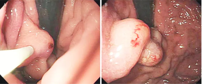
내시경적 지혈을 위해 붉은 반점을 동반한 위정맥류에 히스토아크릴 5 m L와 리피오돌 5 m L를 섞은 히스토아크릴-리피오돌 혼합액 총 10 mL를 위정맥류
Fig. 1. Endoscopic variceal obturation was performed by injecting a total of 10 mL mixed solution of Histoacryl® and lipiodol. A: Injecting catheter was punctured distal to bleeding site on gastric varix. B: After injection of 10 mL mixed solution of Histoacril® and lipiodol, pale dilated gastric varix was observed with extravasation of lipiodol from bleeding site.
A B
크기가 팽창하고 붉은 반점 을 통해 히스토아크릴 혼합액이 새어나올 정도로 주사하였다(Fig. 1). 시술 후 관찰한 내시경소견 상 더 이상의 출혈은 관찰되지 않았으며, 히스토아크릴을 주입한 위 정맥류는 크기가 팽 만 하 였 으 며 , 창 백 한 점 막 및 출 혈 부 위 로 히스토아크릴을 관찰할 수 있었다. 이후 환자는 재출혈 없이 5일 후 퇴원하였다.
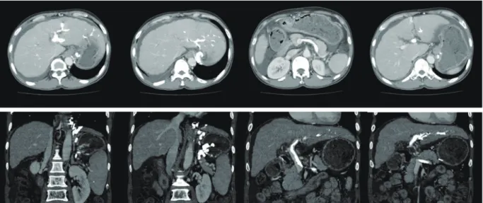
환자는 퇴원 후 3주간 간헐적 발열이 발생하였으며 지방병원 입원 후 발열의 원인을 조사하였으나 원인을 밝혀내지 못하였다. 지속된 발열 및 복부 팽만으로 환자는 입원하였으며 입원 당시 혈압 95/50 mmHg, 맥박 97회/분, 체온 37.8℃, 호흡 18회/분이었으며 시행한 혈액검사에서 백혈구 14,330/mm3, ESR 89 mm/h, C-Reactive Protein (CRP) 7.7 mg/dL, AST 16 IU/L, ALT 6 IU/L, total bilirubin 0.66 mg/dL, total protein 4.0 g/dL, albumin 1.8 g/dL, BUN 8.6 mg/dL Cr 0.84 mg/dL, PT 13.5 sec, aPTT 32.5 sec로 백혈구 증다증 및 ESR, CRP 상승소견을 보였다. 내원 후 시행한 복부 조영증강 컴퓨터 단층촬영 상 비장정맥과 간문맥이 히스토아크릴로 판단되는 고음영(high d e n s i t y) 물질로 막혀있었으며 복수가 새로이 관찰되었다(Fig. 2). 복수천자검사 상 자발성 세균성 복막염 소견은 보이지 않았으며 흉부 컴퓨터 전산화
단층촬영, 객담검사, 요검사, 혈액배양 검사 등에서 발열을 일으킬만한 다른 원인은 발견되지 않았다.
입원 후 광범위 3세대 세팔로스포린 항생제 치료와 이뇨제 복용 시행 후 38℃ 이상의 고열은 소실되었으나 37.4℃ 정도의 미열은 지속되는 상태로 퇴원하였다.
환자는 퇴원 후에도 4개월간 수차례 발열 및 복부팽만으로 외래 진료 및 입원하여 항생제 치료 및 복 수 천 자 를 지 속 하 였 으 나 발 열 은 간 헐 적 으 로 지속되었으며, 치료 6개월 후 이뇨제와 반복적인 복수천자로 인한 고질소혈증(44.3 mg/dL)과 혈청 크레아티닌 상승(2.3 mg/dL)이 발생하였다. 환자는 히스토아크릴에 의한 감염성 색전으로 비장정맥과 간문맥 폐색, 이로 인한 간문맥 고혈압의 악화로 내과적 치료에 더 이상 반응이 없는 난치성 복수로 생체 간이식을 시행하였으며 현재 성공적인 간이식 후 경과 관찰 중이다.
고 찰
위정맥류는 위장 내 확장된 점막하 정맥으로 문맥압 항진증 환자의 약 20%에서 발생하며, 2년내 출혈률은 25%로 알려져 있다[1]. 위정맥류 출혈은
Fig. 2. In abdominal computed tomogram followed-up one month later, both intrahepatic portal vein and extrahepatic portal vein was completely occluded by high density embolic materials which originated from gastric varices.
식도정맥류에 비해 발생빈도는 낮으나 한번 출혈하면 대량 출혈을 일으키며 사망률이 더 높을 뿐 아니라, 지혈에 성공한 경우에도 재출혈이 많이 발생하는 것으로 알려져 있다[5,6]. 위정맥류는 위정맥류의 분 류 에 따 라 치 료 법 을 달 리 하 는 데 위 소 만 부 정맥류(Gastroesophageal varix type 1)의 경우 식도정맥류의 치료와 같이 내시경적 정맥류 결찰술을 시행하며, 내시경적 정맥류 폐색술도 시행할 수 있다[7-9].
내 시 경 적 정 맥 류 폐 색 술 은 내 시 경 을 이 용 하 여 히스토아크릴과 같은 조직접착제를 정맥류 내로 주입하는 방법으로 위저부 정맥류(Gastroesophageal varix type 2)와 단독위정맥류(Isolated gastric varix)의 경우 내 시 경 적 정 맥 류 폐 색 술 을 우 선 적 으 로 시행한다[10,11]. 위신단락(Gastrorenal shunt)이 동반된 경우에는 풍선-폐쇄 역행 경정맥 폐쇄술 (Balloon occluded retrograde transvenous obliteration, BRTO)을 시행할 수 있는데 BRTO는 경경정맥 간내문맥 전신 단락술(t r a n s j u g u l a r intrahepatic portosystemic shunt, TIPS)이나 내시경적 정맥류 폐색술에 비해 재발이 드물고 거의 대부분의 위정맥류를 소실시킬 수 있다는 장점이 있어 위정맥류 출혈의 이차 예방에도 이용된다[12,13].
히스토아크릴은 조직접합체로서 혈액과 접촉하면 순간적으로 중합체로 변환되며 이 중합체는 혈관 내 색 전 물 이 되 어 정 맥 류 를 폐 색 시 키 기 때 문 에 순간적으로 지혈이 된다. 이후 이물반응에 의한 급성 혈관내막괴사가 일어나고 혈관벽의 괴사와 섬유화가 진행되어 정맥류를 폐쇄시킨다. 히스토아크릴은 주입 1주 후부터 중합체가 빠져나가기 시작하고 2-3개월 경과 후에는 정맥류에서 탈락되어 대부분 자연 배출되는데 급성 위정맥류 출혈에서 93.3 - 100%의 우수한 초기 지혈 성공률을 보고하고 있다[11,14,15]. 그러나 내시경적 정맥류 결찰술에 비해 내시경적 정맥류 폐색술은 내시경 수기가 어려우며, 초기에 지혈 효 과 는 우 수 하 나 반 복 적 인 주 입 으 로 정 맥 류 의 완전소실을 달성하기 어렵고 발열, 흉통 등의 경미한 합병증부터 식도협착, 재출혈, 궤양, 천공 등도 발생할 수 있으며, 드물게 뇌색전증, 폐색전증, 문맥 및 비장정맥 혈전증, 비장경색 등과 같은 전신적이고 심각한 합병증까지 초래될 수 있어 제한적으로
시술되고 있다[16-18].
히스토아크릴 주입에 의한 색전증 발생을 방지하기 위해서는 히스토아크릴 1회 주사량이 0.5 ml 이하로 하며, 주입량이 총 1.5-2 mL를 초과하지 않도록 주의하여야 한다. 이외 치료강도, 투여속도, 정맥류의 부분적 폐색과 측부 혈관의 발달여부 등이 히스토아크릴 색전증과 연관이 있다고 알려져 있다[18,19]. 본 증례에서는 응급실 도착 후 적절한 시간 내(3.5시간) 응급 내시경적 지혈술을 시행하였으나, 히스토아크릴 - 리 피 오 돌 혼합액의 주입량이 10 mL로 많은 양이 주입되었고 급 성 출 혈 의 처 치 는 성 공 하 였 으 나 , 이 에 대 한 합 병 증 으 로 간 문 맥 폐 색 및 감 염 성 색 전 이 발생하였다. 따라서 이러한 합병증의 위험도를 낮추기 위해서는 히스토아크릴을 이용한 내시경적 정맥류 폐색술은 경험이 많은 내시경 의사에 의해 시행되어야 하며 일정한 간격으로 복부 전산화 단층촬영으로 추적관찰을 시행하면서 반복적으로, 적절한 양의 히스토아크릴 주입이 이루어져야 한다. 또한 많은 양의 히스토아크릴이 투여된 경우에 시술 후 발열, 복수 등 간문맥 고혈압 합병증의 악화가 발견된 경우, 주입한 히스토아크릴에 의한 감염성 간문맥 색전을 고려하여야 한다.
Conflict of Interest
The authors report no conflict of interest in this work.
참 고 문 헌
1. Sarin SK, Lahoti D, Saxena SP, Murthy NS, Makwana UK. Prevalence, classification and natural history of gastric varices: a long-term follow-up study in 568 portal hypertension patients. Hepatology 1992;16:1343- 9.
2. Soehendra N, Nam VC, Grimm H, Kempeneers I.
Endoscopic obliteration of large esophagogastric varices with bucrylate, Endoscopy 1986;18:25-6.
3. Kind R, Guglielmi A, Rodella L, Lombardo F, Catalano F, et al. Bucrylate treatment of bleeding gastric varices:
12 years’ experience. Endoscopy 2000;32:512-9.
4. Binmoeller KF, Soehendra N. New haemostatic techniques: histoacryl injection, banding/endoloop ligation and haemoclipping. Baillieres Best Pract Res Clin Gastroenterol 1999;13:85-96.
5. de Franchis R, Primignani M. Natural history of portal hypertension in patients with cirrhosis. Clin Liver Dis 2001;5:645-63.
6. Sarin SK. Long-term follow-up of gastric variceal sclerotherapy: an eleven-year experience. Gastrointest Endosc 1997;46:8-14.
7. Lee TH, Shih LN. Clinical experience of endoscopic banding ligation for bleeding gastric varices.
Hepatogastroenterology 2008;55:766-9.
8. Kim JW, Baik SK, Kim KH, Kim HJ, Jo KW, Hong JH, et al. Effect of endoscopic histoacryl injection for treatment of gastric variceal bleeding. Korean J Hepatol 2006;12:394-403.
9. Joo HS, Jang JY, Eun SH, Kim SK, Jung IS, Ryu CB, et al. Long-term results of endoscopic histoacryl (N-butyl- 2-cyanoacrylate) injection for treatment of gastric varices-a 10-year experience. Korean J Gastroenterol 2007;49:320-6.
10. Akahoshi T, Hashizume M, Shimabukuro R, Tanoue K, Tomikawa M, Okita K, et al. Long-term results of endoscopic Histoacryl injection sclerotherapy for gastric variceal bleeding: a 10-year experience.
Surgery 2002;131:S176-81.
11. Seewald S, Ang TL, Imazu H, Naga M, Omar S, Groth S, et al. A standardized injection technique and regimen ensures success and safety of N-butyl-2- cyanoacrylate injection for the treatment of gastric fundal varices. Gastrointest Endosc 2008;68:447-54.
12. Kim ES, Park SY, Kwon KT, Lee DS, Park MJ, Chung ZK, et al. The clinical usefulness of balloon occluded retrograde transvenous obliteration in gastric variceal bleeding. Korean J Hepatol 2003;9:315-23.
13. Baik GH, Kim DJ, Lee HG, Min SK, Kong SJ, Kim JB, et al. Therapeutic efficacy of balloon-occluded retrograde transvenous obliteration in the treatment of gastric varices in cirrhotic patients with gastrorenal shunt. Korean J Gastroenterol 2004;43:196-203.
14. Huang YH, Yeh HZ, Chen GH, Chang CS, Wu CY, Poon SK, et al. Endoscopic treatment of bleeding gastric varices by n-butyl-2-cyanoacrylate (HistoacrylⓇ) injection: long-term efficacy and safety.
Gastrointest Endosc 2000;52:160-7.
15. Ogawa K, Ishikawa S, Naritaka Y, Shimakawa T, Wogatsuma Y, Katsube A, et al. Clinical evaluation of endoscopic injection sclerotherapy using n-butyl-2- cyanoacrylate for gastric variceal bleeding. J Gastroenterol Hepatol 1999;14:245-250
16. Song CW, Park TW, Kim TY, Lee DS, Kim SW, Byun BH, et al. A Case of pulmonary embolism induced by complication of histoacryl injection therapy in gastric varix bleeding. Clin Endosc 1999;19:59-66.
17. Cho SW, Shim CS, Lee MS, Lee MS, Lee JS, Lee ML, et al. Case reports: a case of portal and splenic vein thrombosis developed by complication of histoacryl injection therapy in gastric varix. Clin Endosc 1994;14:437-41.
18. Kim SH, Lee IS, You HY, Cha HM, Song HJ, Kim SW, et al. A case of splenic infarction after histoacryl(R) injection in gastric varix bleeding Clin Endosc 2001;23:494-8.
19. Kim YD. Management of acute variceal bleeding. Clin Endosc 2014;47(4):308-14