www.krspine.org
Clinical Outcomes of Percutaneous Endoscopic Lumbar Discectomy for Adjacent Lumbar Disc Herniation after Lumbar Posterolateral Fusion by Radiologic Evaluations
Young-Chul Ko, M.D., Dong-Jun Ha, M.D., Man-Jun Park, M.D., Jung-Wook Huh, M.D., Sook-Hyun Park, M.D., Tae-Hong Park, M.D., Joon-Hyung Park, M.D.
J Korean Soc Spine Surg 2017 Dec;24(4):221-230.
Originally published online December 31, 2017;
https://doi.org/10.4184/jkss.2017.24.4.221
Korean Society of Spine Surgery
Asan Medical Center 88, Olympic-ro 43 Gil, Songpa-gu, Seoul, 05505, Korea Tel: +82-2-483-3413 Fax: +82-2-483-3414
©Copyright 2017 Korean Society of Spine Surgery pISSN 2093-4378 eISSN 2093-4386
The online version of this article, along with updated information and services, is located on the World Wide Web at:
http://www.krspine.org/DOIx.php?id=10.4184/jkss.2017.24.4.221
This is an Open Access article distributed under the terms of the Creative Commons Attribution Non-Commercial License (http://
creativecommons.org/licenses/by-nc/4.0) which permits unrestricted non-commercial use, distribution, and reproduction in any medium, provided the original work is properly cited.
Spine Surgery
© Copyright 2017 Korean Society of Spine Surgery
Journal of Korean Society of Spine Surgery. www.krspine.org. pISSN 2093-4378 eISSN 2093-4386
This is an Open Access article distributed under the terms of the Creative Commons Attribution Non-Commercial License (http://creativecommons.org/licenses/by-nc/4.0/) which permits unrestricted non-commercial use, distribution, and reproduction in any medium, provided the original work is properly cited.
221
J Korean Soc Spine Surg. 2017 Dec;24(4):221-230. https://doi.org/10.4184/jkss.2017.24.4.221
Original Article
서론
척추의 인접 분절 질환은 유합된 추체와 인접한 상부 또 는 하부의 운동 가능 분절에서 발생하는 모든 퇴행성 변 화 및 비정상적 과정을 포괄하는 개념으로 Hilibrand 등1) 은 인접 분절 병리(adjacent segment pathology)를 “인접 분 절 퇴행(adjacent segment degeneration)”과 “인접 분절 질환 (adjacent segment disease)”으로 분류하고 유합술 후 발생하 는 인접 분절의 영상학적 변화 소견과 임상 증상이 연관될 때 인접 분절 질환으로 정의하였다. 증상이 있는 인접 분절
Clinical Outcomes of Percutaneous Endoscopic Lumbar Discectomy for Adjacent Lumbar Disc Herniation after Lumbar Posterolateral Fusion by Radiologic Evaluations
Young-Chul Ko, M.D., Dong-Jun Ha, M.D., Man-Jun Park, M.D., Jung-Wook Huh, M.D., Sook-Hyun Park, M.D., Tae-Hong Park, M.D., Joon-Hyung Park, M.D.
Department of Orthopedic Surgery Busan Medical Center, Busan, Korea
Study Design: A retrospective study.
Objective: To analyze the effectiveness of percutaneous endoscopic lumbar discectomy (PELD) for adjacent lumbar disc herniation
through radiologic evaluations.Summary of Literature Review: PELD minimizes posterior structural damage, allowing rapid rehabilitation.
Subjects and Methods: This study was conducted on 45 patients who were followed up for 1 year after PELD for adjacent lumbar disc
herniation from March 2014 to February 2016. The modified Macnab criteria, the modified Suezawa and Schreiber score (MSS score), and visual analogue scales for the back (VAS-B) and legs (VAS-L) were evaluated. The disc height ratio and segmental angulation change were compared before posterolateral fusion and before PELD. Moreover, spinal stenosis was confirmed on magnetic resonance imaging (MRI) before PELD.Results: Based on the modified Macnab criteria, 53.3% patients received an evaluation of at least “good,” and the mean MSS score
improved from 4.77 to 6.99 at 1 year after the operation. The mean VAS-B score decreased from 7.02 to 4.67, and the mean VAS-L score decreased from 8.15 to 4.24 at 1 year after the operation. The mean disc height ratio was 87.1%, and the mean segmental angulation change was 6.5°, with a greater change in the “fair” or “poor” group, and the rate of spinal stenosis on MRI was also higher in the “fair”or “poor” group.
Conclusion: The clinical outcomes of PELD, which is accompanied by degenerative changes on simple radiographic images such as disc
space narrowing and increased segmental angulation or spinal stenosis on MRI, may not be satisfactory. Therefore, decisions regarding surgery should be made carefully in such cases.Key words: Lumbar posterolateral fusion, Adjacent segment disease, Adjacent lumbar disc herniation, Percutaneous endoscopic lumbar
discectomy, Radiologic evaluationReceived: July 11, 2017 Revised: July 24, 2017 Accepted: November 10, 2017 Published Online: December 31, 2017 Corresponding author: Dong-Jun Ha, M.D
Department of Orthopedic Surgery, Busan Medical Center, 359 WorldCup- daero, Yeonje-gu, Busan, Korea
TEL: +82-51-607-2550, FAX: +882-51-607-2551 E-mail: [email protected]
질환의 발생 빈도는 5.2~18.5%까지 보고되고 있으며2,3) 인 접 분절에서 역학적 요구의 증가, 운동성의 증가로 인하여 추간판 간격 협소 등의 단순 추간판 퇴행성 변화, 척추관 협 착증, 척추 전후방 전위증, 추간판 탈출증 등의 다양한 형태 로 나타날 수 있다.
인접 분절의 요추부 추간판 탈출증은 인접 분절 질환에서 보일 수 있는 병리소견 중 하나로 증상 지속 시 수술적 치료 의 적응이 되며, 단순 요추부 추간판 탈출증에서와 마찬가 지로 개방적 추간판 절제술이 가장 보편적이고 표준적인 치 료 방법으로 시행될 수 있다. 그러나 개방적 추간판 절제술 은 비교적 긴 수술시간, 많은 출혈량, 술 중 신경근 손상, 술 후 척수 주위 반흔 및 섬유화 등의 합병증 같은 문제점이 발 생할 수 있어4-7) 이러한 단점을 보완하기 위해 다양한 최소 침습적 추간판 제거술이 대두되었다. 그 중 경피적 내시경적 요추부 추간판 절제술은1973년 Kambin에 의하여 경피적 후측방 수핵 절제술이 소개된 후로 수년 간 기술적으로 발 전 하였으며, 요추부 추간판 탈출증의 주요한 치료방법으로 서 자리잡고 있다.8) 경피적 내시경적 요추부 추간판 절제술 은 후관절 절제 없이 Kambin 삼각을 통하여 후측방으로 접 근하게 되어9) 근육 및 인대 등의 후방 구조물 손상을 최소화 함으로써 보다 빠른 재활이 가능하고 재원 기간이 단축되어 일상 생활로의 복귀를 앞당길 수 있는 장점이 있다.10-12)
단순 요추부 추간판 탈출증에서 경피적 내시경적 추간판 절
제술의 효과에 대해서는 이미 여러 연구에서 우수한 임상적 결 과가 보고된 바 있으나, 요추부 후측방 유합술 후 인접 분절에 서 발생한 요추부 추간판 탈출증에서 경피적 내시경적 추간판 절제술의 임상적 효과에 대한 연구는 아직 부족한 실정이다. 이 에 저자들은 요추부 후측방 유합술 후 인접 분절의 요추부 추간 판 탈출증에서 경피적 내시경적 추간판 절제술을 시행하였을 때 삶의 질 지표 및 통증 점수의 개선 등 임상적 결과와 술 전 방사선학적 소견과의 상관관계를 통하여 그 효용성을 분석해 보고자 한다.
대상 및 방법
1. 연구 대상
2014년 3월에서 2016년 2월까지 요추부 후측방 유합술과 함께 척추경나사 및 금속봉 고정술 후 발생한 인접 분절의 요추부 추간판 탈출증으로 경피적 내시경적 추간판 절제술 을 시행 후 최소 1년 추시한 45명의 환자들을 대상으로 하였 다. 인접 분절의 요추부 추간판 탈출증은 요추부 퇴행성 척 추질환으로 후측방 유합술을 시행 받고 증상이 완화 및 소 실된 후 다시 증상이 발생하여 추시 촬영한 자기공명영상에 서 유합 인접 분절의 요추간판 탈출 소견이 새롭게 발견된 경우로 정의하였으며, 그 중 수술적 적응은 방사통 등의 증 상이 있으면서 최소 8주간의 보존적 치료에도 불구하고 증
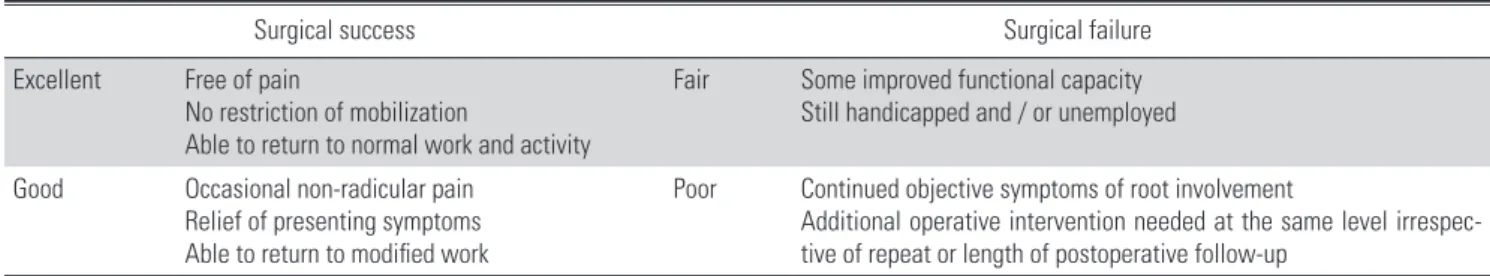
Table 1. Modified macnab criteria
Surgical success Surgical failure
Excellent Free of pain
No restriction of mobilization
Able to return to normal work and activity
Fair Some improved functional capacity Still handicapped and / or unemployed Good Occasional non-radicular pain
Relief of presenting symptoms Able to return to modified work
Poor Continued objective symptoms of root involvement
Additional operative intervention needed at the same level irrespec- tive of repeat or length of postoperative follow-up
Table 2. MSS score
Symptoms Score 2 Score 1 Score 0
Low back pain None Activity related At rest
Sciatica None With SLRT At rest
Sensory deficit None Dysesthesia or paresthesia Hypesthesia or anesthesia
Motor weakness None Full function with slight resistance Two reflexes impaired
Reflex changes None One reflex impaired Two reflexes impaired
MSS: Modified Suezawa and Schreiber, SLRT: Straight leg raising test.
Outcome Analysis of PELD for Adjacent Lumbar Disc Herniation by Radiologic Exam Journal of Korean Society of Spine Surgery
www.krspine.org
223
상이 지속되는 환자들을 대상으로 하였다. 인접 분절의 불안정성이 동반되어있는 환자들은 대상에서 제외하였으며, 굴 신 방사선 사진 상 시상면 전위 3~4 mm 이상 또는 각 변형 10~15도 이상의 소견을 보일 때 분절 불안정성으로 정의하 였다.13)
술 후 임상 결과는 삶의 질 지표와 통증 점수로 평가하였 다. 삶의 질 지표로 Modified Macnab criteria14)와 Modified Suezawa and Schreiber Clinical Score (MSS score)15)를 사용하였고, Modified Macnab criteria 상 “Good” 또는
“Excellent”로 평가된 환자들을 만족 군, “Fair” 또는 “Poor”
로 평가된 환자들을 불만족 군으로 분류하였다(Table 1, 2).
그리고 통증 점수로 Visual Analogue Scale (VAS)를 사용하 였으며, 요통의 VAS-Back (VAS-B)과 하지 방사통의 VAS- Legs (VAS-L)로 나누어 평가하였다. Modified Macnab criteria는 술 후 6개월, 술 후 1년에 평가하였으며 MSS score 와 VAS는 술 전, 술 후 6개월, 술 후 1년에 평가하였다. 전체 환자 중 경피적 내시경적 추간판 절제술 후 증상이 완화되 었다가 다시 재발한 하지 방사통 등 증상이 심하여 Modified Macnab criteria 상 “Poor”로 평가되는 2예가 있었으며 이 들은 술 후 8주 이내에 개방적 추간판 절제술을 재 시행하였 다. 본 연구에서 삶의 질 지표 및 통증 점수는 술 후 6개월 및 1년 째 평가한 것으로 상기 2예는 개방적 추간판 절제술 시 행 후 양호한 예후를 보여, 경피적 내시경적 추간판 절제술 후 Modified Macnab criteria 상 “Poor”로 평가하여 불만족 군으로 분류하되 술 후 6개월 및 1년 째 MSS score 및 VAS 의 추시에서는 제외하였다.
방사선학적 소견과 임상 결과의 관계를 알아보기 위하여 후측방 유합술 직전과 경피적 내시경적 추간판 절제술 직전 에 비교한 단순 측면 방사선 사진 상 인접 분절의 Modified Mochida’s method16)에 따른 disc height ratio와 굴신 방사 선 사진 상 인접 분절의 각 형성 변화를 측정하였다(Fig. 1, 2) 그리고 경피적 내시경적 추간판 절제술 전 촬영한 자기공 명영상에서 척추관 협착 소견의 동반 여부를 확인하였다. 중 심관 협착증은 자기공명영상 축상면에서 중심관 내의 경막 단면(dural sac cross sectional area)이 100 mm2 이하이거나 T2 강조영상에서 마미 전방의 경막내 뇌척수액 부분이 소 실되어 잘 보이지 않는 경우로 정의하였고, 신경공 협착증 은 자기공명영상 시상면에서 신경근 주위 지방조직의 소실 소견을 보이거나 압박으로 인한 신경근의 모양 변형을 보 일 때 진단하였으며, 외측함요부 협착증은 자기공명영상 축 상면에서 척추체 후면과 상관절돌기 사이의 거리가 3 mm 미만인 경우로 정의하였다.17-19) Modified Macnab criteria 에 따른 환자 군은 빈도분석 기초 통계를 실시하였고, MSS score, VAS, disc height ratio, 분절 각 형성 변화량은 반복 측 정 분산분석을 시행하였으며, 만족 군과 불만족 군 간의 disc height ratio 및 분절 각 형성 변화량의 차이는 독립 T검정을 사용하였다.
2. 수술 방법
전례 국소마취 하에 Wilson frame 위에서 복와위로 수술 진 행하였고 단삽입구 관절경을 이용하여 경추간공 접근법으로 한 명의 정형외과 전문의가 수술하였다. 피부 절개는 허리 정 중선에서 약 10~12 cm 떨어진 부위에 시행 후 바늘을 삽입 하였으며, C형 영상 증폭기 촬영 하 전후면 상 척추경의 내측 면, 측면 상 척추체의 후면에 바늘이 위치하는 것을 확인 후
Fig. 1. Mochida method for measuring the disc height ratio. Radiographic measurement of the disc height ratio: a/A (at the final follow-up)×[a/A (before the operation)], a=disc height, A=vertebral height.
A
a
(Lateral view)
Extension
Flexion
A B
Fig. 2. Measuring the segmental angulation. The angle between the line on the lower endplate of the upper vertebra and the line on the upper endplate of the lower vertebra is measured on fl exion and extension ra-the lower vertebra is measured on fl exion and extension ra-lower vertebra is measured on flexion and extension ra- diographs. In addition, the difference (B-A) is calculated.
Fig. 3. (A) A 62-year-old female patient with spinal stenosis at L4-5: simple anteroposterior (AP) and lateral radiographs before lumbar posterolateral fu-62-year-old female patient with spinal stenosis at L4-5: simple anteroposterior (AP) and lateral radiographs before lumbar posterolateral fu- at L4-5: simple anteroposterior (AP) and lateral radiographs before lumbar posterolateral fu- L4-5: simple anteroposterior (AP) and lateral radiographs before lumbar posterolateral fu-: simple anteroposterior (AP) and lateral radiographs before lumbar posterolateral fu- simple anteroposterior (AP) and lateral radiographs before lumbar posterolateral fu-anteroposterior (AP) and lateral radiographs before lumbar posterolateral fu-AP) and lateral radiographs before lumbar posterolateral fu-) and lateral radiographs before lumbar posterolateral fu- and lateral radiographs before lumbar posterolateral fu- sion. (B) Flexion-extension lateral radiographs before lumbar posterolateral fusion. (C) At 63 months postoperatively, simple AP and lateral radiographs before percutaneous endoscopic lumbar discectomy show almost no interval change of L3-4 disc height. (D) Flexion-extension lateral radiographs show an L3-4 segmental angulation change of 5.4°. (E) Left-side central disc herniation of the L3-4 segment without spinal stenosis on magnetic resonance imaging taken before percutaneous endoscopic lumbar discectomy (PELD). (F) The disc material removed after PELD; the patient’s prognosis was good.
A
D
B
E F
C
Fig. 4. (A) A 71-year-old female patient with spinal stenosis at L4-S1: simple anteroposterior (AP) and lateral radio-71-year-old female patient with spinal stenosis at L4-S1: simple anteroposterior (AP) and lateral radio- at L4-S1: simple anteroposterior (AP) and lateral radio- L4-S1: simple anteroposterior (AP) and lateral radio-: simple anteroposterior (AP) and lateral radio- simple anteroposterior (AP) and lateral radio-anteroposterior (AP) and lateral radio- and lateral radio- graphs before lumbar posterolateral fusion. (B) Flexion-extension lateral radiographs before lumbar posterolateral fusion. (C) At 78 months postoperatively, simple AP and lateral radiographs before percutaneous endoscopic lumbar discectomy show decreased L3-4 disc height. (D) Flexion-extension lateral radiographs show an L3-4 segmental angulation change of 9.8°. (E) Left-side posterolateral disc herniation of the L3-4 segment with spinal stenosis on magnetic resonance imaging taken before percutaneous endoscopic lumbar discectomy (PELD). (F) Removed disc material after PELD. (G) Recurrent disc herniation in the same region after PELD (dotted line).
A
D
G
B
E F
C
Outcome Analysis of PELD for Adjacent Lumbar Disc Herniation by Radiologic Exam Journal of Korean Society of Spine Surgery
www.krspine.org
225
Kambin 삼각을 통하여 추간판 공간으로 진입시켰다. 이후indigocarmine 염색제로 추간판 조영 후 유도강선을 진입시키 고 바늘을 제거 하였으며, 유도강선을 따라 절개창을 통해 도관 (obturator)으로 피부를 이완시키면서 섬유륜을 뚫을 때까지 점 진적으로 진입시켰다. Walking channel과 irrigation channel이 있는 단삽입구 관절경을 추간판 공간으로 삽입 후 관절경 영상 을 보면서 disc forcep으로 추간판 절제술을 시행하였다. 절제술 후 신경근의 긴장도 감소로 인한 박동성 움직임 및 경막낭의 자 유로운 부유를 직접 확인하였을 때 추간판의 충분한 제거가 이 루어졌다고 판단하였다.
결과
환자의 평균 연령은 64.2세로 남자가 25명, 여자는 15명이 었다. 기존 유합 분절 수는 1분절이 24예, 2분절이 13예, 3분 절이 3예였으며, 후측방 유합술 후 인접 분절에 추간판 탈출 증이 발생하여 경피적 내시경적 추간판절제술을 시행하기 까지 걸린 평균 시간은 7.8년이었다. 전례에서 기존 유합 분 절의 상위 인접 분절에서 추간판 탈출증이 발생하였으며, 돌
출형이 39예, 탈출형이 1예, 격리형은 없었다. 평균 수술 시 간은 86.4분, 평균 재원 기간은 1주일, 일상 복귀까지 소요되 는 기간은 평균 32.4일 이었다.
Modified Macnab criteria 상 술 후 1년 째 전체 환자 45명 중 24명인 53.3%의 환자들이 “Good” 이상의 만족 군으로 평가되었고, 21명인 46.7%의 환자들이 “Fair” 또는 “Poor”
로 불만족 군으로 평가되었다(Table 3). 전체 환자의 평균 MSS score는 술 전 4.77에서 술 후 6개월 6.34, 술 후 1년 6.99로 향상되었고, Modified Macnab criteria 상 만족 군에
Table 3. Modified macnab criteria evaluation
POD* 6months POD* 12months p-value
Excellent 2 (4.4%) 4 (8.8%) <0.05
Good 22 (48.8%) 20 (44.4%) <0.05
Fair 15 (33.3%) 15 (33.3%) <0.05
Poor† 6 (13.3%) 6 (13.3%) <0.05
*POD: Post percutaneous endoscopic lumbar discectomy operative day.
Table 4. MSS score evaluation (mean)
Preoperation POD‡ 6months POD‡ 12months p-value
All patient (43) 4.77 6.34 6.99 <0.05
Satisfied patient* (24) 4.79 7.67 7.83 <0.05
Dissatisfied patient† (19) 4.75 5.01 6.15 <0.05
MSS: Modified Suezawa and Schreiber,*Satisfied patient : Patients who were evaluated “Excellent” or “Good” by Modified Macnab criteria, †Dissatis fied patient: Patients who were evaluated “Fair” or “Poor” by Modified Macnab criteria and who undergone revision open discectomy were excepted, ‡ POD: Post percutaneous endoscopic lumbar discectomy operative day.
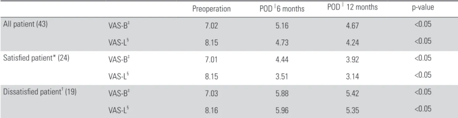
Table 5. VAS evaluation (mean)
Preoperation POD∥6 months POD∥ 12 months p-value
All patient (43) VAS-B‡ 7.02 5.16 4.67 <0.05
VAS-L§ 8.15 4.73 4.24 <0.05
Satisfied patient* (24) VAS-B‡ 7.01 4.44 3.92 <0.05
VAS-L§ 8.15 3.51 3.14 <0.05
Dissatisfied patient† (19) VAS-B‡ 7.03 5.88 5.42 <0.05
VAS-L§ 8.16 5.96 5.35 <0.05
*Satisfied patient: Patients who were evaluated “Excellent” or “Good” by Modified Macnab criteria, †Dissatisfied patient: Patients who were evaluated
“Fair” or “Poor” by Modified Macnab criteria and who undergone revision open discectomy were excepted, ‡VAS-B: Visual analogue scale-Back, §VAS-L:
Visual analogue scale-Leg, ∥POD: Post percutaneous endoscopic lumbar discectomy operative day.
서 평균 MSS score는 술 전 4.79에서 술 후 1년 7.83으로 증 가, 불만족 군에서 평균 MSS score는 술 전 4.75에서 술 후 1 년 6.15로 증가하였다(Table 4).
전체 환자의 평균 VAS-B는 술 전 7.02에서 술 후 6개 월 5.16, 술 후 1년 4.67로 감소되었고, Modified Macnab criteria 상 만족 군에서 평균 VAS-B는 술 전 7.01에서 술 후 1년 3.92로 감소, 불만족 군에서 평균 VAS-B는 술 전 7.03 에서 술 후 1년 5.42로 감소하였다(Table 5). 전체 환자의 평 균 VAS-L은 술 전 8.15에서 술 후 6개월 4.73, 술 후 1년 4.24로 감소되었고, Modified Macnab criteria 상 만족 군에 서 평균 VAS-L은 술 전 8.15에서 술 후 1년 3.14로 감소, 불 만족 군에서 평균 VAS-L은 술 전 8.16에서 술 후 1년 5.35 로 감소하였다(Table 5).
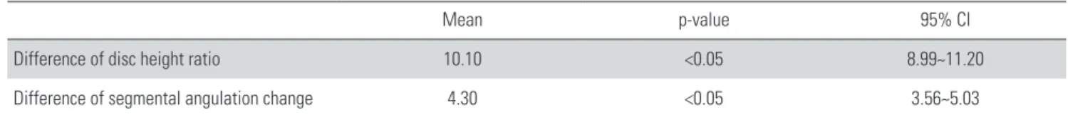
후측방 유합술 직전과 경피적 내시경적 추간판 절제술 직전 에 비교한 단순 측면 방사선 사진 상 인접 분절의 disc height ratio는 전체 환자에서 평균 87.1%로 감소한 경향을 보였고, Modified Macnab criteria 상 만족 군에서 평균 92.2%, 불만족 군에서 평균 82.1%로 불만족 군에서 추간판 간격의 더 큰 감소 를 나타내었다(Table 6). 후측방 유합술 직전과 경피적 내시경 적 추간판 절제술 직전에 비교한 굴신 방사선 사진 상 인접 분 절의 각 형성 변화는 전체 환자에서 평균 6.5도였고, Modified Macnab criteria 상 만족 군에서 평균 4.4도, 불만족 군에서 평 균 8.7도로 불만족 군에서 각 형성의 더 큰 변화량을 나타내었 다(Table 6). 만족 군과 불만족 군 간의 disc height ratio의 평균 차이는 10.10, 분절 각 형성 변화량의 평균 차이는 4.30으로 모 두 유의한 수준을 나타내었다(Table 7). 경피적 내시경적 추간
판 절제술 시행 전 촬영한 자기공명영상에서 인접 분절의 척추 관 협착 소견이 동반되어있는 환자는 전체 환자 45명 중 18명 으로 중심성 협착증이 8예, 신경공 협착증이 10예였다(Table 8). Modified Macnab criteria 상 만족 군 24명 중에서 5명인 20.8%, 불만족 군 21명 중에서 13명인 61.9%에서 척추관 협 착 소견이 동반되어, 불만족 군에서 더 높은 비율로 척추관 협 착 소견이 동반되어 있었다(Table 8). 그리고 중심관 협착증이 동반된 환자 8명 중 Modified Macnab criteria 상 불만족 군은 5명, 신경공 협착증이 동반된 환자 10명 중 Modified Macnab criteria 상 불만족 군은 8명으로 신경공 협착증이 동반된 환자 에서 불만족 군의 비율이 더 높았다(Table 8).
Table 7. Difference of disc height ratio and segmental angulation change between satisfied patient and dissatisfied patient (mean)
Mean p-value 95% CI
Difference of disc height ratio 10.10 <0.05 8.99~11.20
Difference of segmental angulation change 4.30 <0.05 3.56~5.03
CI: Confidence interval.
Table 8. Disc herniation accompanied by spinal stenosis on MRI before PELD
Stenosis on MRI before PELD Central canal stenosis Foraminal stenosis
All patient (45) 18 8 10
Satisfied patient* (24) 5 3 2
Dissatisfied patient† (21) 13 5 8
MRI: Magnetic resonance imaging, PELD: Percutaneous endoscopic lumbar discectomy, *Satisfied patient: Patients who were evaluated “Excellent” or
“Good” by Modified Macnab criteria, †Dissatisfied patient: Patients who were evaluated “Fair” or “Poor” by Modified Macnab criteria and who under- gone revision open discectomy were included.
Table 6. Disc height ratio and segmental angulation change on simple radiography measured by comparing before PLF and before PELD (mean)
Disc height ratio Segmental angulation change
All patient (45) 87.1% 6.5°
Satisfied patient* (24) 92.2% 4.4°
Dissatisfied patient† (21) 82.1% 8.7°
p-value <0.05 <0.05
PL: Posterolateral fusion, PELD: Percutaneous endoscopic lumbar dis- cectomy, *Satisfied patient: Patients who were evaluated “Excellent” or
“Good” by Modified Macnab criteria, †Dissatisfied patient: Patients who were evaluated “Fair” or “Poor” by Modified Macnab criteria and who undergone revision open discectomy were included.
Outcome Analysis of PELD for Adjacent Lumbar Disc Herniation by Radiologic Exam Journal of Korean Society of Spine Surgery
www.krspine.org
227
고찰
요추부 후측방 유합술 후 인접 분절에서 발생한 요추부 추 간판 탈출증은 단순 요추부 추간판 탈출증의 치료방침과 마 찬가지로 보존적 치료를 먼저 시도해볼 수 있으며 보존적 치료에도 불구하고 증상이 지속되거나 신경학적 증상이 진 행될 때 수술적 치료를 고려할 수 있다. 경피적 내시경적 요 추부 추간판 절제술은 최소 침습적 추간판 제거술 중 하나 로 국소 마취 하 수술이 가능하며 후방 조직을 최대한 보존 함으로써 술 후 통증이 적고 재원 기간이 짧다. 20,21) 특히 요 추부의 수술 병력이 있는 상태에서 재수술을 요하는 경우 반흔 조직을 통하지 않고 접근함으로써 후방 구조물 및 척 추 주위 근육의 반복된 손상을 줄일 수 있다는 장점이 있
다.22,23) Li 등24)은 단순 요추부 추간판 탈출증 환자의 수술적
치료로서 경피적 내시경적 추간판 절제술과 개방적 추간판 절제술의 임상 결과를 비교한 메타분석연구에서 술 중 실혈, 수술 시간, 재원 기간, 일상생활로의 복귀 기간 등이 경피적 내시경적 추간판 절제술을 시행한 환자 군에서 더 유리한 결과를 나타내었음을 보고하였다. 그러나 경피적 내시경적 추간판 절제술은 추간판 조직의 불완전한 제거, 높은 재발 율, 비교적 어려운 술기 습득 등의 제한점도 존재한다.25-27)
Gotecha 등28)은 단순 요추부 추간판 탈출증 환자 120예에 서 경피적 내시경적 추간판 절제술 시행 후 6개월 뒤 추시한 삶의 질 평가 지표에서 Modified Macnab criteria 상 89.3%
에서 “Good” 이상의 임상 결과를 나타내었으며 MSS score 는 술 전 평균 3.47에서 술 후 평균 7.92로 향상되었음을 보 고하였다. Xia 등21)은 재발한 요추부 추간판 탈출증 43예에 서 경피적 내시경적 추간판 절제술 시행 후 평균 31개월 추 시 관찰한 연구에서 Modified Macnab criteria 상 81.4%가
“Good” 이상의 임상 결과를 나타내었음을 보고하였다. 반 면에 본 연구에서는 술 후 1년째 Modified Macnab criteria 상 53.3%의 환자들이 “Good” 이상, 평균 MSS score는 술 전 4.77에서 술 후 1년 째 6.99로 나타나 단순 요추부 추간판 탈출증을 대상으로 한 기존 연구 결과보다 삶의 질 지표의 개선 폭이 작은 것으로 나타났다.
Eun 등29)은 단순 요추부 추간판 탈출증에서 경피적 내시 경적 추간판 절제술 시행 후 평균 11년 추시 관찰한 연구 에서 통증 점수가 술 전 VAS-B 8.45±1.52, VAS-L 7.40±
3.04에서 술 후 VAS-B 2.53±1.98, VAS-L 1.82±1.92로 감 소하였음을 보고하였으며, Hoogland 등22)은 재발한 요추부 추간판 탈출증 262예에서 경피적 내시경적 추간판 절제술 시행 후 2년 추시 관찰한 연구에서 허리와 하지 통증의 의 미있는 감소를 보고하였다. 후측방 유합술 후 인접 분절에
서 발생한 요추부 추간판 탈출증에서 경피적 내시경적 추간 판 절제술을 시행한 본 연구에서는 술 전 평균 VAS-B 7.02, VAS-L 8.15에서 술 후 1년 째 평균 VAS-B 4.67, VAS-L 4.24로 허리 및 하지의 통증 감소 효과는 단순 요추부 추간 판 탈출증을 대상으로 한 기존 연구 결과에 비해 크지 않았 다.
기존 연구의 임상 결과에 비하여 본 연구에서 경피적 내 시경적 추간판 절제술 후 삶의 질 지표 개선 및 통증 감소 효 과가 적었던 이유로 술 전 방사선학적 검사 상 동반된 퇴행 성 변화 소견이 임상 결과에 기여하였을 것으로 사료된다.
추간판 간격의 감소 및 분절의 각 형성 등은 후방 관절로 이 행되는 하중을 증가시켜 척추관의 퇴행성 관절증 및 협착증 을 야기시킬 수 있다.30) 본 연구의 대상 환자들은 후측방 유 합술 직전과 경피적 내시경적 추간판 절제술 직전에 비교한 단순 측면 방사선 소견 상 인접 분절에서 disc height ratio가 감소하고 굴신 방사선 소견 상 인접 분절에서 각 형성 변화 가 증가하는 등 대체적으로 인접 분절의 퇴행성 변화 소견 을 나타내었다. 그리고 경피적 내시경적 추간판 절제술 전 촬영한 자기공명영상에서 추간판 탈출 소견과 함께 황색인 대 및 후관절의 비후 등 척추관 협착 소견이 동반되어있는 경우가 많았다. 인접 분절의 disc height ratio 및 각 형성 변 화량은 Modified Macnab criteria 상 불만족 군에서 더 큰 변화를 보였으며, 자기공명영상 상 동반된 척추관 협착 소견 도 불만족 군에서 더 높은 비율로 나타나, 이는 후측방 유합 술 후 인접 분절에 일련의 퇴행성 변화 과정이 진행되면서 추간판 퇴행성 변화 및 척추관 협착 소견 등 복합적인 병변 양상을 나타냈기 때문으로 사료된다. 또한 술 중 출혈로 인 한 불충분한 시야 확보 등으로 인해 추간판이 불완전 제거 된 3례가 있었으며 이들 모두 불만족의 결과를 보여, 술자의 술기 관련 요인 역시 결과에 영향을 미쳤으리라 사료된다.
본 연구의 제한점으로는 후측방 유합술 후 발생한 인접 분절 의 추간판 탈출증 환자들만을 대상으로 하였으며, 개방적 추간 판 절제술 등의 다른 수술 방법과 경피적 내시경적 추간판 절제 술의 임상결과를 비교한 것이 아니라 경피적 내시경적 추간판 절제술을 시행한 환자들만을 대상으로 연구를 실시하여, 다른 대조군과의 비교 분석이 이루어지지 못했다는 것이다. 그리고 수술 방법에 있어 관절경 삽입구 개수 및 접근 방법에 따른 정 밀한 평가가 이루어지지 않았으며, 요추부 유합술 후 인접 분절 의 요추부 추간판 탈출증에서 경피적 내시경적 추간판 절제술 의 임상 결과를 분석한 기존 연구가 아직 부족하여 동일한 연구 대상에 대한 비교 및 고찰이 좀 더 필요할 것으로 사료된다.
결론
요추부 후측방 유합술 후 인접 분절의 추간판 탈출증에서 단 순방사선학적 검사 상 추간판 간격의 감소 및 각 형성의 증가로 표현되는 퇴행성 변화 소견이 있거나 자기공명영상에서 척추 관 협착증이 동반되어 있는 경우 경피적 내시경적 추간판 절제 술을 통한 임상 결과는 만족스럽지 못할 수 있으므로 수술 적응 증의 선택에 있어 주의가 요구된다.
REFERENCES
1. Hilibrand AS, Robbins M. Adjacent segment degeneration and adjacent segment disease: the consequences of spinal fusion?. Spine J. 2004 Nov-Dec;4(6 Suppl):190-4. DOI:
10.1016/j.spinee.2004.07.007 .
2. Frymoyer JW, Hanley EN, Howe J, et al. A comparison of radiographic findings in fusion and nonfusion pa- tients ten or more years following lumbar disc surgery.
Spine (Phila Pa 1976). 1979 Sep-Oct;4(5):435-40. DOI:
10.1097/00007632-197909000-00008.
3. Booth KC, Bridwell KH, Eisenberg BA, et al. Minimum 5-year results of degenerative spondylolisthesis treated with decompression and instrumented posterior fusion.
Spine (Phila Pa 1976). 1999 Aug 15;24(16):1721-7. DOI:
10.1097/00007632-199908150-00014 .
4. Brotchi J, Pirotte B, De Witte O. Prevention of epidural fi- brosis in a prospective series of 100 primary lumbo-sacral discectomy patients: follow-up and assessment at re- operation. Neurol Res. 1999;21 (1 Suppl ) :47-50. DOI:
10.1080/01616412.1999.11741027.
5. Maroon JC, Abla A, Bost J. Association between peri- dural scar and persistent low back pain after lumbar dis- cectomy. Neurol Res. 1999;21 (1 Suppl ):43-6. DOI:
10.1080/01616412.1999.11741026 .
6. Matsui H, Kitagawa H, Kawaguchi Y, et al. Physiologic changes of nerve root during posterior lumbar discectomy.
Spine (Phila Pa 1976). 1995 Mar 15;20(6):654-9. DOI:
10.1097/00007632-199503150-00004.
7. Patel N, Pople IK, Cummins BH. Revisional lumbar mi- crodiscectomy: an analysis of operative findings and clini- cal outcome. Br J Neurosurg. 1995;9(6):733-7. DOI:
10.1080/02688699550040684.
8. Choi G, Lee SH, Lokhande P, et al. Percutaneous endo-
scopic approach for highly migrate intracranial disc her- niations by foraminoplastic technique using rigid work- ing channel endoscope. Spine (Phila Pa 1976). 2008 Jul 1;33(15):E508-15. DOI: 10.1097/BRS.0b013e31817bfa1a.
9. Kambin P, Brager M. Percutaneous posterolateral discecto- my. Anatomy and mechanism. Clin Orthop Relat Res. 1987 Oct;(223):145-54. DOI: 10.1097/00003086-198710000- 00016.
10. Kambin P, Sampson S. Posterolateral percutaneous suction- excision of herniated lumbar intervertebral discs. Report of interim results. Clin Orthop Relat Res. 1986 Jun;(207):37- 43. DOI: 10.1097/00003086-198606000-00008.
11. Casper GD, Mullins LL, Hartman VL. Laser-assisted disc decompression: a clinical trial of the holmium: YAG laser with side-firing fiber. J Clin Laser Med Surg. 1995 Feb;13(1):27-32. DOI: 10.1089/clm.1995.13.27.
12. Mayer HM, Brock M. Percutaneous endoscopic lumbar discectomy (PELD). Neurosurg Rev. 1993;16(2):115-20.
DOI: 10.1007/bf00258242.
13. Park P, Garton HJ, Gala VC, et al. Adjacent segment disease after lumbar or lumbosacral fusion: review of the literature.
Spine (Phila Pa 1976). 2004 Sep 1;29(17):1938-44. DOI:
10.1097/01.brs.0000137069.88904.03.
14. Macnab I. Negative disc exploration. An analysis of the causes of nerve-root involvement in sixty-eight patients.
J Bone Joint Surg Am. 1971 Jul;53(5):891-903. DOI:
10.2106/00004623-197153050-00004 .
15. Suezawa Y, Schreiber A. Complex indications in surgery of the lumbar spinal canal. Z Orthop Ihre Grenzgeb. 1987 May-Jun;125(3):308-19. DOI:10.1055/s-2008-1044732.
16. Lee SH, Chung SE, Ahn Y, et al. Comparative radiologic evaluation of percutaneous endoscopic lumbar discectomy and open microdiscectomy: A matched cohort analysis. Mt Sinai J Med. 2006 Sep;73(5):795-801.
17. Sirvanci M, Bhatia M, Ganiyusufoglu KA, et al. Degen- erative lumbar spinal stenosis: correlation with Oswes- try Disability Index and MR imaging. Eur Spine J. 2008 May;17(5):679-85. DOI: 10.1007/s00586-008-0646-5.
18. Lee GY, Lee JW, Choi HS, et al. A new grading system of lumbar central canal stenosis on MRI: an easy and reliable method. Skeletal Radiol. 2011 Aug;40(8):1033-9. DOI:
10.1007/s00256-011-1102-x.
19. Lee S, Lee JW, Yeom JS, et al. A practical MRI grading sys-
Outcome Analysis of PELD for Adjacent Lumbar Disc Herniation by Radiologic Exam Journal of Korean Society of Spine Surgery
www.krspine.org
229
tem for lumbar foraminal stenosis. AJR Am J Roentgenol.2010 Apr;194(4):1095-8. DOI: 10.2214/AJR.09.2772.
20. Lee DY, Ahn Y, Lee SH. Percutaneous endoscopic lumbar discectomy for adolescent lumbar disc herniation: surgical outcomes in 46 consecutive patients. Mt Sinai J Med. 2006 Oct;73(6):864-70.
21. Xia XP, Chen HL, Cheng HB. Prevalence of adjacent seg- ment degeneration after spine surgery: a systematic re- view and meta-analysis. Spine (Phila Pa 1976). 2013 Apr 1;38(7):597-608. DOI: 10.1097/BRS.0b013e318273a2ea.
22. Hoogland T, van den Brekel-Dijkstra K, Schubert M, et al. Endoscopic transforaminal discectomy for recurrent lumbar disc herniation: a prospective, cohort evaluation of 262 consecutive cases. Spine (Phila Pa 1976). 2008 Apr 20;33(9):973-8. DOI: 10.1097/BRS.0b013e31816c8ade.
23. Chen HC, Lee CH, Wei L, et al. Comparison of percu- taneous endoscopic lumbar discectomy and open lumbar surgery for adjacent segment degeneration and recurrent disc herniation. Neurol Res Int. 2015;2015:791943. DOI:
10.1155/2015/791943.
24. Li X, Han Y, Di Z, et al. Percutaneous endoscopic lumbar discectomy for lumbar disc herniation. J Clin Neurosci.
2016 Nov;33:19-27. DOI: 10.1016/j.jocn.2016.01.043.
25. Cheng J, Wang H, Zheng W. Reoperation after lumbar disc surgery in two hundred and seven patients. Int Orthop.
2013 Aug;37(8):1511-7. DOI: 10.1007/s00264-013- 1925-2.
26. Wang H, Huang B, Li C. Learning curve for percutaneous endoscopic lumbar discectomy depending on the surgeon’s training level of minimally invasive spine surgery. Clin Neu- rol Neurosurg. 2013 Oct;115(10):1987-91. DOI: 10.1016/
j.clineuro.2013.06.008.
27. Ahn Y, Kim CH, Lee JH, et al. Radiation exposure to the surgeon during percutaneous endoscopic lumbar discec- tomy: a prospective study. Spine (Phila Pa 1976). 2013 Apr 1;38(7):617-25. DOI: 10.1097/BRS.0b013e318275ca58.
28. Gotecha S, Ranade D, Patil SV, et al. The role of transfo- raminal percutaneous endoscopic discectomy in lumbar disc herniations. J Craniovertebr Junction Spine. 2016 Oct- Dec;7(4):217-23. DOI: 10.4103/0974-8237.193267.
29. Eun SS, Lee SH, Sabal LA. Long-term follow-up results of percutaneous endoscopic lumbar discectomy. Pain Physi- cian. 2016 Nov-Dec;19(8):E1161-6.
30. Yang KH, King AI. Mechanism of facet load transmission as a hypothesis for low back pain. Spine (Phila Pa 1976). 1984 Sep;9(6):557-65. DOI: 10.1097/00007632-198409000- 00005.
요추부 후측방 유합술 후 발생한 인접 분절의 요추부 추간판 탈출증에서 방사선학적 검사를 통한 경피적 내 시경적 요추부 추간판 절제술의 임상 결과 분석
고영철 • 하동준 • 박만준 • 허정욱 • 박숙현 • 박태홍 • 박준형 부산의료원 정형외과
연구 계획: 후향성 연구
목적: 요추부 후측방 유합술 후 발생한 인접 분절의 추간판 탈출증에서 술 전 방사선학적 검사 및 지표를 통하여 경피적 내시경적 추간판 절제술의 효용 성을 알아본다.
선행문헌의 요약: 경피적 내시경적 추간판 절제술은 후방 구조물 손상을 최소화하여 빠른 재활이 가능하다.
대상 및 방법: 2014년 3월에서 2016년 2월까지 인접 분절의 추간판 탈출증으로 경피적 내시경적 추간판 절제술을 시행 후 1년 추시한 45명의 환자들을 대상으로 하였다. Modified Macnab criteria, Modified Suezawa and Schreiber Score (MSS Score), 허리의 Visual Analogue Scale-Back (VAS-B), 하지의 Visual Analogue Scale-Legs (VAS-L)으로 임상결과를 평가하였다. 방사선학적 지표로 후측방 유합술 직전과 경피적 내시경적 추간판 절제술 직전에 비 교한 인접 분절의 disc height ratio와 각 형성 변화를 측정하고, 경피적 내시경적 추간판 절제술 전 촬영한 자기공명영상에서 척추관 협착 소견의 동반 여 부를 확인하였다.
결과: Modified Macnab criteria 상 술 후 1년째 53.3%의 환자들이 “Good” 이상으로 평가되었으며, 평균 MSS score는 술 전 4.77에서 술 후 1년째 6.99 로 향상되었다. 평균 VAS-B는 술 전 7.02에서 술 후 1년째 4.67로, 평균 VAS-L은 술 전 8.15에서 술 후 1년째 4.24로 감소되었다. 평균 disc height ratio 는 87.1%, 평균 분절 각 형성 변화는 6.5도로 Modified Macnab criteria 상 “Fair” 이하의 불만족 환자 군에서 변화량이 더 컸으며, 술 전 자기공명영상에서 척추관 협착 동반 비율도 불만족 환자 군에서 더 높았다.
결론: 유합술 후 인접 분절의 추간판 탈출증에서 단순방사선학적 검사 상 추간판 간격의 감소 및 각 형성의 증가 등 퇴행성 변화 소견이 있거나 자기공명 영상에서 척추관 협착증이 동반되어 있는 경우 경피적 내시경적 추간판 절제술을 통한 임상 결과는 만족스럽지 못할 수 있으므로 수술 적응증의 선택에 있어 주의가 요구된다.
색인 단어: 요추부 후측방 유합술, 인접 분절 질환, 인접 분절 요추부 추간판 탈출증, 경피적 내시경적 요추부 추간판 절제술, 방사선학적 검사 약칭 제목: 인접 분절 요추간판 탈출증에서 방사선 검사를 통한 경피적 내시경적 추간판 절제술의 임상 결과 분석
접수일: 2017년 7월 11일 수정일: 2017년 7월 24일 게재확정일: 2017년 11월 10일 교신저자: 하동준
부산시 연제구 월드컵대로 359 부산의료원 정형외과
TEL: 051-607-2550 FAX: 051-607-2551 E-mail: [email protected]
![Fig. 1. Mochida method for measuring the disc height ratio. Radiographic measurement of the disc height ratio: a/A (at the final follow-up)×[a/A (before the operation)], a=disc height, A=vertebral height.](https://thumb-ap.123doks.com/thumbv2/123dokinfo/5313607.163734/4.892.179.361.736.1025/mochida-measuring-radiographic-measurement-height-operation-height-vertebral.webp)