가토에서 분말형 황산칼슘이 신연 골형성의 경화에 미치는 효과
오창욱・김풍택・박병철・박일형・경희수・백승훈
경북의대 정형외과학 교실
<국문초록>
목 적 : 황산칼슘(calcium sulfate salt)을 조기 및 조속 신연(early & fast distraction)된 가토 경골에 주사하 여 가골 경화에 대한 촉진효과를 알아보고자 하였다.
대상 및 방법 : 뉴질랜드 백색가토(New Zealand white rabbit) 24마리를 대상으로 소형 외고정 신연기로 고정한 후 경골을 절골하였다. 3일 간의 휴지기(latency period)후 4일간 총 8 m m의 조기 및 조속 신연술 을 시행하였다. 제 7일에 황산칼슘과 c a r b o x y m e t h y l c e l l u l o s e ( C M C )혼합액을 실험군의 신연부에 주사하였 다. 제 1대조군은 C M C용액만을 주사하였고, 제 2대조군은 단순신연술만을 시행하였다. 매주마다 방사 선 촬영으로 신연부 가골의 시간에 따른 변화 등을 비교하였다. 제 3주와 6주에 골밀도를 측정하였고 골밀도비( % B M D )를 산출하였다. 술후 4 2일에 가토를 희생하여 골형성 정도를 비교하였다.
결 과 : 방사선 촬영상 실험군에서 약 3주부터 초기가골이, 6주째 절단부를 연결하는 풍부한 흰색 음영 이 관찰되었으나, 두 대조군에서는 골단부 일부에서 미숙한 골형성만이 관찰되었다. 6주째 측정한 실 험군의 골밀도비는 대조군보다 저명하게 증가된 양상을 보였으며(p<0.01), 조직학적 검사에서도 실험군 에서 우수한 골형성을 보였다.
결 론 : 신연 가골에 황산칼슘을 비침습적으로 주사하여 골 경화를 촉진시킬 수 있을 것으로 사료된 다.
색인 단어 : 가토경골, 조기 조속 신연술, 분말형 황산칼슘, 경화촉진
※통신저자 : 오 창 욱
대구광역시 중구삼덕동 2가 50번지 경북의대정형외과학 교실 TEL : (053) 420 - 5630
FAX : (053) 422 - 6605 E-mail : [email protected]
※ 본 논문의 요지는 2001년도 대한골절학회 추계학술대회에서발표되었음 .
서 론
일리자로프에 의해 고안된 신연 골형성술 (distraction osteogenesis)은외상, 종양적출등으로인한 골결손 (bone defect) 및 사지부동 (limb length d i s c r e p a n c y )을 치료하기 위한 골형성에 매우 효과적 인방법으로 인정되고있다4 , 8 , 9 , 1 0 ). 그러나 광범위한골 결손이나 지연유합의 경우 경화( c o n s o l i d a t i o n )기간의 증가로인한외고정기간의연장과같은문제를야기
하여2 , 3 , 7 ), 신연술에서골형성을촉진시키기 위한방법
들이 모색되고 있으며, 특히 성장인자(growth factor), 탈무기골기질(demineralized bone matrix) 및 골수유래 원시세포(marrow-derived progenitor cells) 이식 등이좋 은결과를보고하고있다5 , 6 , 1 6 ).
본 연구의 목적은 조기조속 신연시킨 가토경골에 황산칼슘(Calcium sulfate salt)을주사하여가골경화에 대한촉진효과를연구하고, 임상적응용의 가능성을 알아보고자하였다.
연구 대상 및 방법
본 연구는경북의대의학연구소(Institutional Animal Care and Use Committee)에의해승인되었다.
1. 실험재료
경북의대 의학연구소에서 사육된 2 0 0 0~2 5 0 0 g m 의 백색 뉴질랜드 가토 2 4마리를 대상으로 3군으로 분할하였다.
2. 실험방법및신연술
1 0 m g / k g의ketamine HCl 및 3mg/kg RompunR( X y l a z i n e h y d r o c h l o r i d e )를 근주하여 마취하고 술전 및 술후에 세 팔 로 스 포 린 계 항생제 ( C e l o s l i nR, Ceftezole sodium(1g/vial), 0.1cc of 100,000 u/kg)를 주사하였으며, 수술부위는털을깎고베타딘액으로소독하였다. 경 피적으로 절골부 양측에 각각 2개의 나사핀(직경 1 . 5 m m )을 경골의 전내측에 삽입하고 특수제작한 일 측방의(monolateral) 막대형소형외고정신연기( U &IR, K o r e a )로고정하였다. 작은 내측 종절개를 통해 경골 간부를 노출시키고 골막을 박리하여 최대한 보호하
면서 0 . 0 4 5인치 Kirschner wire로 5~6회의 천공 후 소 형 절골기로 경골을 절골한 다음, 절골부 양단을 접 촉시키고피하조직및피부를봉합하였다. 각군은 3 일간의휴지기(latency period)후, 12시간당1 m m씩, 4일 간총 8 m m의 조기조속 신연술(early & fast distraction) 을시행하였다.
3. 주입방법
황산칼슘은 미국 Wright medical technology사의 O s t e o s e tR( 9 8 % C a S O4・H2O, 0.5%CaCO3・M g C O3, 0 . 3 % C a C O3)를사용하였다. 주입을위해황산칼슘 알 약을 분말로 갈아서 일정비율(200mg 황산칼슘/ 1 c c C M C )로 C M C용액과 혼합하였다. CMC는 c e l l u l o s e와 polycarboxymethyl ether의 sodium salt로현탁액및유화 제로 이용될 수 있다. 술후 7일째에 제 1군에서혼합 액 1 c c를 신연중심부에 주사하였고 제 2군에서는 신 연중심부에1 c c의 C M C용액만을 주입하였다. 제 3군 은주사없이단순신연술만을시행하였다.
주입부주위의 피부를 소독하고 마취후 형광투시 경하에 18 gauge 주사침을 신연 중심부에 정확하게 삽입하여 1분에 걸쳐 서서히 현탁액을 주입하였다.
술후몇시간동안합병증병발여부를관찰하였다.
4. 관찰및평가방법
방사선 촬영은 매 1주마다 전후면 및 측면촬영을 시행하여신연부에형성된가골의시간에따른변화, 피질골의 형성시기 등을 비교하였으며, 판독자간의 오차를 줄이기 위하여 3명의 정형외과 의사가 별개 로판독하였다.
골밀도측정은 마취하에 이중 에너지 방사선촬영 기(DEXA, dual energy X-ray absorptiometry, LunarR, U S A )를 이용하여 각각 제 3주와 6주에 절단 근위부 (P) 및 원위부(D), 신연가골 중심부( C )의 R O I ( R e g i o n s
Of Interest)를측정하였다. 개체간오차를줄이기위하
여 골밀도비( C/ (P+D)/2 )를산출하여 S P S S로통계학 적검증을시행하였다.
조직학적 관찰을위하여 술후 4 2일에 가토를희생 하였고절골근위및원위부를포함한신생골부위를 종적으로채취하였다. 표본제작을 위해 10% 중화 포 르말린(neutral buffered formalin)액에약 1주간담근후, 10% 질산(nitric acid)과 10% 구연산나트륨( s o d i u m
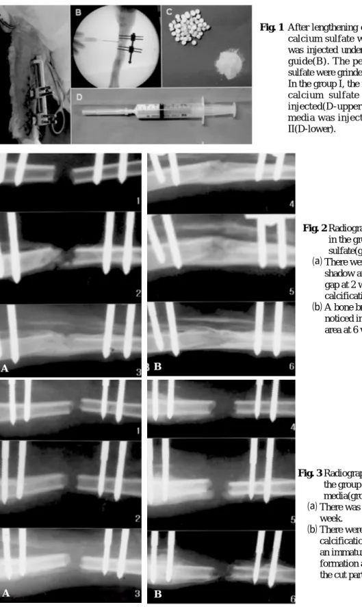
Fig. 1 After lengthening of rabbit tibia(A), calcium sulfate with CMC media was injected under the fluoroscopic guide(B). The pellets of calcium sulfate were grinded into powder(C).
In the group I, the mixed material of calcium sulfate and CMC was injected(D-upper) and only CMC media was injected in the group II(D-lower).
Fig. 3 Radiographs of animals in the group of CMC media(group Ⅱ).
⒜ There was no callus at 2 week.
⒝ There were a tiny
calcification at 4 week and an immature callus formation at the end-side of the cut part at 6 week.
Fig. 2 Radiographs of animals in the group of calcium sulfate(group I).
⒜ There were a faint calcific shadow at the distraction gap at 2 week and marked calcification at 3 week.
⒝ A bone bridging was noticed in the distracted area at 6 week.
A B
A B
c i t r a t e )으로 4 8시간동안탈석회하였다. 이후신연부를 외과용 수술칼( s c a l p e l )로 절편하고 세척하여 고정액 을제거한후농축에탄올로탈수하였다. hematoxylin- eosin 염색후측정부위에따른주관적요소를줄이기 위하여, 가급적중심부에서가까운부위를절편한다 음, 골모세포 출현, 신연골의 피질골 형성 및 골가교 형성, 골수강 생성, 골지주(bony trabecula) 형성 및 두 께등을저배율(x5) 및고배율( x 4 0 )에서비교하였다.
결 과
추시관찰 중 사망한 3마리의가토(제1군: 1마리, 제 2군: 1마리, 제3군: 1마리)와 핀 삽입부에서 분쇄골절 이발생한 3마리의가토(제2군: 1마리, 제3군: 2마리)는 결과에서제외하였다.
1. 방사선적관찰소견
제 1군에서약 1주만에황산칼슘의 음영이소실되 면서, 4주째초기가골이발견된반면제 2군및 3군에 서는 발견되지 않았다. 6주째 제 1군의 모든 가토에 서신연부양단을연결하는풍부한흰색음영이관찰 된반면, 제 2군및 3군의모든가토에서골유합의소 견은관찰되지않았고, 골단부의일부에서미숙한골 형성이 관찰되었다. 제 1군의 일부 가토에서는 성숙 골유합이관찰되기도하였다.
Fig. 6 The value of BMD percentage in the group I increased significantly than those in the group
Ⅱ & Ⅲ, at 6 week.
A
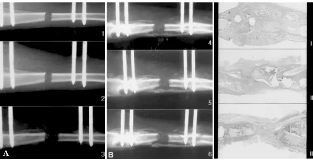
Fig. 4 Radiographs of animals in the group of control (group Ⅲ).
⒜ There was no callus at 2 week.
⒝ A little spotted calcification at 4 week and an irregular calcification in the distracted area at 6 week.
Fig. 5 Histologic findings with H-E stain (x5) at 6 week. There was new trabecular bone with numerous osteoblasts around the group of calcium sulfate(I), continuing the distraction gap. However, there was a little new bone with abundant fibrous tissue at the cut-end in both groups of CMC media(Ⅱ) and simple distraction(Ⅲ).
2. 골밀도측정소견
3주째 측정한제 1군의골밀도비( 1 6 . 1 % )는두대조 군의 골밀도비(제 2군: 6.9%, 제 3군: 7.1%)보다 증가 된 양상을 보였다. 6주째 측정한 제 1군의 골밀도비 ( 5 8 . 4 % )는제 2군(10.2%) 및제 3군( 1 0 . 4 % )보다유의하 게 높게 관찰되었다(p<0.01). 그러나 두 대조군의 골 밀도비사이에는유의한차이가없었다.
3. 조직학적소견
두대조군에서절골부양단주위로골소주형성이비 교적 미약하고 교원질 섬유조직이 풍부한 미숙골의 조직소견이 관찰되었으나 모든 가토에서 두 절골단 사이의 연결없이 전체적으로 불완전한 골형성이 관 찰되었다. 반면 황산칼슘을 주입한 모든 가토에서 신 연부의 왕성한 골형성이 관찰되었고 신생골은 절단 부와 유사한 골소주가 관찰되었으나 두께는 얇았다.
주입된황산칼슘은모두흡수되어관찰되지않았다.
고 찰
사지연장술(limb lengthening)에서외고정기를이용 한 점진적인 신연골 형성술(distraction osteogenesis)은 일리자로프1 2 , 1 3 )에 의해개발된이후보편적으로 사용 되고있다. 그러나, 광범위한골결손이나지연유합의 경우, 경화( c o n s o l i d a t i o n )기간의 증가로 인한 외고정 기간의 연장이 필요하며, 이는 핀감염, 골수염, 관절 강직 및일상생활로의복귀지연과같은임상적인문 제를 야기할 수 있다3 , 2 0 ). 따라서 가골형성 및 경화를 촉진시킨다면이러한문제들을해결할수있으나, 전 기자극, 저강도초음파혹은경구용활성비타민 D와 같은간접적이고비침습적인방법들1 7 , 1 8 )은제한된결 과 만 을 보 고 하 고 있다 . 반면 변형성장인자 (transforming growth factor), 탈 무 기 골 기 질 (demineralized bone matrix), 골수유래 원시세포 (marrow-derived progenitor cells) 이식과같은몇몇침습
적인연구5 , 6 , 1 5 , 1 6 )들은좋은결과를 보여주고있으나세
포추출내지는 제조과정의어려움이있고, 골경화를 촉진시키기 위한 자가골 이식(autogeneous bone graft) 또한 좋은 치료이나, 공여부 합병증 및 채취양 제한
과같은단점이있다.
황산칼슘은 생물적 친화성을 가진 골전도 물질이 자 무기질화에 필요한 칼슘의 농축 재료가 된다1 1 , 1 4 ). Sidqui 등1 9 )은 골아세포( o s t e o b l a s t )가 황산칼슘에 부착 하고파골세포( o s t e o o c l a s t )가이를흡수하여황산칼슘 이골형성촉진에좋은대체물이된다고보고하였다.
기계적인 응력하에서 신연부는 매우 활발한 골생성 과함께신속하게분화하므로황산칼슘은이러한환 경하에서상승적으로작용할 수있을것으로사료된 다. 또한 황산칼슘은 변형성장인자나 골형성 단백 (bone morphogenic protein)보다 저비용으로 경제적인 구입이용이한장점이있다. 그러나황산칼슘의시판 형태는알약형으로, 이를삽입하기위한또다른침습 적수술수기를 필요로하고이는결과적으로 신연부 의골형성저하를야기할수있으므로, 저자들은황산 칼슘분말을 C M C용액에 혼합하여 주사형태로주입 이용이하도록하였는데, Al Ruhaimi1 )는이미 분말형 태로서의 황산 칼슘효과를 확인한 바 있다. 비록 무 해한 용해제로 알려져있으나 골형성에 미칠수 있는 C M C용액의가능한역효과까지검증하였고, 실제두 대조군내에서 비슷한 결과를 보였으므로 역효과는 없다고추정된다.
실험군의 골밀도비는 두 대조군보다 유의하게 높 아 다른연구들과 비슷한결과를 보였으며5 ), 다만골 밀도비의절대값에서약간의차이를보인것은아마 도신연술의방법에기인한것으로사료된다.
Hematoxylin-eosin 염색은 석화화된 조직을 판별할 수는없으므로, 형성가골의정확한양을파악하는데 는 다소 미흡할 수 있으나 실험군과 대조군 사이의 골소주형성에서확연한차이를관찰할수있었다. 생 역학적검사를 시행하지는 않았으나 이는 두 대조군 에서골화가완전치않았으므로큰의미는없다고사 료된다.
결 론
신연 가골에 황산칼슘을 비침습적으로 주입하면 골경화를촉진시켜외고정장치를빨리제거할수있 을 것으로 사료된다. 한편, 가토실험에서의 이러한 발견이 임상으로의 적용여부는 좀더 연구가 필요할 것으로사료된다.
REFERENCES
1) Al Ruhaimi KA: Effect of calcium sulphate on the rate of osteogenesis in distracted bone. Int J Oral Maxillofac Surg. 30:228-33, 2001.
2) Aronson J, and Shen X: Experimental healing of distraction osteogenesis comparing metaphyseal with diaphyseal sites. Clin Orthop, 301: 25-30, 1994.
3) Choi IH, Sohn CS, Chung CY, Cho TJ, Lee JW and Lee DY: Optimum ratio of distraction in double level tibial lengthening. Clin Orthop, 368: 240-246, 1999.
4) De Bastiani G, Aldegheri R, Renzi-Brivio L and Trivella G: Limb lengthening by callus distraction(callotasis). J Pediatr Orthop, 7: 129-134, 1 9 8 7 .
5) Hagino T and Hamada YJ: Accelerating bone formation and earlier healing after using demineralized bone matrix for limb lengthening in rabbits. J Orthop Res, 17: 232-237, 1999.
6) Hamanishi C, Yoshii T, Totani Y and Tanaka S:
Bone mineral density of lengthened rabbit tibia is enhanced by transplantation of fresh autologous bone marrow cells. An experimental study using dual X- ray absorptiometry. Clin Orthop, 303: 250-255, 1994.
7) Fischgrund J, Paley D, and Suter C: Variables affecting time to bone healing during limb lengthening. Clin Orthop, 301: 31-37, 1994.
8) Ilizarov GA: The tension-stress effect on the genesis and growth of tissues: Part I. The influence of stability of fixation and soft tissue preservation. Clin Orthop, 238: 249-281, 1989.
9) Ilizarov GA: The tension-stress effect on the genesis and growth of tissues: Part II. The influence of the rate and frequency of distraction. Clin Orthop, 239:
263-285, 1989.
10) Ilizarov GA, and Ledyaev VI: The replacement of long tubular bone defects by lengthening distraction osteotomy of one of the fragments. Clin Orthop, 280: 7-10, 1992.
11) Kelly CM, Wilkins RM, Gitelis S, Hartjen C, Watson JT, Kim PT: The use of a surgical grade calcium sulfate as a bone graft substitute: results of a multicenter trial. Clin Orthop, 382: 42-50, 2001.
12) Kojimoto H, Yasui N, Goto T, Matsuda S and Shimomura Y: Bone lengthening in rabbits by callus distraction. The role of periosteum and endosteum. J Bone Joint Surg, 70-B: 543-549, 1988.
13) Li G, Simpson AH, Kenwright J and Triffitt JT:
Effect of lengthening rate on angiogenesis during distraction osteogenesis. J Orthop Res, 17: 362-367, 1 9 9 9 .
14) Pecora G, Andreana S, Margarone JE 3rd, Covani U, and Sottosanti JS: Bone regeneration with a calcium sulfate barrier. Oral Surg Oral Med Oral Pathol Oral Radiol Endod, 84: 424-429, 1997.
15) Rauch F, Lauzier D, Travers R, Glorieux F, Hamdy R: Effects of locally applied transforming growth factor-beta 1 on distraction osteogenesis in a rabbit limb-lengthening model. Bone, 26: 619-624, 2 0 0 0 .
16) Richards M, Huibregtse BA, Caplan AI, Goulet JA and Goldstein SA: Marrow-derived progenitor cell injections enhance new bone formation during distraction. J Orthop Res, 17: 900-908, 1999.
17) Sato W, Matsushita T, Nakamura K: Acceleration of increase in bone mineral content by low-intensity ultrasound energy in leg lengthening. J Ultrasound Med, 18: 699-702, 1999.
18) Shimazaki A, Inui K, Azuma Y, Nishimura N, Yamano Y : Lowintensity pulsed ultrasound accelerates bone maturation in distraction osteogenesis in rabbits. J Bone Joint Surg, 82-B:
1077-1082, 2000.
19) Sidqui M, Collin P and Vitte C : Osteoblast adherence and resorption activity of isolated osteoclasts on calcium sulfate hemihydrate.
Biomaterials, 16: 1327-1332, 1995.
20) White SH, and Kenwright J : The importance of delay in distraction of osteotomies. Orthop Clin North Am, 22:569-579, 1991.
Effect of injected calcium-sulfate on the consolidation of distraction osteogenesis in rabbit model
Chang-Wug Oh, Poong-Taek Kim, Byung-Chul Park, Il-Hyung Park, Hee-Soo Kyung and Seung-Hoon Baek
Department of Orthopedic Surgery, Collage of Medicine, Kyungpook National University, Taegu, Korea
Purpose : To investigate whether injection of calcium sulfate salt powder could be used to facilitate consolidation of early & fast distraction osteogenesis.
Materials and Methods : Group I was experimental group and Groups Ⅱ and Ⅲ were controls. After 3 days of latency period, a small distractor was distracted for a total of 8 mm for 4 days. Calcium sulfate salt powder suspended in carboxymethylcellulose(CMC) solution was injected, whereas CMC media alone was injected in one control group and without intervention in the other control group.
Plain radiographs were taken on every weeks. We assesed the bone mineral density(BMD) at 3 and 6 weeks and %BMD was calculated. The rabbits were sacrificed at 6 weeks for histologic examination.
R e s u l t s : In radiography, the distracted area was consolidated in the experimental group but not in control groups. The % BMD of the experimental group was significantly greater than that of control groups at 6 weeks(p<0.01). In histologic examination, greater amount of newly formed bone was noted in the distraction zone of the experimental group, compared to two control groups.
Conclusion : Implantation of calcium sulfate powder can accelerate consolidation in distraction osteogenesis in rabbits.
Key Words : rabbit tibia model, early & fast distraction osteogenesis, injected calcium-sulfate powder, consolidation
Address reprint requests to
50, 2 Ga, Samduk Dong, Kyungpook National University Hospital, Department of Orthopedic Surgery, Chung-gu, Taegu, Korea TEL : (053) 420 - 5630
FAX : (053) 422 - 6605 E-mail : [email protected]
Abstract