INTRODUCTION
The Korean orthographic system consists of both phono- grams (Hangul) and ideograms (Hanja). Hangul is a phonetic alphabet comprised of consonants and vowels that are grouped together to form syllables that generally exhibit regular corre- spondences between graphemes and phonemes. On the other hand, Hanja is derived from complex Chinese characters with distinct meanings. In this respect, Hanja and Hangul are simi- lar to Kanji (ideograms) and Kana (phonograms), respectively, of the Japanese language. Many Japanese language studies have described dissociations between the neural substrates involved
in the reading of Kanji and Kana. For example, difficulty in Kana reading has been associated with lesions of the left angu- lar gyrus, adjacent lateral occipital gyri, deep perisylvian tem- poroparietal area, and posterior superior temporal gyrus, whereas lesions involving the fusiform gyrus and left posterior inferior temporal cortex have been identified in individuals with difficulty in Kanji reading.1-4
The results of some Korean studies on the dissociation be- tween Hangul and Hanja reading have concurred with previ- ous Japanese findings.5-7 However, the exact brain regions in- volved in the processing of Hangul and Hanja reading have not been firmly established.8-10
The authors conducted serial functional magnetic reso- nance imaging (fMRI) to identify the areas of the brain asso- ciated with Hanja reading by investigating a patient exhibit- ing Hangul/Hanja reading dissociation after an acute ischemic stroke.
Ideographic Alexia without Involvement of the Fusiform Gyrus in a Korean Stroke Patient: A Serial Functional
Magnetic Resonance Imaging Study
Jiwon Yang,1 Nambeom Kim,2 Hyon Lee,1 Kee Hyung Park1
1Department of Neurology, Gil Medical Center, Gachon University, Incheon, Korea
2Neuroscience Research Institute, Gachon University, Incheon, Korea
Background Korean orthography is composed of Hanja (ideograms) and Hangul (phonograms). Based on previous studies, the fusiform gyrus has been associated with ideogram reading. We examine serial functional magnetic resonance imaging (fMRI) images in a patient ex- hibiting dissociation of Hanja and Hangul reading to identify brain areas associated with Hanja reading.
Case Report fMRI were taken of a 63-year-old man showing profound Hanja alexia with normal Hangul reading after an acute stroke in- volving the left frontal and parietal lobes, who later spontaneously recovered his Hanja reading ability. Scans were taken while performing Hanja and Hangul reading tasks on three occasions. As a result, in spite of having profound Hanja alexia, partial activation of the fusiform gyrus was observed on the first fMRI. Serial fMRI scans showed activation of the bilateral middle frontal gyri that increased in parallel with the patient’s recovery of Hanja reading.
Conclusions The frontal lobe, not only fusiform gyrus, may play role in reading Hanja, although more evidence is needed.
Key Words alexia, fusiform gyrus, ideogram, phonogram.
Received: September 1, 2016 Revised: September 13, 2016 Accepted: September 13, 2016
Correspondence: Kee Hyung Park, MD, PhD, Department of Neurology, Gil Medical Center, Gachon University, 21 Namdong-daero, 774beon-gil, Nam- dong-gu, Incheon 21565, Korea
Tel: +82-32-460-3346, Fax: +82-32-460-3344, E-mail: [email protected]
cc This is an Open Access article distributed under the terms of the Cre- ative Commons Attribution Non-Commercial License (http://creative- commons.org/licenses/by-nc/3.0) which permits unrestricted non-com- mercial use, distribution, and reproduction in any medium, provided the ori- ginal work is properly cited.
DND
Print ISSN 1738-1495 / On-line ISSN 2384-0757
Dement Neurocogn Disord 2016;15(3):82-87 / http://dx.doi.org/10.12779/dnd.2016.15.3.82
CASE REPORT
DND
CASE REPORT
A 63-year-old right handed man was admitted because of memory impairment. His past medical history included hy- pertension and angina of ten years duration, for which he had been placed on regular antihypertensive medication. The pa- tient denied a history of diabetes mellitus, episodes of stroke, or neuropsychological symptoms. He had been educated for 12 years and had learned Hanja at school. His wife said he had no problems reading or writing Hanja before admission. Dur- ing neurological examinations, he was fully conscious and had a Korean Mini-Mental State Examination score of 22/30. Defi- cits were observed mainly in the domains of attention, calcula- tion, and immediate memory recall. Interestingly, we found that he was unable to read or write Hanja, but could read Han- gul. Hangul writing was partially impaired. In his wife’s state- ment, he did not have any problems reading Hanja and Han-
gul before this stroke event. Conventional brain MRI revealed an infarct involving the left frontal and parietal lobe including the angular gyrus. Fluorodeoxyglucose positron emission to- mography scans showed decrease uptake in these infarcted re- gions (Fig. 1). There were no additional hypometabolic re- gions. fMRI and simple language tests were performed two weeks, six weeks, and six months after stroke onset to identify activated brain regions associated with Hanja and Hangul reading in each scan. An implicit reading task was performed during each fMRI scan. Three sets of test were prepared for the fMRI experiment. Each set included ten two-syllable Hanja and Hangul words and each test included 20 words: 10 Hanja and 10 Hangul words, and a total of 30 Hanja and Hangul words were tested in one visit (Supplementary Table 1 in the online- only Data Supplement). After the fMRI, an independent read- ing and writing task was performed to obtain response times and hit-rates. Written consent was obtained from the study
Fig. 1. Diffusion weighted images (upper row) and corresponding FDG-PET CT images (lower row) revealed destructive lesions (white ar- row) in the area of left frontal and left parietal lobe. FDG-PET: fluorodeoxyglucose-positron emission tomography.
right left
Jiwon Yang et al.
Dissociation of Hanja and Hangul Reading
subject, and the study protocol was approved by the Institu- tional Review Board (GIRBD 0024-2012) of Gil Medical Cen- ter. Detailed fMRI scan protocol and analysis method are de- scribed in the Supplementary data (Supplementary data in the online-only Data Supplement).
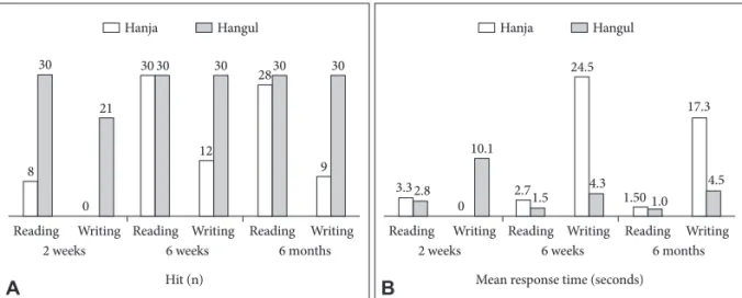
Hit-rates for Hangul reading were perfect (30/30) at every session for language tasks. The mean response time for Hangul reading improved from 2.8 seconds per letter [standard devia- tion (SD) 2.1 sec] to 1.03 seconds per letter (SD 0.2 sec) over the 6-month experimental period. In contrast, Hanja reading, Hanja writing and Hangul writing was markedly impaired ini- tially. However, the number of correct hits increased from 8 to 28, and the mean response time for Hanja reading shortened from 3.3 to 1.5 seconds after six months. Hangul agraphia also improved to almost normal; however, Hanja agraphia persist- ed. The overall results are provided in Fig. 2.
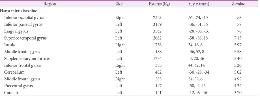
fMRI scans during Hanja reading showed activation of the occipital regions bilaterally including the lingual gyrus, ex-
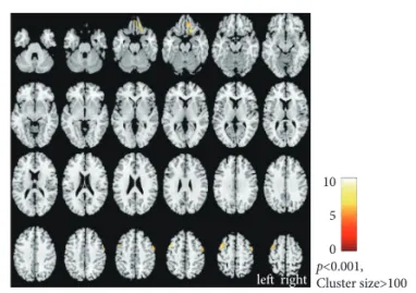
tending to the parietal cortex bilaterally from the prefrontal area, and both insula at the initial time point and six months after the onset of symptoms (Fig. 3, Table 1 and 2). Although the fusiform gyrus was partially activated bilaterally (Fig. 3A, Supplementary Table 2 in the online-only Data Supplement), the patient could not read Hanja. At six months after symptom onset, his ability to read Hanja greatly improved; however fMRI revealed no significant changes in the fusiform gyrus re- gion (Fig. 3B). Instead, both middle frontal gyri showed signif- icantly greater activation after six months (Fig. 4, Table 3).
DISCUSSION
Several models have been proposed to explain the mecha- nisms related to reading and brain localization. The dual route of phonogram-ideogram processing is one such model, which has been predominantly studied for Japanese.1
Two different orthographic systems exist in the Japanese
Fig. 2. Language tasks presented as a bar chart at time of 2 weeks, 6 weeks and 6 months after the symptom onset. A: Total number of hits (correct) during the reading and writing of Hanja and Hangul. B: Mean response time for the same test.
Hit (n) Mean response time (seconds)
Hanja Hangul Hanja Hangul
A B
Reading Writing Reading Writing Reading Writing Reading Writing Reading Writing Reading Writing
2 weeks 2 weeks
8 30
3.32.8 2.71.5 4.3
30 30
9 30 2830
30
12 21
0
10.1
1.50
17.3
1.0 4.5 24.5
0
6 weeks 6 months 6 weeks 6 months
Fig. 3. The activation areas for “Hanja reading” minus “baseline” (A) at 2 weeks and (B) at 6 months after the symptom onset (Threshold at p<0.001, uncorrected, cluster size >100).
A B
p<0.001,
Cluster size>100 p<0.001,
Cluster size>100 10
5 0
10 5 0
left right left right
DND
language [i.e., Kanji (ideogram) and Kana (phonogram)]. Al- though the correlation between Kanji and Kana is not straight- forward, it is generally believed that the anatomical substrates mediating ideograms and phonograms differ. Thus Japanese alexia patients sometimes show dissociative disturbances when reading Kanji or Kana. Lesion studies have shown that angular gyrus involvement more frequently leads to difficulty reading Kana, whereas patients with lesions in the posterior inferior temporal lobe, including the fusiform gyrus, are likely to develop difficulty reading Kanji. These studies also suggest that the anatomical pathways mediating ideograms and pho- nograms differ, that is, the ventral pathway (posterior portions of the middle and inferior temporal gyri) is involved in read- ing ideograms2,4,11,12 and the dorsal pathway (inferior parietal lobe) is used in reading phonograms.1,3,13 Similar findings have been reported on the dissociative reading disturbances seen in lesion case studies regarding Korean orthography. Two pa-
tients with Hanja alexia had a lesion in the left posterior inferi- or temporal lobe,6,7 while one case of Hangul alexia was associ- ated with a lesion in the inferior parietal lobule, insula and cingulate gyrus.7 Another fMRI study in Korean speaking vol- unteers showed that the right fusiform gyrus and adjacent temporo-occipital region seem to be more specifically involved in processing Hanja script.8
Our patient also showed a dissociative disturbance between reading Hanja and Hangul. MRI scans showed two distinct ischemic lesions in the left middle frontal gyrus and left inferi- or parietal lobe with preservation of the posterior inferior tem- poral cortex, which is supposed to be the anatomical substrate for ideogram reading. Nevertheless, he initially presented with severe Hanja alexia and normal Hangul reading. The areas with increased signal on fMRI during Hanja reading were mainly occipital and frontal regions bilaterally rather than the inferior occipito-temporal region, such as the fusiform gyrus.
Table 1. Activated areas of “Hanja reading” compared to “baseline” at 2 weeks after stroke onset
Region Side Extents (KE) x, y, z (mm) Z-value
Hanja minus baseline
Inferior occipital gyrus Right 7548 36, -74, -10 >8
Inferior parietal gyrus Left 3139 -36, -51, 56 >8
Lingual gyrus Left 3562 -28, -86, -16 >8
Superior temporal gyrus Left 2682 -58, -38, 18 7.15
Insula Right 758 34, 18, 8 5.97
Middle frontal gyrus Left 188 -36, 52, 8 5.58
Supplementary motor area Left 1734 -4, 20, 46 5.40
Inferior frontal gyrus Right 303 44, 32, 14 5.20
Cerebellum Left 402 -30, -28, -34 5.02
Middle frontal gyrus Right 285 34, 52, 6 4.92
Precentral gyrus Left 147 -50, -2, 46 4.32
Caudate Left 141 -12, -6, -16 3.70
It represents corresponding image series in Fig. 3A. Threshold at p<0.001, uncorrected, cluster size >100. MNI coordinates (x, y, z) were measured in millimeters.
MNI: Montreal Neurological Institute.
Table 2. Activated areas of “Hanja reading” compared to “baseline” at 6 months after stroke onset
Region Side Extents (KE) x, y, z (mm) Z-value
Hanja minus baseline
Middle frontal gyrus Left 3916 -46, 4, 52 >8
Middle occipital gyrus Right 5399 32, -96, 4 >8
Middle occipital gyrus Left 4634 -26, -82, 4 7.69
Inferior frontal gyrus Right 1349 40, 30, 12 6.59
Middle frontal gyrus Right 1392 40, 6, 58 6.46
Superior temporal gyrus Left 345 -58, -36, 18 6.42
Superior frontal gyrus (medial part) Left 1404 -10, 20, 40 6.31
Middle frontal gyrus Left 169 -36, 48, 8 4.85
It represents corresponding image series in Fig. 3B. Threshold at p<0.001, uncorrected, cluster size >100. MNI coordinates (x, y, z) were measured in millimeters.
MNI: Montreal Neurological Institute.
Jiwon Yang et al.
Dissociation of Hanja and Hangul Reading
In addition, as the patient’s Hanja alexia improved, activation increased in both middle frontal gyri.
These findings, 1) initial profound Hanja alexia with a de- structive lesion involving the left frontal lobe and 2) improved Hanja alexia with increased fMRI activation in both middle frontal gyri, suggest the possibility that the frontal lobe, as well as the fusiform gyrus, could contribute to mediating Korean ideogram reading.
Differences exist between the usage, exposure rate, and age of acquisition of the Korean and Japanese ideographic systems.
In contrast to Japanese orthography which heavily incorpo- rates the usage of Kanji, the Korean system depends less on Chinese derived characters, and most Korean words and sen- tences can be communicated without the use of any ideograms (Hanja). Also the same ideograms in Japanese can be pro- nounced in different ways, which is not the case in Korean.5,7 For these reasons, the two language systems may show differ- ent patterns of brain activation when ideograms and phono- grams are read.
It is difficult to designate the exact location associated with Hanja alexia in this study, due to the fact that two ischemic le- sions were concomitantly present. An fMRI study conducted on normal Korean subjects revealed strong activation in the left lateralized middle frontal cortex during Chinese character reading.9,10 Chinese investigators observed a wide area of acti-
vation associated with Chinese letter reading including the left frontal and temporal cortices, the right visual system including the fusiform gyrus, the right parietal lobe and cerebellum.14-16 Different regions where associated with reading Chinese char- acters by Korean (Hanja) and Chinese native speakers; howev- er, both groups showed activation in the left frontal area.
Taken together, these studies indicate that different cerebral regions might be associated with reading Chinese characters by Korean (Hanja), Japanese (Kanji) and Chinese native speak- ers. In the case of Korean orthography, different results have been reported regarding the areas involved in processing ideo- grams, which are similar to Japanese6-8 and Chinese9,10 popula- tions. Our case study supports that the frontal lobe might play a role in reading Korean ideograms.
Increasing activation in the bilateral middle frontal gyri in parallel with Hanja reading improvements may also represent the activation of language restorative processes from perile- sional areas or compensatory processes from contralateral neu- ral circuits post stroke.17 However, because this study was con- ducted on one subject, we cannot comment on the statistical significance of our findings.
Our study has some limitations. First, the presence of two lesions in the left cerebral hemisphere makes it difficult to con- clude which is causative of his dissociative alexia. The analysis of additional subjects with single lesions associated with ideo- graphic alexia would be needed to answer these questions.
Second, there were no fMRI data on a control group to deter- mine the normal substrates dissociating the two systems.
Third, detailed error analysis of mistaken words was not fully performed. However, our subject showed either fully normal reading or no responses attempted and error analysis was in- adequate. Finally, functional and neurophysiological changes after ischemic stroke, for example diaschisis, need to be con- sidered when interpreting the fMRI results.
In conclusion, we found that not only fusiform gyrus, but also the frontal lobe may be involved in reading Hanja (Kore- an ideograms) after analyzing serial fMRI scans of a patient presenting with dissociative Hanja alexia after an acute isch- emic stroke.
Fig. 4. The middle frontal gyrus bilaterally showed greater activa- tion after six months in parallel with Hanja reading improvement (Threshold at p<0.001, uncorrected, cluster size >100).
p<0.001, Cluster size>100
10 5 0 left right
Table 3. The different activated areas for Hanja reading between two times of 2 weeks and 6 months after stroke onset
Region Side Extents (KE) x, y, z (mm) Z-value
(Hanja > baseline) at 6 months minus (Hanja > baseline) at 2 weeks
Middle frontal gyrus Left 167 -44, 8, 52 5.35
Superior frontal gyrus (orbital part) Right 170 12, 38, -24 5.09
Middle frontal gyrus Right 124 50, 0 ,56 3.93
It represents corresponding image series in Fig. 4. Threshold at p<0.001, uncorrected, cluster size >100. MNI coordinates (x, y, z) were measured in millimeters.
MNI: Montreal Neurological Institute.
DND
Conflicts of Interest
The authors have no financial conflicts of interest.
Acknowledgements
This work was supported by the National Research Foundation of Korea Grant funded by the Korean Government (NRF-2013S1A5A2A03045081).
REFERENCES
1. Iwata M. Neural mechanism of reading and writing in the Japanese language. Funct Neurol 1986;1:43-52.
2. Kawamura M, Hirayama K, Hasegawa K, Takahashi N, Yamaura A.
Alexia with agraphia of kanji (Japanese morphograms). J Neurol Neu- rosurg Psychiatry 1987;50:1125-1129.
3. Sakurai Y, Asami M, Mannen T. Alexia and agraphia with lesions of the angular and supramarginal gyri: evidence for the disruption of se- quential processing. J Neurol Sci 2010;288:25-33.
4. Soma Y, Sugishita M, Kitamura K, Maruyama S, Imanaga H. Lexical agraphia in the Japanese language. Pure agraphia for Kanji due to left posteroinferior temporal lesions. Brain 1989;112 ( Pt 6):1549-1561.
5. Kim H, Na DL. Dissociation of pure korean words and Chinese-deriv- ative words in phonological dysgraphia. Brain Lang 2000;74:134-137.
6. Kwon JC, Lee HJ, Chin J, Lee YM, Kim H, Na DL. Hanja alexia with agraphia after left posterior inferior temporal lobe infarction: a case study. J Korean Med Sci 2002;17:91-95.
7. Kwon M, Kim JS, Lee JH, Sim H, Nam K, Park H. Double dissocia- tion of Hangul and Hanja reading in Korean patients with stroke. Eur Neurol 2005;54:199-203.
8. Lee KM. Functional MRI comparison between reading ideographic and phonographic scripts of one language. Brain Lang 2004;91:245-
9. Yoon HW, Cho KD, Chung JY, Park H. Neural mechanisms of Kore-251.
an word reading: a functional magnetic resonance imaging study.
Neurosci Lett 2005;373:206-211.
10. Yoon HW, Chung JY, Kim KH, Song MS, Park HW. An fMRI study of Chinese character reading and picture naming by native Korean speakers. Neurosci Lett 2006;392:90-95.
11. Kawahata N, Nagata K, Shishido F. Alexia with agraphia due to the left posterior inferior temporal lobe lesion--neuropsychological anal- ysis and its pathogenetic mechanisms. Brain Lang 1988;33:296-310.
12. Sakurai Y, Yagishita A, Goto Y, Ohtsu H, Mannen T. Fusiform type alexia: pure alexia for words in contrast to posterior occipital type pure alexia for letters. J Neurol Sci 2006;247:81-92.
13. Sakurai Y, Terao Y, Ichikawa Y, Ohtsu H, Momose T, Tsuji S, et al.
Pure alexia for kana. Characterization of alexia with lesions of the inferior occipital cortex. J Neurol Sci 2008;268:48-59.
14. Wu CY, Ho MH, Chen SH. A meta-analysis of fMRI studies on Chi- nese orthographic, phonological, and semantic processing. Neuroim- age 2012;63:381-391.
15. Tan LH, Spinks JA, Gao JH, Liu HL, Perfetti CA, Xiong J, et al.
Brain activation in the processing of Chinese characters and words: a functional MRI study. Hum Brain Mapp 2000;10:16-27.
16. Chee MW, Weekes B, Lee KM, Soon CS, Schreiber A, Hoon JJ, et al. Overlap and dissociation of semantic processing of Chinese char- acters, English words, and pictures: evidence from fMRI. Neuroim- age 2000;12:392-403.
17. Pillai JJ. Insights into adult postlesional language cortical plasticity provided by cerebral blood oxygen level-dependent functional MR imaging. AJNR Am J Neuroradiol 2010;31:990-996.