Received: 3 July 2012, Accepted: 11 October 2012 Corresponding author: In Joo Kim
Division of Endocrinology and Metabolism, Department of Internal Medicine, Pusan National University Hospital, Pusan National University School of Medicine, 179 Gudeok-ro, Seo-gu, Busan 602-739, Korea
Tel: +82-51-240-7224, Fax: +82-51-254-3127, E-mail: [email protected]
Pancytopenia Associated with Hypopituitarism in an Acromegaly Patient: A Case Report and a Review of the Literature
Jung Hee Koh, Yong Jae Lee, Ji Hyun Kang, Bo Kwang Choi, Yun Kyung Jeon, Sang Soo Kim, Bo Hyun Kim, In Joo Kim Department of Internal Medicine, Pusan National University School of Medicine, Busan, Korea
We present the case of a patient with acromegaly who had pancytopenia with hypopituitarism secondary to the excision of a pitu- itary macroadenoma and radiation therapy. A 28-year-old man presented with pancytopenia and serum electrolyte abnormalities.
He was diagnosed with acromegaly and underwent surgery and gamma-knife radiotherapy for a pituitary macroadenoma at the age of 22 years. A recent brain magnetic reso nance imaging showed an empty sella, and the basal hormonal profile demonstrated deficiencies of pituitary hormones except thyrotropin. As presenting pancytopenia, his bone marrow biopsy showed hypocellular marrow. The total number of hemocytes increased after hydrocortisone replacement. Hypopituitarism was a possible cause of pan- cytopenia, and glucocorticoids had crucial effects on converting pancytopenia to normal in this case. (Endocrinol Metab 27:308- 313, 2012)
Key Words: Acromegaly, Hypopituitarism, Pancytopenia
INTRODUCTION
Acromegaly is not a common disease and its annual estimated incidence is 3-4 cases per 1 million people worldwide [1]. Hypopi- tuitarism is reported to occur in over 50% of patients with acromeg- aly 5-10 years after radiation therapy [2].
Aplastic anemia is a clinical condition that results from a marked diminution of marrow blood cell production. Its diagnosis is usu- ally based on the presence of pancytopenia accompanied by hy- pocellular marrow without abnormal or malignant cells or fibrosis.
It is sometimes related to connective tissue disease, idiosyncratic responses to certain pharmaceuticals, exposure to toxic chemicals, and systemic diseases, such as infection [3]. While hormonal defi- ciency is rarely thought of a cause of pancytopenia with hypocel- lular bone marrow, the mechanisms of hormonal effects on hema- topoiesis have not yet been fully understood and there have been only a few cases with bone marrow hypoplasia and pancytopenia which were related to hormone insufficiencies. We experienced a case of an acromegalic patient with pancytopenia who developed in hypopituitarism after surgical treatment and radiotherapy but re-
covered completely with hydrocortisone replacement therapy.
CASE REPORT
A 28-year-old man was referred from a private clinic with a his- tory of mandibular hypertrophy and peripheral thickening in the hands and feet, 6 years ago. His insulin like growth factor 1 (IGF-1) level increased to 1,197.6 ng/mL (normal, 114-492). Oral glucose tol- erance test results revealed that growth hormone (GH) levels were not suppressed (63 ng/mL), so the patient was diagnosed with acro- megaly. Pituitary macroadenoma had been found on magnetic reso- nance imaging (MRI) (Fig. 1A), and he underwent surgery through the transsphenoidal approach for pituitary macroadenoma removal the same year. One month after surgery, the IGF-1 level was 1,318.1 ng/mL, thyroid function tests were normal, and the prolactin level was 15.5 ng/mL (normal, 0-20). The adrenocorticotropic hormone (ACTH) level was 33.8 pg/mL (normal, 10-60) in the morning. MRI was performed 2 months after surgery and showed a remnant ade- noma (Fig. 1B). Then, he underwent gamma-knife radiosurgery (GKS). After GKS, he was transferred to the endocrinology depart-
This is an Open Access article distributed under the terms of the Creative Commons Attribution Non-Commercial License (http://creativecommons.org/licenses/by-nc/3.0/) which permits unrestricted non-commercial use, distribution, and reproduction in any medium, provided the original work is properly cited.
Copyright © 2012 Korean Endocrine Society
Fig. 1. A T1-weighted sagittal (upper) and coronal views (lower) of a pituitary macroadenoma by magentic resonance imaging which was taken before and after surgery. (A) A 40-mm macroadenoma invading the cavernous sinus before surgery. (B) Two months after pituitary surgery, a residual mass was seen around the cav- ernous sinus on both sides. (C) Three years after radiotherapy, the remnant mass decreased in both cavernous sinuses and pituitary gland was enhanced on the cor- onal view (arrow). (D) Six years after pituitary surgery, the pituitary gland was atrophied, and the sella was empty and filled with cerebrospinal fluid.
A B C D
GH (ng/mL) IGF-1 (ng/mL)
50
40
30
20
10
1,400 1,200 1,000 800 600 400 200
6 yr
before 4 yr
before 5 yr
before Surgery
1st admission GH IGF-1 Radiotherapy
20 mg
Sandostatin® LAR® SQ monthly
20 mg 30 mg
3 yr
before 2 yr
before 6 yr before
Fig. 2. Clinical course of the patient (changes in insulin like growth factor [IGF]-1 and growth hormone [GH] levels).
serial decrease and Sandostatin LAR dosage was tapered from 12 months ago (Fig. 2). Two months after Sandostatin LAR discontinu- ation, his complete blood count (CBC) test showed a slight decrease in leukocytes (3.2 × 10⁹/L), anemia (hemoglobin, 10.8 g/dL), and normal platelet counts (167 × 109/L).
The patient visited the emergency department 6 years after radio- ment and still had a high level of IGF-1 (1,298.7 ng/mL). Thus, we
prescribed bromocriptine 2.5 mg per day for 1 month, which was changed to Sandostatin LAR (Octreotide acetate, Novatis Pharm., Basel, Switzerland) 20 mg monthly. After 8 months, the IGF-1 levels were still high, so we increased the dose of Sandostatin LAR to 30 mg monthly after using 20 mg. The IGF-1 levels finally showed a
nausea, and vomiting. On admission, his vital signs were as follows:
blood pressure, 100/60 mmHg; pulse rate, 80 beat/min; and body temperature, 36.5°C. He had not been medicated during the past 4 weeks. CBC test results showed pancytopenia: leukocytes, 1.5 × 10⁹/L; neutrophils, 0.72 × 10⁹/L; hemoglobin, 11.0 g/dL; and plate- lets, 67 × 109/L. Peripheral blood smear (PBS) also revealed pancy- topenia with both a low reticulocyte produrction index (0.13) and a relatively low reticulated platelet count (4.1%; normal, 0.5-5.5%).
Hyponatremia was also noted: sodium, 109.9 mmol/L; potassium, 3.80 mmol/L; and chloride, 81.4 mmol/L. Hyponatremia was not corrected 2 days after hypertonic saline administration. The basal hormone status on admission, revealed cortisol deficiency-cortisol, 3.18 μg/dL (normal, 5-25) and ACTH, 17.15 pg/mL (normal, 10-60).
IGF-1 was 45.78 ng/mL (normal, 114-492). Thyroid function test re- sults were suggestive of euthyroid sick syndrome-triiodothyronine (T3), 72.3 ng/dL (normal, 80-170); free thyroxine (T4), 1.81 ng/dL (normal, 0.80-2.10); and thyroid stimulationg hormone (TSH), 3.03 µIU/L (normal, 0.3-5.0). Hypogonadism was also noted: luteinizing hormon, 0 mIU/mL (normal, 0.4-5.7); follicular stimulating hor- mone, 1.8 mIU/mL (1.5-12.4); and testosterone, 0.45 ng/mL (normal, 2.5-18.3). Combined anterior pituitary function tests demonstrated hypopituitarism in multiple axes except for TSH. MRI of the pitu- itary gland revealed an empty sella (Fig. 1D). He was administered oral hydrocortisone (10 mg) daily from the fourth admission day onward. Hypertonic saline was infused until the fifth admission day and was discontinued when the sodium level was corrected to 120.2 mmol/L. The sodium level was further corrected to 130.4 mmol/L on the third day of hydrocortisone replacement therapy.
The number of leukocytes also increased to 4.36 × 10⁹/L on the fifth day of the therapy. Because neutropenia continued for 3 years at the level of 1.1-1.5 × 10⁹/L, he underwent bone marrow biopsy which revealed a hypocellular marrow (Fig. 3).
Five months after discharge, the patient visited the emergency department again due to general weakness, diarrhea, nausea, and vomiting. He reported that he had not taken any medications, in- cluding hydrocortisone after discharge from the hospital. Complete blood cell counts showed pancytopenia: leukocytes, 1.18 × 10⁹/L;
neutrophils, 0.86 × 10⁹/L; hemoglobin, 9.1 g/dL; and platelets, 106
× 109/L. On PBS, normocytic anemia and neutropenia was noted, the reticulocyte production index was 0.21, mean cell volume was 82 fL (normal, 80-94), and mean cell hemoglobin was 28.5 pg (nor- mal, 27-32). The iron profiles were as follows: iron, 50 μg/dL (nor-
mal, 80-200); total iron binding capacity, 216 μg/dL (normal, 252- 456); and ferritin 327.2 ng/mL (normal, 15-332). Vitamin B12 and folate levels were normal: 856.12 pg/mL (normal, 200-950) and 7.04 ng/mL (normal, 3-17), respectively. The serum erythropoietin level was 38.2 mIU/mL (reference, 3.5-16.2). Hyponatremia and hypoka- lemia were also noted: sodium, 118 mmol/L; potassium, 3.45 mmol/
L. To evaluate hyponatremia, we measured serum osmolarity which was 251 mOsm/kg and urine osmolarity which was 231 mOsm/kg.
These results were suggestive of hypotonic hyponatremia with dys- function of urinary concentration. Biochemical tests revealed nor- mal liver and kidney functions. In addition, C-reactive protein (CRP) and procalcitonin were measured to rule out infection: CRP, 0.51 mg/dL (normal, 0-0.5); procalcitonin, 0.06 ng/mL (normal, ≤ 0.05).
Stool test results obtained at 2 weeks were negative for leukocytes and cultures. In addition, abdominal computed tomography was normal.
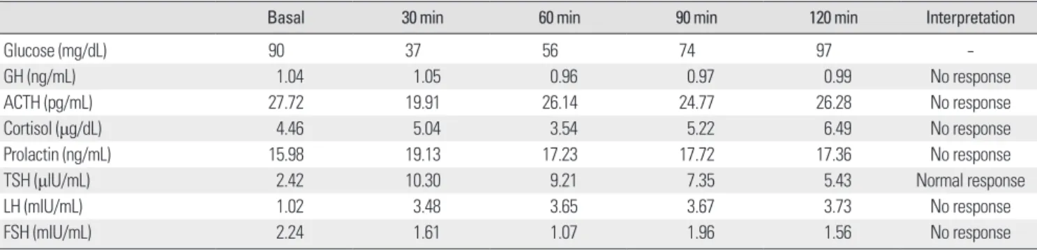
In order to ascertain a role of pituitary hormone on pancytope- nia, basal pituitary hormone tests were performed. Serum cortisol levels were lower than reference values-4.42 μg/dL at 8:00 AM and 4.38 μg/dL at 4:00 PM (normal, 5-25). Thyroid function test re- sults were normal: T3, 71 ng/dL; free T4, 1.21 ng/dL; and TSH, 2.42 μIU/L. The testosterone level was 0.4 ng/mL (normal, 2.45-18.3).
Combined anterior pituitary stimulation tests demonstrated hypo- pituitarism in all axes except for TSH (Table 1).
Again, the patient received replacement therapy with oral hydro- cortisone 10 mg daily. One week after hydrocortisone replacement, the number of hemocytes increased: leukocytes, 3.17 × 10⁹/L; he- Fig. 3. High-power view of a bone marrow section. Decreased hematopoiesis with hypocellurarity are noted (H&E stain, × 100).
moglobin, 9.2 g/dL; and platelets, 316 × 109/L. The serum sodium level was 137.1 mmol/L, which was in the normal range. He was restored to a good general condition. He was prescribed hydrocor- tisone (10 mg) daily and discharged from the hospital. His last lab- oratory results were almost within the normal range 3 month after hydrocortisone replacement therapy: sodium, 141.7 mmol/L; potas- sium, 4.19 mmol/L; leukocytes, 3.76 × 10⁹/L; hemoglobin, 12.2 g/dL;
and platelets, 258 × 109/L (Fig. 4).
DISCUSSION
Pancytopenia rarely occurs in bone marrow failure syndrome secondary to hypopituitarism. There have been several reports of pancytopenia that are related to hormone deficiency. We reviewed nine cases by using Medline search (Table 2). Six of these cases were associated with Sheehan syndrome [4-9], one was associated with macroprolactinoma [10], and one with hypothalamic glioma and one with suprasellar germinoma [11,12]. To the best of our knowledge, this is the first case of pancytopenia secondary to hy- popituitarism.
It is not clear which hormones are important to induce or improve pancytopenia in cases of pancytopenia due to hypopituitarism.
Most patients recover by simultaneous replacement of cortisol and thyroid hormone [4-7,9]. Lee [12] described a case of an 11-year-old girl who developed postablative hypothyroidism and prolonged pancytopenia after radiotherapy for a suprasellar germinoma while receiving cortisol replacement. Hence, the authors believed that hypothyroidism was a major contributing factor to pancytopenia in patients with panhypopituitarism. Conversely, Laway et al. [8] re- ported a patient with Sheehan’s syndrome who had pancytopenia and hyponatremia. The patient was treated with hydrocortisone and showed a complete recovery from pancytopenia. They con- cluded that glucocorticoids play a crucial role in reversing pancyto- penia in Sheehan’s syndrome [8]. Badawi et al. [11] reported a young male patient with long-term panhypopituitarism and pancytopenia attributable to poor adherence to androgen replacement therapy, which resolved after institution of testosterone therapy. They em- phasized the importance of testosterone in the treatment of hypo- pituitarism-induced pancytopenia.
Even though hormonal effects of hormone levels on myelopoie- Table 1. Results of combined anterior pituitary hormone stimulation tests
Basal 30 min 60 min 90 min 120 min Interpretation
Glucose (mg/dL) 90 37 56 74 97 -
GH (ng/mL) 1.04 1.05 0.96 0.97 0.99 No response
ACTH (pg/mL) 27.72 19.91 26.14 24.77 26.28 No response
Cortisol (μg/dL) 4.46 5.04 3.54 5.22 6.49 No response
Prolactin (ng/mL) 15.98 19.13 17.23 17.72 17.36 No response
TSH (μIU/mL) 2.42 10.30 9.21 7.35 5.43 Normal response
LH (mIU/mL) 1.02 3.48 3.65 3.67 3.73 No response
FSH (mIU/mL) 2.24 1.61 1.07 1.96 1.56 No response
ACTH, adrenocorticotropin; FSH, follicular stimulating hormone; GH, growth hormone; LH, luteinizing hormone; TSH, thyroid stimulating hormone.
WBC (109/µL) Hb (g/L) Platelet (109/L)
5.0
4.0
3.0
2.0
1.0
140
120
100
80
400
300
200
100
1 wk
before 1 wk
later 1 day
before WBC Hb Platelet
1 molater 3 mo later
Fig. 4. Changes in hematologic findings through hydrocortisone replacement therapy.
Hb, hemoglobin; WBC, white blood cell.
Hydrocortisone replacement start
Authors of cases No. Hormonal abnormalities Causes of hypopituitarism Replacement ofhormone to improve pancytopenia Kim et al. [4] 1 Hypocortisolism, hypothyroidism and
hypogonadism Sheehan syndrome Prednisolone and L-thyroxine Ozdogan et al. [5] 1 Hypocortisolism, hypothyroidism,
hypogonadism and GH deficiency Sheehan syndrome Prednisolone and L-thyroxine Gokmen Akoz et al. [6] 1 Hypocortisolism, hypothyroidism,
hypogonadism and GH deficiency Sheehan syndrome Prednisolone and L-thyroxine Badawi et al. [11] 1 Hypocortisolism, hypothyroidism,
hypogonadism and GH deficiency Hypothalamic glioma Testosterone Laway et al. [7] 3 Hypocortisolism, hypothyroidism,
hypogonadism and GH and PRL deficiency
Sheehan syndrome Prednisolone and L-thyroxine Lee [12] 1 Hypocortisolism, hypothyroidism,
GH deficiency and central DI Suprasellar germinoma L-thyroxine Laway et al. [8] 1 Hypocortisolism, hypothyroidism,
hypogonadism and GH and PRL deficiency Sheehan syndrome Prednisolone Holmes et al. [10] 1 Hypocortisolism, hypothyroidism,
hypogonadism and hyper PRL Macroprolactinoma Hydrocortisone, L-thyroxine and testosterone Fatma et al. [9] 1 Hypocortisolism, hypothyroidism,
hypogonadism and PRL deficiency Sheehan syndrome Hydrocortisone and L-thyroxine
Present case 1 Hypocortisolism and hypogonadism Acromegaly Hydrocortisone
DI, diabetes incipitus; GH, growth hormone; PRL, prolactine.
sis and thrombopoiesis are not clear, erythropoiesis is affected by metabolic needs and pituitary hormones play an important role in the treatment of erythropoiesis. A previous study reported that ane- mia occurred in 32% of patients with hypopituitarism and that the minimum recorded hemoglobin level was 9.3 g/dL [13]. Laway et al. [14] demonstrated that, anemia, leukopenia, thrombocytopenia, bicytopenia, and pancytopenia were significantly higher in patients who were diagnosed with Sheehan’s syndrome than in those who were not. Among pituitary hormones, thyroid hormone and testos- terone have been shown to significantly stimulate erythroid pro- genitors, increase erythropoietin production and potentiate eryth- ropoietin action [15,16]. Steroid hormones directly stimulate eryth- ropoiesis [13]. In addition, GH and IGF-1 are known to have a direct effect on erythroid and myeloid precursor progenitor cells [17,18].
Our patient showed mild pancytopenia for 3 years and pancyto- penia worsen during two events of electrolyte imbalance. During each event, the patient had the similar symptoms, such as nausea, vomiting and diarrhea, and the symptoms improved with physio- logic doses of hydrocortisone. Thus, he was diagnosed as having hypopituitarism with adrenal crises, which was probably caused by the stress of gastroenteritis. Uniquely, the patient showed insuf- ficiencies of pituitary hormones except thyrotropin. Even testoster- one level was low, though it had been low since he diagnosed ac-
romegaly. We ruled out testosterone deficiency because testoster- one levels had been low for a long time and there were no symp- toms associated with testosterone deficiency. At first, we recom- mended testosterone replacement therapy, but he refused it. More- over, after only administration of hydrocortisone, the patient showed a full recovery from pancytopenia. Therefore, this case supports the hypothesis that glucocorticoids are effective in treating pancy- topenia induced by hypopituitarism.
In conclusion, hypopituitarism itself might be a possible cause of pancytopenia, although it rarely develops the aforementioned he- matologic abnormalities due to an excess of GH in acromegaly.
Physicians should consider hormone insufficiency a cause of pan- cytopenia in patients who have undergone prior surgical treatment or radiotherapy for pituitary gland lesions.
요 약
말단비대증으로치료이후뇌하수체기능저하증을 동반한범혈
구감소증을보인환자에서당질코르티코이드보충이후범혈구감소
증의호전을보인증례를보고하는바이다. 본증례는 28세남자환 자로, 범혈구감소증과혈청전해질장애를나타내었다. 환자는 23세
때뇌하수체선종으로인한말단비대증을진단받고외과적선종제
거수술과감마나이프방사선치료를받은상태였다. 뇌하수체자기
공명영상에서공터키안증후군소견과함께갑상선자극호르몬을제
외한다른뇌하수체전엽호르몬의감소를동반하고있었다. 동시에
지속적인범혈구감소증을보여시행한골수검사에서저세포성골
수소견을보였다. 히드로코르티손투여후혈구세포의수는모두
증가하였다. 본증례를통해뇌하수체기능저하증은범혈구감소증
의원인이될수있으며, 당질코르티코이드의보충이범혈구감소증
의회복에결정적역할을시사하고있다.
REFERENCES
1. Holdaway IM, Rajasoorya C: Epidemiology of acromegaly. Pituitary 2:29- 41, 1999
2. Melmed S, Colao A, Barkan A, Molitch M, Grossman AB, Kleinberg D, Clemmons D, Chanson P, Laws E, Schlechte J, Vance ML, Ho K, Giustina A; Acromegaly Consensus Group: Guidelines for acromegaly management:
an update. J Clin Endocrinol Metab 94:1509-1517, 2009
3. Segel GB, Lichtman MA: Aplastic anemia: acquired and inherited. In:
Kaushansky K, Williams WJ. Williams Hematology. 8th ed. pp569-590, New York, McGraw-Hill Medical, 2010
4. Kim DY, Kim JH, Park YJ, Jung KH, Chung HS, Shin S, Yun SS, Park S, Kim BK: Case of complete recovery of pancytopenia after treatment of hy- popituitarism. Ann Hematol 83:309-312, 2004
5. Ozdogan M, Yazicioglu G, Karadogan I, Cevikol C, Karayalcin U, Undar L:
Sheehan’s syndrome associated with pancytopenia due to marrow aplasia:
full recovery with hormone replacement therapy. Int J Clin Pract 58:533- 535, 2004
6. Gokmen Akoz A, Atmaca H, Ustundag Y, Ozdamar SO: An unusual case of pancytopenia associated with Sheehan’s syndrome. Ann Hematol 86:
307-308, 2007
7. Laway BA, Bhat JR, Mir SA, Khan RS, Lone MI, Zargar AH: Sheehan’s syndrome with pancytopenia: complete recovery after hormone replace- ment (case series with review). Ann Hematol 89:305-308, 2010
8. Laway BA, Mir SA, Bhat JR, Lone MI, Samoon J, Zargar AH: Hemato- logical response of pancytopenia to glucocorticoids in patients with Shee- han’s syndrome. Pituitary 15:184-187, 2012
9. Fatma M, Mouna E, Nabila R, Mouna M, Nadia C, Mohamed A: Sheehan’s syndrome with pancytopenia: a case report and review of the literature. J Med Case Rep 5:490, 2011
10. Holmes GI, Shepherd P, Walker JD: Panhypopituitarism secondary to a macroprolactinoma manifesting with pancytopenia: case report and litera- ture review. Endocr Pract 17:e32-e36, 2011
11. Badawi MA, Salih F, Al-Humaidi AA, El Khalifa MY, Elhadd TA: Bone marrow hypoplasia responsive to testosterone therapy in a patient with panhypopituitarism: need for adherence to androgen replacement. Endocr Pract 14:229-232, 2008
12. Lee AC: Pancytopenia secondary to hypopituitarism may just be due to hypothyroidism alone. Ann Hematol 89:1181, 2010
13. Nishioka H, Haraoka J: Hypopituitarism and anemia: effect of replace- ment therapy with hydrocortisone and/or levothyroxine. J Endocrinol In- vest 28:528-533, 2005
14. Laway BA, Mir SA, Bashir MI, Bhat JR, Samoon J, Zargar AH: Prevalence of hematological abnormalities in patients with Sheehan’s syndrome: re- sponse to replacement of glucocorticoids and thyroxine. Pituitary 14:39- 43, 2011
15. Moriyama Y, Fisher JW: Effects of testosterone and erythropoietin on ery- throid colony formation in human bone marrow cultures. Blood 45:665- 670, 1975
16. Dainiak N, Hoffman R, Maffei LA, Forget BG: Potentiation of human erythropoiesis in vitro by thyroid hormone. Nature 272:260-262, 1978 17. Merchav S, Tatarsky I, Hochberg Z: Enhancement of erythropoiesis in vi-
tro by human growth hormone is mediated by insulin-like growth factor I.
Br J Haematol 70:267-271, 1988
18. Sohmiya M, Kato Y: Effect of long-term administration of recombinant human growth hormone (rhGH) on plasma erythropoietin (EPO) and hae- moglobin levels in anaemic patients with adult GH deficiency. Clin Endo- crinol (Oxf) 55:749-754, 2001