Partial denture metal framework may harbor potentially pathogenic bacteria
Cristiane Machado Mengatto1*, Leonardo Marchini2, Luciano Angelo de Souza Bernardes3, Sabrina Carvalho Gomes1, Alecsandro Moura Silva4, Célia Marisa Rizzatti-Barbosa5
1Department of Conservative Dentistry, Federal University of Rio Grande do Sul, School of Dentistry, Porto Alegre, Rio Grande do Sul, Brazil
2Department of Preventive and Community Dentistry, University of Iowa, College of Dentistry, Iowa City, Iowa, USA
3Department of Science and Technology, State University of Santa Cruz, School of Computer Science, Ilheus, Bahia, Brazil
4Department of Dental Materials and Prosthodontics, State University Julio de Mesquita Filho, School of Dentistry, Sao Jose dos Campos, Sao Paulo, Brazil
5Department of Prosthodontics and Periodontology, State University of Campinas, Piracicaba Dental School, Piracicaba, Sao Paulo, Brazil
PURPOSE. The aim of this study was to characterize and compare bacterial diversity on the removable partial denture (RPD) framework over time. MATERIALS AND METHODS. This descriptive pilot study included five women who were rehabilitated with free-end mandibular RPD. The biofilm on T-bar clasps were collected 1 week (t1) and 4 months (t2) after the RPD was inserted (t0). Bacterial 16S rDNA was extracted and PCR amplified.
Amplicons were cloned; clones were submitted to cycle sequencing, and sequences were compared with GenBank (98% similarity). RESULTS. A total of 180 sequences with more than 499 bp were obtained. Two phylogenetic trees with 84 (t1) and 96 (t2) clones represented the bacteria biofilm at the RPD. About 93% of the obtained phylotypes fell into 25 known species for t1 and 17 for t2, which were grouped in 5 phyla: Firmicutes (t1=82%; t2=60%), Actinobacteria (t1=5%; t2=10%), Bacteroidetes (t1=2%; t2=6%), Proteobacteria (t1=10%;
t2=15%) and Fusobacteria (t1=1%; t2=8%). The libraries also include 3 novel phylotypes for t1 and 11 for t2. Library t2 differs from t1 (P=.004); t1 is a subset of the t2 (P=.052). Periodontal pathogens, such as F. nucleatum, were more prevalent in t2. CONCLUSION. The biofilm composition of the RPD metal clasps changed along time after RPD wearing. The RPD framework may act as a reservoir for potentially pathogenic bacteria and the RPD wearers may benefit from regular follow-up visits and strategies on prosthesis-related oral health instructions.
[J Adv Prosthodont 2015;7:468-74]
KEY WORDS: Framework; Denture plaque; Microorganisms; 16S rDNA gene; Removable partial denture
INTRODUCTION
There has been a longstanding controversy over the influ-
ence of wearing removable partial denture (RPD) on oral health. While many studies report that the RPD alters nei- ther caries prevalence nor periodontal status in the long- term1-4 others argue that there is a detrimental effect of RPD on abutment teeth.5-7 Furthermore, the literature agrees that dentures, both partial and complete, are considered reser- voirs for various microorganism species, particularly oppor- tunistic pathogens associated with systemic and oral diseas- es, such as caries, dentoalveolar abscesses, noma lesions, endocarditis, aphthous ulcers, denture stomatitis, and peri- odontal disease.8-18 For example, caries-associated bacteria, such as Streptococcus mutans19,20 and Bifidobacteria spp.14, and periodontal pathogens11,17,21 were identified in the complete denture plaque and oral cavity of patients even after the extraction of all teeth. However, no study has ever investi- gated the microorganisms adhering to the surface of the RPD framework. Thus, the microbial species adhering to
Corresponding author:
Cristiane Machado Mengatto
Department of Conservative Dentistry, School of Dentistry, Federal University of Rio Grande do Sul, Rua Ramiro Barcelos #2492, Porto Alegre, Rio Grande do Sul, Zipcode 90035-003, Brazil
Tel. 55 51 9991 4176: e-mail, cristiane.mengatto@ufrgs.br
Received June 5, 2015 / Last Revision September 15, 2015 / Accepted October 20, 2015
© 2015 The Korean Academy of Prosthodontics
This is an Open Access article distributed under the terms of the Creative Commons Attribution Non-Commercial License (http://creativecommons.
org/licenses/by-nc/3.0) which permits unrestricted non-commercial use, distribution, and reproduction in any medium, provided the original work is properly cited.
This research was supported by the The State of Sao Paulo Research Foundation, Brazil (Grant# 01058-3).
the inner surface of the cobalt-chromium alloy clasp remain unknown.
Culture-dependent efforts were made to analyze chang- es in oral bacterial composition of denture wearers.5,6,20-23 However, these studies have been limited by the difficulties of in vitro growth techniques. Advances in molecular tech- niques have allowed the identification of many species of oral microorganisms, including those that cannot be culti- vated.8,10,15,16,24,25 A recent in vitro study showed that poten- tially pathogenic microorganisms from human saliva adhere more largely at framework alloys than at acrylic resin sur- face and that cobalt-chromium alloy has a unique pattern of microorganism biofilm formation compared to other investigated surfaces.26 Other authors evaluated the microbial diversity of supra and subgingival plaque of RPD wearers, using denaturing gradient gel electrophoresis (DGGE), a culture-independent method.7 They observed that health- associated genera tended to decrease, whereas the disease- associated species including Streptococcus mutans tended to increase. Thus, it is possible that the oral environment may be altered by RPD treatments.
To the best of our knowledge, no study determined bacterial diversity on RPD framework in vivo through cul- ture-independent methods. Therefore, the objectives of this preliminary descriptive study are 1) to characterize bac- terial community adhered to the RPD clasps and 2) identify changes in bacterial diversity at two time points: 1 week and 4 months after RPD placement. Our hypothesis is that the bacterial diversity on the RPD framework would change over time.
MATERIALS AND METHODS
The dental records of 130 partially dentate patients referred to the Piracicaba School of Dentistry, State University of Campinas, Sao Paulo, Brazil, were assessed. Individuals were eligible if they were completely edentulous at the maxilla and used previous complete denture and partially edentate at the mandible with no previous RPD (n=54).
Twenty eight individuals did not respond to the telephone contact. The remaining (n=26) were clinically examined and
included if they were systemically healthy, without peri- odontitis (clinical attachment loss no more than 3 mm with no bleeding on probing), no smokers, with no presence of restorations, active caries, and crowns, no need of pre-pros- thetic surgery, no prescription of systemic antibiotics or chemical plaque control in the last six months before the study commencement. Only patients without mandibular posterior teeth and at least 6 mandibular teeth were to be included in order to standardize RPD framework design.
A total of five women (60 to 74 years, mean age 67.6) composed the sample. The study was approved by the insti- tutional ethical review board and the subjects signed informed consent.
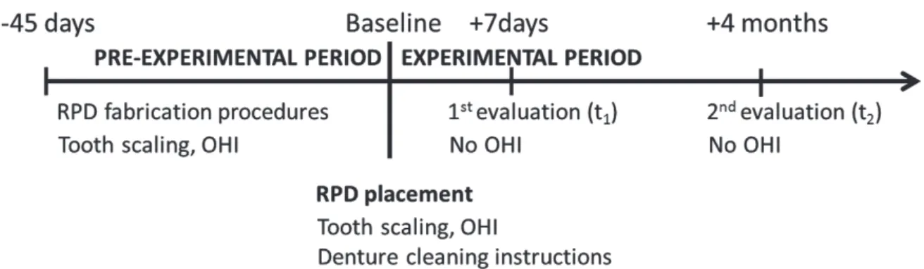
During a pre-experimental period (approximately 45 days before RPD delivery) supragingival scaling and polish- ing were performed and oral hygiene instructions (OHI) were given. The OHI included how to properly floss and how to brush the RPD and the teeth with soft toothbrush and specific toothpaste (Colgate Triple Action, Colgate Palmolive Industry Com., Sao Paulo, Brazil). Subjects were instructed to remove the dentures for cleaning after each meal and before sleeping. The Fig. 1 shows the fluxogram of the study.
The clinical procedures to fabricate maxillary complete dentures and mandibular RPDs were performed by a single professional and followed a strict protocol for the produc- tion and placement of the dentures.27 The cobalt-chromium RPD design was standardized and fabricated by the same dental technician. The clasps used were T-bar (Roach) retainers for direct abutment teeth (canines). The clasps were about 1 mm distant from the gingival edge. The den- ture bases and/or retainers were connected by a lingual plate that was seated on cingulum rests.
At t0 (day 0, baseline), the new maxillary complete den- ture and mandibular RPD were delivered to patients. Oral hygiene was checked and instructions were repeated at the delivery appointment. Patients were oriented to abstain from any form of chemical control during the study. They also received reinstructions on how to perform tooth brushing as well as denture cleaning.
Biofilm was sampled after 1 week (t1) and 4 months (t2)
Fig. 1. Timeline of the study after enrolment process, interventions, and assessments performed on participants.
(RPD) removable partial denture; (OHI) oral hygiene instructions.
of RPD delivery. All subjects consumed no food or bever- ages 30 minutes prior to the biofilm collection. First, the RPD was removed from the patient’s mouth, washed with saline solution for 10 seconds, and left dry on paper towel for 1 minute. Then, the biofilm adhered on the inner sur- face of the T-bar clasps was collected with sterile curettes, pooled in a test tube containing 100 μL of DNA extraction solution, and stored at -20°C. rDNA extraction and ethanol precipitation steps followed a previously described proto- col.15 Concentrated rDNA was suspended in 20 μL 10 mM Tris-HCl pH 7.5, 1 mM EDTA solution and PCR-amplified by using universally conserved primers D88 and E94.16 Briefly, 1 μL of each sample was added to 49 μL of reac- tion mixture containing 5 μL 10 × PCR Buffer (Promega Corporation, Madison, WI, USA), 1.25 unit Taq DNA Polymerase (Promega Corporation), 20 pmol of each prim- er, and 0.2 mM of each deoxyribonucleotides. PCR amplifi- cation was performed in a DNA thermal cycler (PTC-100, MJ Research, Watertown, MA, USA), as reported previous- ly.15 The PCR products were purified by using spin columns (Wizard Clean-up System, Promega Corporation).
A total of 3 μL of the purified PCR products were cloned by using the plasmid vector (TOPO TA Cloning® Kit for Sequencing, Invitrogen Life Technologies, Carlsbad, CA).
Transformation used chemically competent cells (One Shot® TOP10 Chemically Competent E. coli, Invitrogen Life Technologies). The next steps for cloning and plasmid DNA purification followed previously described procedures.15
Purified plasmid DNA was sequenced using primer D88.16 Sequencing reactions were performed in a DNA thermal cycler (MJ Research PTC-100, MJ Research, Inc., Waltham, MA, USA) using DYEnamic ET Terminator Cycle Sequencing Kit (Amersham Biosciences Corp., Piscataway, NJ, USA). Once chain termination was com- plete, DNA sequencing was carried out in an ABI Prism 3100 Genetic Analyzer (Life Technologies, Carlsbad CA, USA). The sequencing protocol was 95°C, for 30 seconds, followed by 40 cycles of 95°C, 20 seconds; 50°C, 15 sec- onds, and 60°C, 2 minutes; with a final hold step at 4°C.
The resultant 250 sequences presenting 499 or more nucle- otides of good quality, which was ascertained by inspecting the chromatograms, were pooled and grouped into two 16S rDNA clone libraries, named t1 and t2. Chimeric sequences were identified using Bellerophon (http://foo.maths.uq.edu.
au/huber/bellerophon.pl) and discarded. A total of 180 reminiscent sequences, including 84 from t1 and 96 from t2, were compared with sequences of known phylotypes from GenBank (http://www.ncbi.nlm.nih.gov) using the BLASTn algorithm. Sequence similarity of 98% or greater was used as the definition of a species-level cluster. Clones that were less than 98% similar to the closest known organisms were supposed to represent novel phylotypes.16 Contiguous sequences were assembled with the Phred/Phrap/Consed software package (http://www.phrap.org), aligned with the CLUSTAL W software. Phylogenetic trees were construct- ed with MEGA 5 software, according to the calculation of a Jukes and Cantor distance matrix using the neighbor-join-
ing method. Bootstrap confidence values for branching nodes were inferred by generating 1,000 resampling trees.
The percentage of coverage was calculated by Good’s method with the formula [1 – (n / N)] × 100, where n is the number of phylotypes in a sample represented by one clone (singletons) and N is the total number of sequences in that sample. The computer program ∫- LIBSHUFF (http://whitman.myweb.uga.edu/libshuff.html) was used to statistically compare the clone libraries between time points (P ≤ .05). Chao28 and ACE29 (Abundance-based Coverage Estimator) statistics were calculated using MOTHUR (www.
mothur.org). The Chao and ACE estimators are statistical approaches used to estimate the species diversity and rich- ness of microbial communities. Chao calculation considers the number of observed species, the number of singletons (species captured once), and the number of doubletons (species captured twice).28 The observed species (Sobs) rep- resent the microorganisms collected and identified. The ACE calculation considers the data from all species with fewer than 10 individuals, rather than just singletons and doubletons.29 Bacteria diversity was considered the depen- dent variable and time was considered independent vari- able. The sequences representing novel phylotypes were deposited at GenBank under the following accession num- bers KF715649 to KF715662.
RESULTS
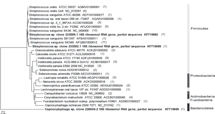
Phylogenetic trees are shown in Fig. 2 and Fig. 3 and reveal 25 known phylotypes for t1 and 17 known phylotypes for t2. The library t1 also included 3 novel phylotypes while t2 had 11 novel phylotypes. The phylotypes accounted for 86% of t1 and 80% of t2 cloned sequences, according to Good’s population coverage.
The ∫-Libshuff analysis indicated that library t1 is contained in t2 (P = .05), but t2 differed from t1 (P = .004). So, community structure of the first library can be considered a subset of the second. When OTU (operational taxonomic unit) definition of 0.02 is considered, the t2 shared approximately 40% of its membership (11 phylotypes) with those of t1, while 17 phylo- types were exclusive of t1 and 17 were unique to t2.
The sequences were assigned to five phyla: Firmicutes (82% of t1 and 60% of t2), Actinobacteria (5% of t1; 10%
of t2), Bacteroidetes (2% of t1; 6% of t2), Proteobacteria (10% of t1; 15% of t2) and Fusobacteria (1% of t1; 8% of t2) (Fig. 2 and Fig. 3). The major phyla were different between the libraries: the Firmicutes were decreased, while the Proteobacteria as well as other minor phyla were increa- sed in t2. Sequences with more than 98% homology to Genbank represented 13 genera in t1 and 9 genera in t2. Seven genera were shared between t1 and t2 libraries (Veillonella, Streptococcus, Capnocytophaga, Fusobacterium, Haemophylus, Neisseria, and Lautropia). The Collector’s curves, estimated by Chao and ACE, represent that the spe- cies richness of the two libraries differs; it was highest in t1 but lower in t2 (Fig. 4).
Fig. 2. Phylogenetic tree with the highest-scored BLAST search results from 84 clones of library t1. The right side of the Fig. shows grayscale bars that represent the distribution of phylotypes among 5 phyla. The scale bar represents
evolutionary distance (5% nucleotide sequence divergence). The organism names in bold letters mark the new phylotypes identified in the project. Final codes correspond to GenBank accession numbers.
Fig. 3. Phylogenetic tree with the highest-scored BLAST search results from 96 clones of library t2. The right side of the Fig. shows grayscale bars that represent the distribution of phylotypes among 5 phyla. The scale bar represents
evolutionary distance (5% nucleotide sequence divergence). The organism names in bold letters mark the new phylotypes identified in the project. Final codes correspond to GenBank accession numbers.
DISCUSSION
The present study revealed that the RPD may act as a reser- voir of bacteria species, many of them putative pathogens mainly associated with periodontal disease and caries.
The literature has posed that RPD may be associated with increased risk of dental caries and periodontal dis- ease.5-7,19,20 However, the bacterial pathogens of such associa- tion are not clearly described. Even though more than 700 bacterial species have been identified in distinct oral habitats such as saliva, supra and subgingival biofilms, tongue, hard and soft palate in healthy or diseased conditions,8,10,15,16,24,25
few studies have aimed at evaluating the bacterial composi- tion and shift related to RPD use. To the best of our knowledge, this pilot study is the first to attend this subject by means of culture-independent molecular methods. The advantages of such methods are highlighted at the literature and the present results illustrate them. Additionally, this is the first clinical study which described the diversity of bio- films adhered to the Co-Cr framework in contact to the enamel tooth surface. Most previous studies about the accumulation of biofilm in prosthesis wearers have based their observations on the acrylic denture base biofilm pres- ence and/or composition.7,14,20,30 However, it is considered that the bacterial species adhered to the Co-Cr surfaces may differ from that of acrylic resin surfaces.26 It was shown not only several species colonizing the RPD clasps but also an expressive shift of microbial species over time. The librar- ies population contains a small number of dominant spe- cies (Streptococcus sanguinis and Veilonella parvula) but presents a large number of unique phylotypes, i.e., a high degree of richness. Streptoccoccus was the most abundant genus in both libraries, comprising about a half of the clones (t1 =
56%; t2 = 49%). Other studies showed that 63 to 86% of the initial colonizing bacteria in dental plaque were Streptoccoccus along with some Veillonella and Actinomyces.31 The present study did not find Actinomyces genus in t1, but it was present in t2.
Most Streptococcus of the t1 fell in 3 species (S. sangui- nis, S. mitis, and S. oralis). The second library (t2) had more varied species of Streptococcus (S. sanguinis, S. oralis, S.
mitis, S. gordonii, S. infantis, and S. pneumonia) some of which may be considered opportunistic pathogens. Previous stud- ies reported that certain Streptococcus species, including S.
sanguinis, S. mitis, S. oralis, and S. gordonii act as primary colo- nizers in bacterial adhesion to tooth surfaces.8,14 Initial adhesion involving Streptococcus and surfaces with similar hydrophobic properties would be facilitated on metal alloys, particularly for small coccus (1 μm), like S. oralis.22 Actinomyces, which presents larger cells (2 to 3 μm) compared to Streptococcus, need greater surface defects for retention or depend on Streptoccoccus first colonizers when the metal surface is polished.32 No sequence representative of S.
mutans was found. The present results agree with previous literature, reporting that S. mutans, shown to be less hydro- phobic than S. sanguinis and S. oralis, is less likely to adhere to alloy surfaces.22
Rarefaction curves show that the t1 library represents a small part of the overall population and t2 library seems to be a development of t1 community. The different genus found on the present study indicates a complex microbial community on the metal clasp surface, formed by bacteria of Actinomyces, Yellow, Green, Purple, and Orange peri- odontal complexes.33 Moreover, the reduction of cocci over time (56% vs. 46%) may be indicative of a more mature plaque in t2. In t2, bacterial species diversity is also increased Fig. 4. Collector’s curves of observed (Sobs) and estimated (Chao and ACE) phylotype richness as a function of the number of clones recovered from libraries t1 and t2. The number of unseen phylotypes is represented by the gap between the observed and estimated phylotypes. After the sampling of about 55 clones for t1 and about 70 clones for t2, the gap between the observed and estimated phylotype richness was relatively constant, indicating repeated sampling of same phylotypes within samples. The increased sampling effort for t1 would have yielded more phylotypes. In contrast, the horizontal shape of the curves in t2 library indicates a trend of diminishing likelihood of finding new phylotypes as sampling continues.
compared to t1 (P = .004). Fusobacteria filum was repre- sented more in t2. Although Fusobacterium spp. have been identified in supragingival plaque of healthy individuals,24 they have been typically associated with periodontal diseases.16 Denture plaque status was associated with the increased detection rate of F. nucleatum in edentulous patients wearing dentures.21 The present results demonstrated that Firmicutes were reduced, while Fusobacteria were increased in t2 com- pared to t1, which suggests that the RPD clasps is a reser- voir of bacteria that shift from non-maleficent to potential- ly-disease-related bacteria over time, as reported previous- ly.7 Whether oral and abutment tooth conditions change from health to periodontal disease will require further investigation.
Many clones were considered novel phylotypes. In t1, two sequences exhibited low similarity to S. sanguinis (93.3%
and 96.8%) and one clone had Capnocytophaga ochracea as closest relative (96.8%). In t2, 4 sequences displayed very low identity with Leptotrichia goodfellowii (89.6%, 90.2%, 92.1%, and 91.1%), commonly isolated from healthy oral cavities.24 Two novel phylotypes had 91.0% and 91.6% iden- tity with Corynebacterium casei. Although Corynebacterium can be typically isolated from dental plaque, it is not associated with oral diseases. Other novel phylotypes in t2 exhibited 96.3% similarity to Eikenella corrodens and less than 91.5%
identity to Catonella morbi. C. morbi resides in the oral cavity and is associated with primary endodontic infections and endocarditis.34 One clone of t2 had 96.4% identity to Selenomonas sputigena, which is typically found in the oral cav- ity in congregation to F. nucleatum and may be associated with aggressive periodontitis.35 Other novel phylotype had 95.7% identity to its closest relative Actynomices odontolyticus, which has been isolated from deep dental caries and is an opportunistic pathogen of immunocompromised patients, involved in bacteremia and pneumonia.36 Obviously, the physiological and clinical relevance of the novel phylotypes detected in this study remain unclear. At one extreme, some of the phylotypes may be transient residents. On the other extreme, these organisms may be critical for maintenance of ecosystem stability and oral health or may be occasional pathogens.
Although 16S rDNA sequencing represents a broad- range analysis, data should be interpreted with caution. A different definition for OTU could provide different results.
Additionally, it cannot be inferred that bacteria that have not been detected are not present on the clasp surface.
Note that some species may be present but below the limit of detection, and thus may be poorly represented in the clone libraries.
In the present study oral hygiene instructions (OHI) were given up to day zero. In this sense, it is conceivable that the absence of OHI between t1 and t2 might have resulted not only in biofilm growth and maturation per se, observed herein, but also in an inflammatory response, even subclinical, that, in turn, also can influence the biofilm composition. This retro-filling process is described at the literature.37,38 Bearing this two-way influence in mind, the
results herein show that RPD wearers retain mature biofilm at claps favoring the maintenance of an environment ade- quate to the maturation of the biofilm as a whole and the long-term impact to the gingival status. On the other hand, it should be noted that the present study established careful oral hygiene instructions and training for subjects up to t1. This situation simulates what usually occurs in the dental office, where patients receive oral hygiene instructions dur- ing RPD fabrication and adjustment appointments, but the next follow-up appointment takes a long time to be sched- uled. The result of this delay is that dentures are used for longer time than they are supposed to be, and the patients lose the ability to perform plaque control.6 Previous studies reported that a maintenance interval longer than 6 months was significant predictor for positive red complex bacteria in RPD wearers.23 The results of the present study demon- strate that, after 4 months, a complex and mature biofilm is formed on RPD clasp surfaces. The establishment of more frequent follow-up appointments for RPD wearers may be a valuable strategy to reduce potential pathogen accumula- tion on RPD clasps, possibly favoring the gingival condi- tion along time.
CONCLUSION
This study concluded that the RPD clasps harbors a highly diverse bacterial population and the bacterial community on the RPD metal clasp develops into a complex mature com- munity, including putative periodontal and opportunistic pathogens.
ORCID
Cristiane Machado Mengatto http://orcid.org/0000-0002-1059- 8419Leonardo Marchini http://orcid.org/0000-0003-1291-6684 Célia Marisa Rizzatti-Barbosa http://orcid.org/0000-0002-8747- 0034
ACKNOWLEDGEMENTS
The authors thank Professor Francisco G. Nobrega for advice and access to laboratory facilities and supplies. The authors are also grateful to the Center for Computational Biotechnology and Biotechnological Information Management (NBCGIB) of the State University of Santa Cruz (UESC) for computa- tional support.
REFERENCES
1. Bergman B, Hugoson A, Olsson CO. A 25 year longitudinal study of patients treated with removable partial dentures. J Oral Rehabil 1995;22:595-9.
2. Mullally BH, Linden GJ. Periodontal status of regular dental attenders with and without removable partial dentures. Eur J Prosthodont Restor Dent 1994;2:161-3.
3. Schwalm CA, Smith DE, Erickson JD. A clinical study of pa-
tients 1 to 2 years after placement of removable partial den- tures. J Prosthet Dent 1977;38:380-91.
4. Zlatarić DK, Celebić A, Valentić-Peruzović M. The effect of removable partial dentures on periodontal health of abut- ment and non-abutment teeth. J Periodontol 2002;73:137-44.
5. do Amaral BA, Barreto AO, Gomes Seabra E, Roncalli AG, da Fonte Porto Carreiro A, de Almeida EO. A clinical follow- up study of the periodontal conditions of RPD abutment and non-abutment teeth. J Oral Rehabil 2010;37:545-52.
6. Vanzeveren C, D’Hoore W, Bercy P. Influence of removable partial denture on periodontal indices and microbiological status. J Oral Rehabil 2002;29:232-9.
7. Zhu X, Wang S, Gu Y, Li X, Yan H, Yan H, Miyoshi S, Shi L.
Possible variation of the human oral bacterial community af- ter wearing removable partial dentures by DGGE. World J Microbiol Biotechnol 2012;28:2229-36.
8. Campos MS, Marchini L, Bernardes LA, Paulino LC, Nobrega FG. Biofilm microbial communities of denture sto- matitis. Oral Microbiol Immunol 2008;23:419-24.
9. Danser MM, van Winkelhoff AJ, de Graaff J, van der Velden U. Putative periodontal pathogens colonizing oral mucous membranes in denture-wearing subjects with a past history of periodontitis. J Clin Periodontol 1995;22:854-9.
10. Diaz PI, Chalmers NI, Rickard AH, Kong C, Milburn CL, Palmer RJ Jr, Kolenbrander PE. Molecular characterization of subject-specific oral microflora during initial colonization of enamel. Appl Environ Microbiol 2006;72:2837-48.
11. Fernandes CB, Aquino DR, Franco GC, Cortelli SC, Costa FO, Cortelli JR. Do elderly edentulous patients with a history of periodontitis harbor periodontal pathogens? Tex Dent J 2012;129:751-61.
12. Hutter G, Schlagenhauf U, Valenza G, Horn M, Burgemeister S, Claus H, Vogel U. Molecular analysis of bacteria in peri- odontitis: evaluation of clone libraries, novel phylotypes and putative pathogens. Microbiology 2003;149:67-75.
13. Ledder RG, Gilbert P, Huws SA, Aarons L, Ashley MP, Hull PS, McBain AJ. Molecular analysis of the subgingival micro- biota in health and disease. Appl Environ Microbiol 2007;73:
516-23.
14. Mantzourani M, Gilbert SC, Fenlon M, Beighton D. Non- oral bifidobacteria and the aciduric microbiota of the denture plaque biofilm. Mol Oral Microbiol 2010;25:190-9.
15. Marchini L, Campos MS, Silva AM, Paulino LC, Nobrega FG. Bacterial diversity in aphthous ulcers. Oral Microbiol Immunol 2007;22:225-31.
16. Paster BJ, Boches SK, Galvin JL, Ericson RE, Lau CN, Levanos VA, Sahasrabudhe A, Dewhirst FE. Bacterial diversity in human subgingival plaque. J Bacteriol 2001;183:3770-83.
17. Sachdeo A, Haffajee AD, Socransky SS. Biofilms in the eden- tulous oral cavity. J Prosthodont 2008;17:348-56.
18. Wade WG, Spratt DA, Dymock D, Weightman AJ. Molecular detection of novel anaerobic species in dentoalveolar ab- scesses. Clin Infect Dis 1997;25:S235-6.
19. Mihalow DM, Tinanoff N. The influence of removable par- tial dentures on the level of Streptococcus mutans in saliva. J Prosthet Dent 1988;59:49-51.
20. Rocha EP, Francisco SB, Del Bel Cury AA, Cury JA.
Longitudinal study of the influence of removable partial denture and chemical control on the levels of Streptococcus mutans in saliva. J Oral Rehabil 2003;30:131-8.
21. Yasui M, Ryu M, Sakurai K, Ishihara K. Colonisation of the oral cavity by periodontopathic bacteria in complete denture wearers. Gerodontology 2012;29:e494-502.
22. Grivet M, Morrier JJ, Benay G, Barsotti O. Effect of hydro- phobicity on in vitro streptococcal adhesion to dental alloys.
J Mater Sci Mater Med 2000;11:637-42.
23. Mine K, Fueki K, Igarashi Y. Microbiological risk for peri- odontitis of abutment teeth in patients with removable par- tial dentures. J Oral Rehabil 2009;36:696-702.
24. Aas JA, Paster BJ, Stokes LN, Olsen I, Dewhirst FE. Defining the normal bacterial flora of the oral cavity. J Clin Microbiol 2005;43:5721-32.
25. Dewhirst FE, Chen T, Izard J, Paster BJ, Tanner AC, Yu WH, Lakshmanan A, Wade WG. The human oral microbiome. J Bacteriol 2010;192:5002-17.
26. Urushibara Y, Ohshima T, Sato M, Hayashi Y, Hayakawa T, Maeda N, Ohkubo C. An analysis of the biofilms adhered to framework alloys using in vitro denture plaque models. Dent Mater J 2014;33:402-14.
27. Carr AB, McGivney GP, Brown DT, McCracken WL. McCracken’s Removable Partial Prosthodontics. 12 ed. St. Louis: Mosby;
2010.
28. Chao A. Non-parametric estimation of the number of class- es in a population. Scand J Stat 1984;11:265-70.
29. Chao A, Lee SM. Estimating the number of classes via sam- ple coverage. J Am Stat Assoc 1992;87:210-7.
30. Marsh PD, Percival RS, Challacombe SJ. The influence of denture-wearing and age on the oral microflora. J Dent Res 1992;71:1374-81.
31. Nyvad B, Kilian M. Microbiology of the early colonization of human enamel and root surfaces in vivo. Scand J Dent Res 1987;95:369-80.
32. Taylor R, Maryan C, Verran J. Retention of oral microorgan- isms on cobalt-chromium alloy and dental acrylic resin with different surface finishes. J Prosthet Dent 1998;80:592-7.
33. Socransky SS, Haffajee AD, Cugini MA, Smith C, Kent RL Jr.
Microbial complexes in subgingival plaque. J Clin Periodontol 1998;25:134-44.
34. Siqueira JF Jr, Rôças IN. Catonella morbi and Granulicatella adiacens: new species in endodontic infections. Oral Surg Oral Med Oral Pathol Oral Radiol Endod 2006;102:259-64.
35. Gonçalves LF, Fermiano D, Feres M, Figueiredo LC, Teles FR, Mayer MP, Faveri M. Levels of Selenomonas species in generalized aggressive periodontitis. J Periodontal Res 2012;
47:711-8.
36. Cone LA, Leung MM, Hirschberg J. Actinomyces odontolyti- cus bacteremia. Emerg Infect Dis 2003;9:1629-32.
37. Haffajee AD, Teles RP, Patel MR, Song X, Veiga N, Socransky SS. Factors affecting human supragingival biofilm composi- tion. I. Plaque mass. J Periodontal Res 2009;44:511-9.
38. Rüdiger SG, Carlén A, Meurman JH, Kari K, Olsson J.
Dental biofilms at healthy and inflamed gingival margins. J Clin Periodontol 2002;29:524-30.