ISSN 2234-3806 • eISSN 2234-3814
166 www.annlabmed.org http://dx.doi.org/10.3343/alm.2014.34.2.166 Ann Lab Med 2014;34:166-169
http://dx.doi.org/10.3343/alm.2014.34.2.166
Letter to the Editor
Diagnostic Hematology
Acute Lymphoblastic Leukemia with Mature B-Cell Phenotype and t(9;11;11)(p22;q23;p11.2): A Case Study and Literature Review
Borahm Kim, M.D.1, Seung-Tae Lee, M.D.1, Hee-Jin Kim, M.D.1, Soo-Hyun Lee, M.D.2, Keon HeeYoo, M.D.2, Hong Hoe Koo, M.D.2, and Sun Hee Kim, M.D.1
Departments of Laboratory Medicine and Genetics1 and Pediatrics2, Samsung Medical Center, Sungkyunkwan University School of Medicine, Seoul, Korea
Patients with infantile ALL are fundamentally different from adult patients in that infants have a worse prognosis, which is attrib- uted to the increased relative incidence of MLL gene rearrange- ments [1, 2]. MLL is located on chromosome 11q23 and is a frequent target of chromosome translocations in hematopoietic malignancies. Patients with ALL and MLL rearrangements usu- ally have distinct characteristics, such as organomegaly, marked leukocytosis, and a high incidence of central nervous system leukemia [3, 4]. Most importantly, regardless of the age at pre- sentation, MLL rearrangement in ALL is associated with a poor response to therapy and a poor prognosis [5].
In contrast to lymphoblastic leukemias with MLL rearrange- ments, B-lineage acute leukemias with surface immunoglobulin expression are generally associated with leukemic manifestation of Burkitt lymphoma. Leukemic cells typically have a deeply ba- sophilic and vacuolated cytoplasm and a t(8;14) translocation or its variants, which can be generally identified by using con- ventional cytogenetic or FISH analysis [6]. However, a series of cases of B-cell lymphoblastic leukemia with surface immuno- globulin expression and without features of Burkitt lymphoma have been reported [7-16]. Among these cases, a small num- ber of patients presented with MLL gene rearrangements, espe-
cially the t(9;11) [10-16]. We describe an infant with ALL with surface immunoglobulin expression and MLL gene rearrange- ments including the t(9;11).
The patient was a 4-month-old boy who presented with fever for 5 days. At admission, the patient had leukocytosis (white blood cells, 117.2×10⁹/L), anemia (Hb, 9.4 g/dL), and throm- bocytopenia (platelets, 54 ×109/L). The results of physical ex- amination were unremarkable. There was no lymphadenopathy or organomegaly. The serum uric acid (10.7 mg/dL) and lactate dehydrogenase (1,186 µmol/L) levels were elevated. Radio- graphic evaluation revealed no thoracic or abdominal masses.
In the sample collected at admission, approximately 90% of the white blood cells were blasts having small to medium size, high nuclear-cytoplasmic ratio, round nuclei with fine chromatin and inconspicuous nucleoli, and pale blue cytoplasm without gran- ules or vacuoles (Fig. 1A). The cellular elements of bone mar- row aspirates and biopsies consisted almost entirely of blasts with a similar morphology. Immunophenotypic analysis by flow cytometry performed on bone marrow aspirate showed blasts expressing CD19, cytoplasmic CD79a, CD10, cytoplasmic CD22, and surface immunoglobulin λ light chain. CD34 and terminal deoxynucleotidyl transferase (TdT) were not expressed
Received: May 8, 2013
Revision received: August 19, 2013 Accepted: November 7, 2013 Corresponding author: Sun Hee Kim
Department of Laboratory Medicine, Samsung Medical Center, 81 Ilwon-ro, Gangnam-gu, Seoul 135-710, Korea
Tel: +82-2-3410-2704, Fax: +82-2-3410-2719 E-mail: [email protected]
© The Korean Society for Laboratory Medicine.
This is an Open Access article distributed under the terms of the Creative Commons Attribution Non-Commercial License (http://creativecommons.org/licenses/by-nc/3.0) which permits unrestricted non-commercial use, distribution, and reproduction in any medium, provided the original work is properly cited.
Kim B, et al.
Mature B-ALL with t(9;11;11)
http://dx.doi.org/10.3343/alm.2014.34.2.166 www.annlabmed.org 167
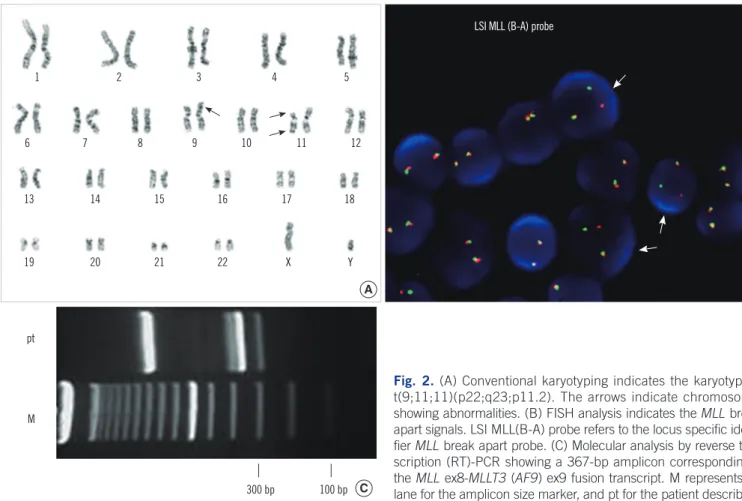
(Fig. 1B). All other markers including myelomonocytic markers were not expressed. Conventional karyotyping of uncultured bone marrow aspirate showed a 3-way t(9;11;11) translocation among 9p22, 11q23, and 11p11.2 in 20 metaphase cells (Fig.
2A). FISH studies performed on bone marrow aspirates re- vealed a MLL translocation (Fig. 2B). In additional studies, there was no evidence of MYC rearrangements. Molecular analysis by reverse transcription (RT)-PCR also revealed a 367-bp sized amplicon corresponding to the MLL ex8-MLLT3 (AF9) ex9 fu- sion transcript (Fig. 2C).
The patient received induction chemotherapy for ALL. A rapid response was observed with the resolution of leukocytosis, and complete remission (CR) was documented after the induction phase. Sibling peripheral blood stem cell transplantation (PB- SCT) was performed after the consolidation phase. Minimal re- sidual disease (MRD) evaluation at this time was negative and no evidence of relapse has been found during 8 months of fol- low-up.
The majority of B-lineage lymphoblastic leukemia cases pres- ent B cells of the pre-pre-B and pre-B stages, without surface immunoglobulin light chain expression. Mature B-cell pheno- type is found in less than 2% of cases [13]. MLL translocation, particularly t(9;11), exists in a small number of these patients.
To date, more than 50 different translocation fusion partners of MLL have been identified [5]. Regardless of the variety of cy- togenetic abnormalities, ALL with MLL gene rearrangement forms a distinct subset of acute leukemias. The three most fre- quent MLL rearrangements are t(4;11), t(9;11), and t(11;19) [17, 18]. However, the t(9;11), although common in de novo AML and therapy-related AML, is only rarely seen in B-cell lym- phoblastic leukemias (B-ALL) [19, 20].
We found 13 previously reported cases of B-ALL with surface light chain expression and MLL rearrangements in children and the characteristics are summarized in Table 1. Among the 14 cases including the present case, 13 patients were children, and 9 had t(9;11). Interestingly, none of these patients showed t(4;11)(q21;q23), the most frequent MLL rearrangement found in B-ALL of children [18]. Rather, the t(9;11) was most fre- quently observed. In the present case, the three-way transloca- tion t(9;11;11)(p22;q23;p11.2) bearing the MLL-AF9 fusion gene was detected by FISH and RT-PCR analyses.
The frequent association between the t(9;11) and surface light chain restriction suggests that ALL with this profile is a dis- tinct subset of MLL-positive B-ALL. Reported cases showed variable response to treatment. Some patients showed poor prognosis with multiple relapses (like most patients with MLL+
A
Fig. 1. (A) Blasts of the patient exhibit non-FAB-L3 morphology. (B) Scatter plots of flow cytometric immunophenotyping show blasts with CD19+, CD10+, CD34-, terminal deoxynucleotidyl transferase (TdT)-, and surface immunoglobulin (sIg) λ+.
Abbreviation: FAB, French-American-British classification.
B 1,000
500
0
103 102 101 100
103 102 101 100
103 102 101 100
103 102 101 100
100 101 102 103 100 101 102 103
100 101 102 103 100 101 102 103
100 101 102 103
PerCP-Cy5-5-A CD10
CD7 CD34
KAPPA
SSC-A CD19
cCD22Count CD33LAMBDA
50 40 30 20 10
0 100 101 102 103 TdT
Kim B, et al.
Mature B-ALL with t(9;11;11)
168 www.annlabmed.org http://dx.doi.org/10.3343/alm.2014.34.2.166 Table 1. Lymphoblastic leukemia with surface light chain immunoglobulin expression and MLL rearrangement including t(9;11)
Case
No. Age/Sex Morphology CD19 CD20 CD22 CD34 TdT CD10 sIg Karyotype FISH Reference
1 11 months/F L1/L2 + + + - NA - κ 46,XX MLL rearrangement 13
2 12 months/F L1 + + + - - - λ NA MLL rearrangement 12
3 23 months/F L1 + + + - NA - λ t(9;11)(p21~22;q23)[4]/46,XX[12] NA 11
4 8 yr/F L1 + + NA NA + - λ t(9;11)(p22;q23) NA 10
5 5 months/M NA + - NA + - - λ NA MLL rearrangement 15
6 12 months/F L1 + + NA - - + λ t(9;11)(p22;q23) NA 15
7 4 months/M L1 + - + - - - λ t(9;11)(p21;q23) NA 15
8 8 months/F Non-L3 + + + - - - λ t(9;11)(p21;q23) NA 15
9 5 months/M Non-L3 + + + - - - λ 46,XX[20] NA* 16
10 13 months/F Non-L3 + NA + - - - κ 46,XX[14] MLL-AF9 rearrangement 16
11 23 months/F Non-L3 + + + - - - λ 46,XX[4]/46,XX, t(9;11) (p21~22;q23)[12]
NA 16
12 8 months/M Non-L3 + - + - - - κ 46,XY[10] MLL-AF9 rearrangement 16
13 67 yr/F L2 + - + + + - κ 46,XX,t(2;11)(p21;q23) NA 14
14 4 months/M L1 + - + - - + λ 46,XY[16]/46,XX,t(9;11;11)
(p22;q23;p11.2)[4]
MLL rearrangement index case
*MLL-AF10 fusion transcript by molecular study.
Abbreviations: TdT, terminal deoxynucleotidyl transferase; sIg, surface immunoglobulin; F, female; M, male; NA, not available.
Fig. 2. (A) Conventional karyotyping indicates the karyotype of t(9;11;11)(p22;q23;p11.2). The arrows indicate chromosomes showing abnormalities. (B) FISH analysis indicates the MLL break- apart signals. LSI MLL(B-A) probe refers to the locus specific identi- fier MLL break apart probe. (C) Molecular analysis by reverse tran- scription (RT)-PCR showing a 367-bp amplicon corresponding to the MLL ex8-MLLT3 (AF9) ex9 fusion transcript. M represents the lane for the amplicon size marker, and pt for the patient described.
1
6
13
19
2
7
14
20
3
8
15
21
4
LSI MLL (B-A) probe
9
16
22
5
10
17
X 11
18
Y 12
pt
M
300 bp 100 bp
A B
C
Kim B, et al.
Mature B-ALL with t(9;11;11)
http://dx.doi.org/10.3343/alm.2014.34.2.166 www.annlabmed.org 169
B-ALL), while others showed CR with no evidence of relapse.
The patient described here received chemotherapy and PBSCT, and has so far shown no evidence of relapse. However, leuke- mias with MLL rearrangement are typically associated with a poor prognosis owing to the recurrent relapses, and Blin et al.
[16] reported a rapid response to chemotherapy contrasted by the high incidence of relapse and poor overall prognosis in pa- tients with this profile. Future identification of patients with this profile will allow us to expand our knowledge regarding prognos- tic significance and optimal treatment for this rare subgroup of patients.
Authors’ Disclosures of Potential Conflicts of Interest
No potential conflicts of interest relevant to this article were re- ported.
REFERENCES
1. Pui CH, Frankel LS, Carroll AJ, Raimondi SC, Shuster JJ, Head DR, et al. Clinical characteristics and treatment outcome of childhood acute lymphoblastic leukemia with the t(4;11)(q21;q23): a collaborative study of 40 cases. Blood 1991;77:440-7.
2. Heerema NA, Sather HN, Ge J, Arthur DC, Hilden JM, Trigg ME, et al.
Cytogenetic studies of infant acute lymphoblastic leukemia: poor prog- nosis of infants with t(4;11) - a report of the Children’s Cancer Group.
Leukemia 1999;13:679-86.
3. Pui CH, Kane JR, Crist WM. Biology and treatment of infant leukemias.
Leukemia 1995;9:762-9.
4. Ayton PM and Cleary ML. Molecular mechanisms of leukemogenesis mediated by MLL fusion proteins. Oncogene 2001;20:5695-707.
5. Krivtsov AV and Armstrong SA. MLL translocations, histone modifica- tions and leukaemia stem-cell development. Nat Rev Cancer 2007;7:
823-33.
6. Swerdllow SH, Campo E, et al. eds. WHO classification of tumours of haematopoietic and lymphoid tissues in 2008. Lyon: IARC Press, 2008:
262-4.
7. Sullivan MP, Pullen DJ, Crist WM, Brecher M, Ramirez I, Sabio H, et al.
Clinical and biological heterogeneity of childhood B cell acute lympho- cytic leukemia: implications for clinical trials. Leukemia 1990;4:6-11.
8. Hammami A, Chan WC, Michels SD, Nassar VH. Mature B-cell acute leukemia: a clinical, morphological, immunological, and cytogenetic study of nine cases. Hematol Pathol 1991;5:109-18.
9. Vasef MA, Brynes RK, Murata-Collins JL, Arber DA, Medeiros LJ. Sur- face immunoglobulin light chain-positive acute lymphoblastic leukemia of FAB L1 or L2 type: a report of 6 cases in adults. Am J Clin Pathol 1998;110:143-9.
10. Lorenzana AN, Rubin CM, Le Beau MM, Nachman J, Connolly P, Sub- ramanian U, et al. Immunoglobulin gene rearrangements in acute lym- phoblastic leukemia with the 9;11 translocation. Genes Chromosomes Cancer 1991;3:74-7.
11. Talmant P, Berger R, Robillard N, Mechineau-Lacroix F, Garand R.
Childhood B-cell acute lymphoblastic leukemia with FAB-L1 morpholo- gy and a t(9;11) involving the MLL gene. Hematol Cell Ther 1996;38:
265-8.
12. Li S and Lew G. Is B-lineage acute lymphoblastic leukemia with a ma- ture phenotype and l1 morphology a precursor B-lymphoblastic leuke- mia/lymphoma or Burkitt leukemia/lymphoma? Arch Pathol Lab Med 2003;127:1340-4.
13. Frater JL, Batanian JR, O’Connor DM, Grosso LE. Lymphoblastic leuke- mia with mature B-cell phenotype in infancy. J Pediatr Hematol Oncol 2004;26:672-7.
14. Kansal R, Deeb G, Barcos M, Wetzler M, Brecher ML, Block AW, et al.
Precursor B lymphoblastic leukemia with surface light chain immuno- globulin restriction: a report of 15 patients. Am J Clin Pathol 2004;121:
512-25.
15. Tsao L, Draoua HY, Osunkwo I, Nandula SV, Murty VV, Mansukhani M, et al. Mature B-cell acute lymphoblastic leukemia with t(9;11) translo- cation: a distinct subset of B-cell acute lymphoblastic leukemia. Mod Pathol 2004;17:832-9.
16. Blin N, Méchinaud F, Talmant P, Garand R, Boutard P, Dastugue N, et al. Mature B-cell lymphoblastic leukemia with MLL rearrangement: an uncommon and distinct subset of childhood acute leukemia. Leukemia 2008;22:1056-9.
17. Huret JL, Dessen P, Bernheim A. An atlas of chromosomes in hemato- logical malignancies. Example: 11q23 and MLL partners. Leukemia 2001;15:987-9.
18. Meyer C, Schneider B, Jakob S, Strehl S, Attarbaschi A, Schnittger S, et al. The MLL recombinome of acute leukemias. Leukemia 2006;20:777- 84.
19. Super HG, Strissel PL, Sobulo OM, Burian D, Reshmi SC, Roe B, et al.
Identification of complex genomic breakpoint junctions in the t(9;11) MLL-AF9 fusion gene in acute leukemia. Genes Chromosomes Cancer 1997;20:185-95.
20. Dobson CL, Warren AJ, Pannell R, Forster A, Lavenir I, Corral J, et al.
The mll-AF9 gene fusion in mice controls myeloproliferation and speci- fies acute myeloid leukaemogenesis. EMBO J 1999;18:3564-74.