ISSN 2234-3806 • eISSN 2234-3814
http://dx.doi.org/10.3343/alm.2016.36.2.117
Direct Identification and Antimicrobial Susceptibility Testing of Bacteria From Positive Blood Culture Bottles by Matrix-Assisted Laser Desorption/Ionization Time-of- Flight Mass Spectrometry and the Vitek 2 System
Sung Jin Jo, M.D.*, Kang Gyun Park, M.T.*, Kyungja Han, M.D., Dong Jin Park, M.D., and Yeon-Joon Park, M.D.
Department of Laboratory Medicine, College of Medicine, The Catholic University of Korea, Seoul, Korea
Background: We evaluated the reliability and accuracy of the combined use of matrix-as- sisted laser desorption/ionization time-of-flight mass spectrometry (MALDI-TOF MS) bac- terial identification and Vitek 2 antimicrobial susceptibility testing (AST) for bacteria from positive blood culture bottles.
Methods: Direct identification and AST were performed in parallel to the standard meth- ods in monomicrobial positive blood culture bottles. In total, 254 isolates grown on aerobic and/or anaerobic bottles were identified with MALDI-TOF Vitek MS (bioMérieux, France), and 1,978 microorganism/antimicrobial agent combinations were assessed. For isolates from anaerobic bottles, an aliquot of the culture broth was centrifuged, washed, and fil- tered through a nylon mesh. For isolates from aerobic/pediatric bottles, a lysis step using 9.26% ammonium chloride solution and 2% saponin solution was included.
Results: The overall correct identification rate was 81.8% (208/254) and that for gram- positive/gram-negative isolates was 73.9%/92.6%, respectively, and it was 81.8%, 87.6%, and 57.9% for isolates from aerobic, anaerobic, and pediatric bottles, respectively. Identi- fication was not possible in 45 cases, and most of these isolates were streptococci (N=14) and coagulase-negative staphylococci (N =11). Misidentification occurred only in one case. Compared with standard methods, direct AST showed 97.9% (1,936/1,978) agree- ment with very major error of 0.25%, major error of 0.05%, and minor error of 1.8%.
Conclusions: This simple and cost-effective sample preparation method gives reliable re- sults for the direct identification and AST of bacteria. For the identification of streptococci and coagulase-negative staphylococci, the method should be further improved.
Key Words: Direct identification, MALDI-TOF MS, Vitek MS, Vitek 2, Positive blood culture
Received: July 14, 2015
Revision received: August 27, 2015 Accepted: November 16, 2015 Corresponding author: Yeon-Joon Park Department of Laboratory Medicine, School of Medicine, The Catholic University of Korea, Seoul St. Mary’s Hospital, 222 Banpo-daero, Seocho-gu, Seoul 06591, Korea
Tel: +82-2-2258-1640 Fax: +82-2-2258-1719 E-mail: [email protected]
*Sung Jin Jo and Kang Gyun Park contributed equally to this paper.
© The Korean Society for Laboratory Medicine This is an Open Access article distributed under the terms of the Creative Commons Attribution Non-Commercial License (http://creativecom- mons.org/licenses/by-nc/3.0) which permits unrestricted non-commercial use, distribution, and reproduction in any medium, provided the original work is properly cited.
INTRODUCTION
Rapid identification and antimicrobial susceptibility testing (AST) of microorganisms causing bloodstream infections is pivotal to guide antimicrobial therapy [1]. Reducing the time before admin- istration of appropriate antimicrobial therapy improves patient outcomes [2]. The current standard method for detecting caus-
ative agents of bloodstream infections is blood culture in com- mercially available blood culture systems using liquid medium [2]. When an automated blood culture system is used, signal- positive culture bottles are taken out of the system, and an aliquot of broth is subcultured onto solid media, which are incubated until visible colonies form, which takes up to two days [3, 4].
Matrix-assisted laser desorption/ionization time-of-flight mass
spectrometry (MALDI-TOF MS) was recently introduced in the clinical microbiology laboratories for the identification of various microorganisms. Although it identifies bacteria and yeasts within a few minutes, it requires isolated colonies, which takes 18-48 hr of incubation [5]. To reduce the time needed for identification and AST, a few investigators tried to inoculate the microorgan- isms from positive blood culture bottles directly into several au- tomated systems and MALDI-TOF MS systems [1-3, 5-12].
However, direct identification from positive blood culture bottles requires sample preparation steps because these samples con- tain proteins/debris derived from human blood and culture broth, which could interfere with the spectra of the microorgan- isms [9]. Thus, lysis solutions (ammonium chloride, ethanol/for- mic acid/acetonitrile, and trifluoroacetic acid) [1, 2, 9, 11, 12], the lysis-filtration method [3, 8], gel separation [5], or commer- cial kits [6] were used for sample preparation. However, some of these sample preparation procedures are laborious and/or expensive [11], and in the case of acetonitrile, samples must be prepared and manipulated in the fume hood because of aceto- nitrile’s strong odor.
Only a few studies have been reported on the use of direct AST [2, 3, 7]. In combination with MALDI-TOF MS, the direct inoculation Vitek 2 AST method could yield results up to 24 hr earlier than the time taken by the standard method [3]. These studies have reported the effectiveness of direct identification of pathogens from positive blood culture bottles in combination with AST. However, these methods either used ethanol/formic acid/acetonitrile [2] or the labor-intensive lysis-filtration method [3], or evaluated only gram-negative isolates [7]. Therefore, in this study, we explored a simpler, faster, and more reliable pro- tocol using an extraction method for direct identification and AST of bacteria from positive blood culture bottles.
METHODS
In total, 254 blood cultures, obtained in the period from January to February 2015, were included in this study. Of these, 235 were samples obtained from adult patients, which included samples from two bottles (aerobic and anaerobic), and 19 were from pediatric patients. In parallel to the standard method, an aliquot from the positive blood culture bottle was taken for direct identification by Vitek MS (bioMérieux, Marcy L’Etoile, France) and full panel AST by Vitek 2 (bioMérieux). Results from direct identification and AST were not reported to the clinicians. This study was exempted by institutional review board of Seoul St.
Mary’s Hospital, Seoul, Korea.
1. Standard identification and AST
Blood samples were collected and inoculated in blood culture bottles (Plus aerobic/F, Lytic/10 Anaerobic/F, Peds Plus/F; Bec- ton Dickinson, Franklin Lakes, NJ, USA) and incubated in the BACTEC FX (Becton Dickinson) blood culture system. Signal- positive blood culture bottles were detected by continuous mon- itoring of CO2 level.
When a blood culture bottle was flagged as positive by the BACTEC system, an aliquot was taken from the positive blood culture bottle for Gram stain and subculture on solid media.
Bottles showing more than one morphotype or yeasts on Gram stain were excluded from the study. Then, 100-µL aliquots were inoculated onto sheep blood agar (Asan BAP I; Asan Pharma- ceutical Co., Ltd. Seoul, Korea) and MacConkey agar (Asan Mac II; Asan Pharmaceutical. Co., Ltd.). Agar plates were incu- bated in the dark in an atmosphere containing 5% CO2 at 37°C for 24 hr. For anaerobic subculture, sheep blood agar was incu- bated at 37°C in anaerobic atmosphere for two days. Colonies of the isolates formed on agar plates were used for identification and AST by the Vitek 2 system.
A standardized inoculum was prepared from the several col- onies formed on agar medium, and the appropriate Vitek 2 cards were inoculated, following the manufacturer’s recom- mendation. The GP, GN, ANC and NH cards (bioMérieux) were used for microbial identification, and the AST-P600, AST-P601, AST-ST01, AST-N224, and AST-N225 cards (bioMérieux) were used for AST of enterococci, staphylococci, streptococci, En- terobacteriaceae, and non-fermentative gram-negative bacteria (GNB), respectively. The resulting minimum inhibitory concen- tration (MIC) values were classified into clinical categories of susceptible, intermediate, or resistant following the CLSI guide- lines [13].
2. Sample preparation for direct identification and AST The sample preparation method was different between anaero- bic samples and aerobic/pediatric samples. As the anaerobic bottle contains the saponin ingredient (0.26% w/v), the lysis step was omitted. For samples from the anaerobic bottle, a 10- mL aliquot from positive blood culture bottles was centrifuged at 4,500g for 3 min, and the pellet was washed twice with 0.45%
NaCl, suspended in 10 mL 0.45% NaCl, and filtered through a nylon mesh (66 µm pore size) to remove cell debris. The filtered sample was centrifuged at 4,500g for 3 min, and the pellet was suspended in 0.45% NaCl. The opacity of this suspension was adjusted to a 0.5 McFarland (McF) standard for direct AST by Vitek 2 and Gram stain. Samples showing mixed morphotypes
or yeasts were excluded from the study.
For identification with Vitek MS, 100 µL of the suspension was centrifuged at 15,000g for 3 min. The pellet was mixed with 20 µL alcohol in the tube and placed on a target plate (Vitek MS- DS, bioMérieux), dried at room temperature, and 1 µL of the CHCA matrix solution (α-cyano-4-hydroxycinnamic acid solution, bioMérieux) was applied to the spot. All isolates were tested once without duplication.
For aerobic/pediatric samples, to lyse red blood cells, a lysis step was included. Briefly, a 10-mL aliquot from a positive blood culture bottle was centrifuged at 4,500g for 3 min and lysis so- lution [9.26% ammonium chloride solution (0.1 mL) and 2%
saponin solution (5 mL)] were added to the pellet, which was followed by vortexing for 10 sec and incubation for 10 min at room temperature. The resulting lysate was centrifuged at 4,500g for 3 min, washed twice with 0.45% NaCl, and processed as described above.
Mass spectra were obtained by using a Vitek MS system. The Vitek MS IVD system v2.0 was used for spectral analysis, and the results were compared with a reference library v2.0 of spec- tra provided by the manufacturer. The spectra were externally calibrated by using Escherichia coli (ATCC 8739). A bottle was considered to have a valid Vitek MS result if the target slide gave a confidence level ≥90%.
3. Statistical analysis
The identification was considered accurate, if the direct method by Vitek MS provided the same results as the standard method by Vitek 2 identification for any of the two bottles (aerobic/an- aerobic). The identification was considered incorrect (misidenti- fication), if Vitek MS provided results that were different from those of the standard method. Isolates were tagged as “no iden- tification,” if the Vitek MS database did not provide identification results. The warning messages “bad-spectrum” or “not enough peaks” appeared in case of a poor-quality deposit. For direct AST, the Vitek cards were selected according to the Gram stain results and direct identification results from the Vitek MS sys- tem. Comparison of AST between the direct and standard meth- ods was expressed in terms of agreement, very major error (false susceptibility), major error (false resistance), or minor error (susceptible/resistance versus intermediate susceptibility).
Chi-square test or Fisher’s exact test was used for statistical comparisons of the performance of aerobic/anaerobic bottles, as appropriate. A P value <0.05 was considered statistically significant.
RESULTS
1. Direct identification by Vitek MS
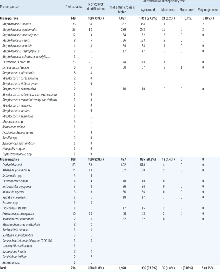
Of 254 (146 gram-positive, 108 gram-negative) monomicrobial positive blood cultures evaluated, 233 isolates were identified to the species level and 21 isolates were identified to the genus level by the standard method using Vitek 2. Table 1 shows the identification results of the direct identification method using Vi- tek MS compared with those results of isolates grown using solid media culture and identified by Vitek 2. Overall, Vitek MS correctly identified 208 (81.8%) isolates, and 45 isolates were not identified. Misidentification occurred for only one sample;
Propionibacterium acnes was misidentified as Clostridium bifer- mentans. Of the 208 correct identification results, 196 isolates (94.2%) were identified to the species level, and 12 isolates were identified to the genus level. According to the Gram stain results, Vitek MS correctly identified 108 (73.9%) of the gram- positive isolates and 100 (92.6%) of the gram-negative isolates.
The proportion of correct identification for Enterobacteriaceae, non-fermentative GNB, and staphylococci was 81/83 (97.6%), 12/15 (80.0%), and 72/85 (84.7%), respectively.
Vitek MS did not provide identification results for 45 isolates, and among them, 29 samples were not identified owing to the
“bad spectrum” warning message, nine samples were scored as
“not-identified,” and seven samples were not identified owing to the “not enough peaks” warning message. Most of these isolates were gram-positive (N=37); 14 streptococci, 11 coagulase-neg- ative staphylococci (CNS), three enterococci, two Staphylococ- cus aureus, two Micrococcus spp., two Bacillus spp., and one each of Actinomyces odontolyticus, Finegoldia magna, and Pep- tostreptococcus spp.
Table 2 shows identification results according to the type of culture bottle. Of the 235 sets of positive blood culture bottles from adult patients, 182 aerobic bottles and 145 anaerobic bot- tles were flagged as positive and identified by the standard method by Vitek 2. No identification discordance was observed between the aerobic and anaerobic bottles from the same pa- tient. The correct identification rate by the direct method was 149/182 (81.8%) and 127/145 (87.6%) for aerobic and anaer- obic bottles, respectively (P =0.742). The correct identification rate of gram-positive isolates in aerobic and anaerobic bottles was 76.2% and 77.4%, respectively (P =0.960). The correct identification rate of gram-negative isolates in aerobic and an- aerobic bottles was 88.8% and 97.3%, respectively (P =0.783).
No identification was observed in 33/182 aerobic bottles and 17/145 anaerobic bottles (P =0.221; Table 2). In addition, 53
Table 1. Comparison of identification and antimicrobial susceptibility testing results between the direct method and the standard method
Microorganism N of isolates N of correct
identifications
Antimicrobial susceptibility test N of antimicrobials
tested Agreement Minor error Major error Very major error
Gram-positive 146 108 (73.9%) 1,081 1,051 (97.2%) 24 (2.2%) 1 (0.1%) 5 (0.5%)
Staphylococcus aureus 36 34 357 354 1 0 2
Staphylococcus epidermidis 23 18 289 272 15 0 2
Staphylococcus haemolyticus 12 9 34 32 2 0 0
Staphylococcus capitis 8 5 136 133 2 0 1
Staphylococcus hominis 4 4 34 33 1 0 0
Staphylococcus saprophyticus 1 1 17 17 0 0 0
Staphylococcus cohnii spp. urealyticus 1 1
Enterococcus faecium 23 21 144 143 1 0 0
Enterococcus faecalis 6 5 60 57 2 1 0
Streptococcus mitis/oralis 8 3
Streptococcus parasanguinis 2 0
Streptococcus viridans group 2 0
Streptococcus pneumoniae 2 1 10 10 0 0 0
Streptococcus gallolyticus ssp. pasteurianus 1 0 Streptococcus constellatus ssp. constellatus 1 0
Streptococcus salivarius 1 0
Streptococcus mutans 1 0
Streptococcus anginosus 1 1
Micrococcus spp. 3 1
Aerococcus urinae 1 1
Propionibacterium acnes 4 3
Bacillus spp. 2 0
Actinomyces odontolyticus 1 0
Finegoldia magna 1 0
Peptostreptococcus spp. 1 0
Gram-negative 108 100 (92.6%) 897 885 (98.6%) 12 (1.4%) 0 0
Escherichia coli 53 53 522 518 4 0 0
Klebsiella pneumoniae 14 13 162 160 2 0 0
Salmonella spp. 3 3
Enterobacter cloacae 4 4 18 18 0 0 0
Enterobacter aerogenes 3 3 36 36 0 0 0
Klebsiella oxytoca 3 3 36 36 0 0 0
Serratia marcescens 1 1 18 17 1 0 0
Pantoea spp. 1 0
Providencia stuartii 1 1 17 15 2 0 0
Pseudomonas aeruginosa 10 10 56 53 3 0 0
Acinetobacter baumannii 2 0 32 32 0 0 0
Stenotrophomonas maltophilia 2 2
Burkholderia cepacia 1 0
Ralstonia mannitolilytica 3 1
Chryseobacterium indologenes (CDC IIb) 1 0
Haemophilus influenzae 1 1
Bacteroides fragilis 2 2
Clostridium tertium 2 2
Neisseria spp. 1 1
Total 254 208 (81.8%) 1,978 1,936 (97.9%) 36 (1.8%) 1 (0.05%) 5 (0.25%)
aerobic bottles and 90 anaerobic bottles were tagged as “no growth.” Moreover, 90 isolates and 53 isolates were detected only in aerobic and anaerobic bottles, respectively. However, the rate of no identification among these 90 and 53 isolates was similar (23/90 isolates; 25.5%, 13/53 isolates; 24.5%). The dis- proportionate growth did not affect the identification rate be- tween the bottles. Of the 19 pediatric bottles, 11 were correctly identified (five Staphylococcus epidermidis, one each of Staphy- lococcus aureus, Staphylococcus saprophyticus, Escherichia coli, Pseudomonas aeruginosa, Neisseria spp., and Micrococ- cus spp.), and eight were not identified (2 Staphylococcus capi- tis, 2 Staphylococcus epidermidis, and one each of Streptococ- cus pneumoniae, Micrococcus spp., Streptococcus constella- tus, and Burkholderia cepacia).
2. Direct AST
In total, 120 isolates (69 gram-positive cocci, 51 gram-negative bacteria) were analyzed for full panel AST by Vitek 2. Then, 1,978 isolate/antimicrobial agent combinations were analyzed.
The direct AST results showed 97.9% (1,936/1,978) agreement with the standard method AST results. The rates for very major error, major error, and minor error were 0.25% (5/1,978), 0.05%
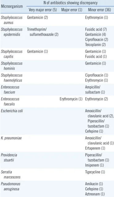
(1/1,978), and 1.8% (36/1,978), respectively (Table 1). The mi- croorganism/antimicrobial agent combinations that did not agree with standard methods are listed in Table 3.
Gram-positive isolates showed an agreement rate of 97.2%
(1,051/1,081), with very major error rate of 0.5% (5/1,081), major error rate of 0.1% (1/1,081), and minor error rate of 2.2%
(24/1,081). Staphylococcus epidermidis was the major cause of the disagreement, and gentamicin (N =9) and fusidic acid (N=8) were an attribute of high error rates. Among the gram-
negative isolates, the overall agreement rate was 98.6% (885/
897), with a minor error rate of 1.4% (12/897). Escherichia coli and Pseudomonas aeruginosa were the major causes of the disagreement, and amoxicillin/clavulanic acid (N =3) were an attribute of high error rates.
DISCUSSION
In this study, we evaluated the reliability and accuracy of per- forming direct inoculation of organisms from positive blood cul- ture bottles in combination with Vitek MS and Vitek 2 to achieve Table 2. Identification rates according to the type of blood culture
bottle
Adult patients
(N=235) Pediatric patients (N=19) Aerobic Anaerobic Pediatric bottle Correct
identification
81.8% (149/182) 87.6% (127/145) 57.9% (11/19)
Gram-positive 76.2% (77/101) 77.4% (55/71) 53.3% (8/15) Gram-negative 88.8% (72/81) 97.3% (72/74) 75% (3/4) No identification 18.1% (33/182) 11.7% (17/145) 42.1% (8/19)
Misidentification - 0.7% (1/145)
No growth 22.6% (53/235) 38.3% (90/235)
Identification results between aerobic and anaerobic bottles were not differ- ent statistically.
Table 3. Microorganism/antimicrobial agent combinations showing discrepancy between the results by the direct method and those by the standard method in antimicrobial susceptibility test by Vitek 2
Microorganism N of antibiotics showing discrepancy Very major error (5) Major error (1) Minor error (36) Staphylococcus
aureus
Gentamicin (2) Erythromycin (1)
Staphylococcus
epidermidis Trimethoprim/
sulfamethoxazole (2) Fusidic acid (7) Gentamicin (4) Ciprofloxacin (2) Teicoplanin (2) Staphylococcus
capitis
Gentamicin (1) Gentamicin (1)
Fusidic acid (1) Staphylococcus
hominis Gentamicin (1)
Staphylococcus haemolyticus
Ciprofloxacin (1) Erythromycin (1) Enterococcus
faecium Ampicillin/
sulbactam (1) Enterococcus
faecalis
Erythromycin (1) Erythromycin (2)
Escherichia coli Amoxicillin/
clavulanic acid (2), Piperacillin/
tazobactam (1) Cefepime (1)
K. pneumoniae Amoxicillin/
clavulanic acid (1) Ertapenem (1) Providencia
stuartii Piperacillin/
tazobactam (1) Imipenem (1) Serratia
marcescens Tigecycline (1)
Pseudomonas aeruginosa
Amikacin (1) Cefepime (1) Aztreonam (1)
rapid identification and AST. During the study period, only one sample containing yeast was identified, and we did not include this case in our study. The proportion of correct identification for Enterobacteriaceae, non-fermentative GNB, and staphylococci was 97.6%, 80.0%, and 84.7%, respectively. Gram-positive iso- lates showed a relatively low correct identification rate in other specimens, by the direct MALDI-TOF method [14]. Compared with the standard method, the direct method showed rapid and reliable results, especially for the gram-negative group. These data coincide with previous data that presented a better identifi- cation of gram-negative isolates (84-99%) when compared with gram-positive isolates (64.8-86.3%) [1, 5, 6, 11, 12].
Excluding the “no identification” results, the concordance rate of identification between the direct method and the stan- dard method was very reliable (99.5%, 207/208), and most of the “no identification” results were attributed to the poor quality of the spots (which led to the appearance of “bad spectrum” or
“not enough peaks” warning messages on the system). This method has limitations for the identification of CNS and strepto- cocci; however, “no identification” results were obtained for these organisms, and they were not identified incorrectly. Other protocols that tried direct MALDI-TOF MS inoculation showed similar results (63.3-83.5%) for the identification of CNS isolates [1, 2, 5, 6, 11, 12]. Prod’hom et al. [1] suggested that the lower yield of MALDI-TOF MS results with streptococci and staphylo- cocci may be attributed to inter-species relatedness and bacte- rial cell wall composition. It is notable that misidentification oc- curred only for Propionibacterium acnes, which is known to be identified inaccurately with MALDI-TOF MS, even with the use of colonies [15].
Anaerobic bottles showed a higher correct identification rate than did aerobic bottles; however, the difference was not statisti- cally significant. Given that sample preparation is simpler with anaerobic bottles, if the organism growth is observed using both aerobic and anaerobic bottles, we recommend using anaerobic bottles. Our simple sample preparation method resulted in a higher correct identification rate than that of the previous study (82.2%), which used centrifugation alone [7]. The relatively lower correct identification rate in pediatric bottles was mainly attributed to gram-positive isolates.
In the present study, the accuracy of AST was excellent with both gram-positive and gram-negative isolates, and the antimi- crobial agreement rate was comparable or superior to that of other studies [2, 3, 7]. The relatively higher very major error rate among gram-negative bacteria observed in the study by Ma- chen et al. [3] was observed with Proteus spp. against various
antibiotics (ampicillin, cefazolin, ceftazidime, and ceftriaxone);
however, in this study, Proteus spp. was not included. Very ma- jor error was found with Staphylococcus spp. alone, and it was against gentamicin and trimethoprim/sulfamethoxazole, which was also reported by Machen et al. [3].
In summary, our simple and cost-effective sample preparation method would be very useful for rapid identification and AST of bacteria from positive blood culture bottles. However, for identi- fication of streptococci and CNS, further improvement in the method is needed.
Authors’ Disclosures of Potential Conflicts of Interest
No potential conflicts of interest relevant to this article were re- ported.
Acknowledgments
This work was supported by a grant of the Korea Health Tech- nology R&D Project through the Korea Health Industry Develop- ment Institute (KHIDI), funded by the Ministry of Health & Wel- fare, Republic of Korea (grant number : HI12C1093) and also by the Technology Innovation Program (No: 10049771, Devel- opment of Highly-Specialized Platform for IVD Medical Devices) funded by the Ministry of Trade, Industry & Energy (MI, Korea).
REFERENCES
1. Prod’hom G, Bizzini A, Durussel C, Bille J, Greub G. Matrix-assisted la- ser desorption ionization-time of flight mass spectrometry for direct bacterial identification from positive blood culture pellets. J Clin Micro- biol 2010; 48:1481-3.
2. Romero-Gómez MP, Gómez-Gil R, Paño-Pardo JR, Mingorance J. Iden- tification and susceptibility testing of microorganism by direct inocula- tion from positive blood culture bottles by combining MALDI-TOF and Vitek-2 Compact is rapid and effective. J Infect 2012; 65:513-20.
3. Machen A, Drake T, Wang YF. Same day identification and full panel antimicrobial susceptibility testing of bacteria from positive blood culture bottles made possible by a combined lysis-filtration method with MAL- DI-TOF VITEK mass spectrometry and the VITEK2 system. PLoS One 2014;9:e87870.
4. Schneiderhan W, Grundt A, Wörner S, Findeisen P, Neumaier M. Work flow analysis of around-the-clock processing of blood culture samples and integrated MALDI-TOF mass spectrometry analysis for the diagno- sis of bloodstream infections. Clin Chem 2013;59:1649-56.
5. Stevenson LG, Drake SK, Murray PR. Rapid identification of bacteria in positive blood culture broths by matrix-assisted laser desorption ioniza- tion-time of flight mass spectrometry. J Clin Microbiol 2009;48:444–7.
6. Kok J, Thomas LC, Olma T, Chen SC, Iredell JR. Identification of bacte- ria in blood culture broths using matrix-assisted laser desorption-ioniza-
tion Sepsityper™ and time of flight mass spectrometry. PLoS One 2011;6:e23285.
7. Ling TK, Liu ZK, Cheng AF. Evaluation of the VITEK 2 system for rapid direct identification and susceptibility testing of gram-negative bacilli from positive blood cultures. J Clin Microbiol 2003;41:4705-7.
8. Fothergill A, Kasinathan V, Hyman J, Walsh J, Drake T, Wang YF. Rapid identification of bacteria and yeasts from positive-blood-culture bottles by using a lysis-filtration method and matrix-assisted laser desorption ionization-time of flight mass spectrum analysis with the SARAMIS da- tabase. J Clin Microbiol 2013;51:805-9.
9. Lavergne RA, Chauvin P, Valentin A, Fillaux J, Roques-Malecaze C, Ar- naud S, et al. An extraction method of positive blood cultures for direct identification of Candida species by Vitek MS matrix-assisted laser de- sorption ionization time of flight mass spectrometry. Med Mycol 2013;
51:652-6.
10. Ferreira L, Sánchez-Juanes F, Porras-Guerra I, García-García MI, Gar- cía-Sánchez JE, González-Buitrago JM, et al. Microorganisms direct identification from blood culture by matrix-assisted laser desorption/ion- ization time-of flight mass spectrometry. Clin Microbiol Infect 2011;17:
546-51.
11. Monteiro J, Inoue FM, Lobo AP, Sugawara EK, Boaretti FM, Tufik S.
Fast and reliable bacterial identification direct from positive blood cul- ture using a new TFA sample preparation protocol and the Vitek®MS system. J Microibiol Methods 2015;109:157-9.
12. Marinach-Patrice C, Fekkar A, Atanasova R, Gomes J, Djamdjian L, Brossas JY, et al. Rapid species diagnosis for invasive candidiasis using mass spectrometry. PLos One 2010;5:e8862.
13. Clinical and Laboratory Standards Institute. Performance standards for antimicrobial susceptibility testing. 24th Informational supplement, M100-S24. Wayne, PA: National Committee for Clinical Laboratory Stan- dards, 2014.
14. Kim Y, Park KG, Lee K, Park YJ. Direct identification of urinary tract pathogens from urine samples using the Vitek MS system based on Matrix-Assisted Laser Desorption Ionization-Time of Flight Mass Spec- trometry. Ann Lab Med 2015;35:416-22.
15. Coltella L, Mancinelli L, Onori M, Lucignano B, Menichella D, Sorge R, et al. Advancement in the routine identification of anaerobic bacteria by MALDI-TOF mass spectrometry. Eur J Clin Microbiol Infect Dis 2013;32:
1183-92.