척추측만증 평가 척도에 관한 문헌 고찰: 비방사선 방법을 중심으로
김동주1, 최성경2, 조효림2, 하유빈3, 최성환4, 박서현4, 이승덕5, 금동호4, 성원석2*, 김은정2*
1동국대학교 한의과대학, 2동국대학교 분당한방병원 침구의학과, 3동국대학교 분당한방병원 한방내과
4동국대학교 분당한방병원 한방재활의학과, 5동국대학교 한의과대학 침구의학과 Original Article
⋅Received:2 February 2021 ⋅Revised:15 February 2021 ⋅Accepted:18 February 2021
⋅Correspondence to:Won-Suk Sung
Department of Acupuncture & Moxibustion Medicine, Dongguk University Bundang Oriental Hospital 268, Buljeong-ro, Bundang-gu, Seongnam-si, Gyeonggi-do, 13601, Republic of Korea
Tel:+82-31-710-3725, E-mail:1984sws@hanmail.net
⋅Correspondence to:Eun-Jung Kim
Department of Acupuncture & Moxibustion Medicine, Dongguk University Bundang Oriental Hospital 268, Buljeong-ro, Bundang-gu, Seongnam-si, Gyeonggi-do, 13601, Republic of Korea
Review Study on the Measurement Tools of Scoliosis:
Mainly on Non-radiological Methods
Kim Dong-Joo1, Seong-Kyeong Choi2, Hyo-Rim Jo2, Yu-bin Ha3, Sung-Hwan Choi4, Seo-Hyun Park4, Seung Deok Lee5, Dong-Ho Keum4, Won-Suk Sung2, Eun-Jung Kim2
1College of Korean Medicine, Dongguk University
2Department of Acupuncture & Moxibustion, Dongguk University Bundang Oriental Hospital
3Department of Internal Korean Medicine, Dongguk University Bundang Oriental Hospital
4Department of Rehabilitation Medicine of Korean Medicine, Dongguk University Bundang Oriental Hospital
5Department of Acupuncture & Moxibustion Medicine, College of Korean Medicine, Dongguk University Objectives: The purpose of this study is to investigate the characteristics, validity, and reliability of non-radiological assessment tools of scoliosis that have been studied so far.
Methods: Electronic databases including Pubmed, Cochrane Library, CNKI, Science On, RISS, OASIS were searched by keywords including ‘scoliosis assessment’, ‘scoliosis screening’, ‘physical examination’, ‘functional measurement’,
‘photography’, and ‘smartphone’.
Results: 32 articles using radiation-free assessments were identified from 1,011 records. The mostly used non-radiological methods were Surface topography, Scoliometer, Ultrasound, Digital Infrared Thermal Imaging, and Photography. The other methods were Gait analysis, 3D depth sensor imaging, and Low intensity electromagnetic scan.
Conclusions: It was found that non-radiological assessment tools might reduce the number of radiographs taken in scoliosis patients. To increase the reliability and validity, further research on the measurement tools of scoliosis will be needed.
Key Words : Scoliosis, Scoliosis measurement tools, Scoliosis assessment, Non-radiological assessment, Clinical practice, Diagnosis
pISSN 1010-0695•eISSN 2288-3339
서 론
척추측만증은 정중앙의 축을 기준으로 하여 척추 가 좌우 측면 방향으로 만곡 또는 편위되어 있는 질 환이다. 관상면상 뿐만 아니라 시상면상에서도 추체 의 회전이 동반되는 경우가 많아 변형이 3차원적인 형태를 나타내는 것이 특징이다.1,2) 척추측만증은 흔 한 척추 질환으로, 건강보험심사평가원의 2019년 자 료에 따르면 환자수는 9만 4천여명이었으며, 그 중 약 43%가 10대 청소년 환자로 가장 높은 비중을 차 지하였다.
척추측만증의 진단과 예후 평가에 있어 가장 보편 적으로 사용되는 척도는 Cobb이 1948년에 제시한 Cobb’s angle이다. X-ray PA view에서 측정하고자 하는 만곡의 가장 상부에 위치한 척추체의 상연의 연장선 A과 가장 하부에 위치한 척추체 하연의 연장 선 B를 그린 후 각각 A, B에 수직선 A’, B’를 그려 A’와 B’가 이루는 교차각을 Cobb’s angle이라 명명 하며 일반적으로 10° 이상인 경우 척추측만증으로 진단한다.3)
방사선 촬영을 통한 Cobb’s angle이 현재 표준적 으로 사용되고 있지만, 반복된 방사선 촬영이 잠재적 으로 암 발생의 위험을 증가시키고 인체에 해로운 영향을 미친다는 연구들이 보고되었다.4-7) 방사선 노 출에 대한 환자들의 두려움 외에도 Cobb’s angle은 3차원상의 변형을 반영하지 못하며 측정자 내, 측정 자 간 편차가 커 장기 모니터링에 있어 신뢰성과 효 율성을 저하시킬 수 있다.8-10) 뿐만 아니라, 한의사는 현재 X-ray나 MRI 등의 의료영상장비를 직접적으로 활용할 수 없어 척추측만증의 진단과 피드백이 쉽지 않은 상황이다.11) 따라서 한의사들이 쉽게 접근할 수 있으며, 환자들이 반복적으로 방사선에 노출되는 것 을 보호하기 위해 다른 척추측만증 평가도구를 살펴 볼 필요가 있다.
이에 본 연구는 척추측만증의 비방사선적 평가도
검토하여 평가 도구별 특성과 타당도, 신뢰도 등을 함께 살펴보고자 한다.
연구대상과 방법
본 연구는 척추측만증의 비방사선적 평가 척도에 대한 문헌 고찰 연구로서, 해당 연구에서 사용한 비 방사선 평가 척도와 연구 방법 및 타당도와 신뢰도 를 포함한 결과를 추출하고, 이후에 평가자의 합의를 통해 평가 척도에 대한 종합적 판단을 yes 또는 no 로 결정하였다.
문헌검색은 2021년 1월 5일까지 국내외 학술 데이 터베이스에 등록 또는 출판된 논문을 대상으로 시행 하였다. 국외 학술 데이터베이스로는 Pubmed, Cochrane Library, CNKI, 국내 학술 데이터베이스로 는 학술연구정보서비스 (RISS), 전통의학정보포털 (OASIS), 과학기술 지식 인프라 (Science ON)를 활 용하였다. 검색어는 scoliosis assessment, scoliosis screening, physical examination, functional measurement, photography, smartphone을 사용해 각 데이터베이스의 특성에 맞추어 조합하였다.
상기된 데이터베이스에서 최초로 검색된 논문은 총 1,011건이었으며, 제목과 초록을 검토하여 척추측 만증과 직접적인 관련이 없는 경우, 척추측만증의 진 단과 관계되지 않는 경우, 문헌고찰 연구 및 학위논 문, 2000년 이전의 논문을 제외하여 89건의 논문이 1차 선정되었다. 이후 2단계에서는 문헌의 전문을 보 면서 적합성을 검토하여 방사선을 이용한 평가 척도 만 포함된 경우, 평가 도구가 포함되지 않은 경우, 전 문을 찾을 수 없을 경우 등을 제외하였다. (Figure 1)
결 과
앞서 정한 기준에 따라 총 1,011편의 논문을 분석 한 결과 32편의 논문을 최종 선정하였다. 출판년도
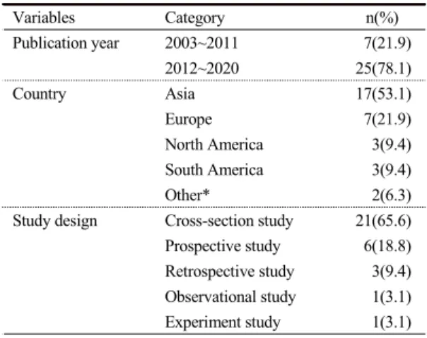
이었으며, 2012~2020년에 보고된 연구는 25편 (78.1%)이었다. 연구가 진행된 국가는 아시아 17편 (53.1%), 유럽 7편 (21.9%), 북미 3편 (9.4%), 남미 3편 (9.4%), 기타 2편 (6.3%) 순이었다. 연구 설계는 단면조사 (cross-section) 연구가 21편 (65.6%)으로 가장 많았으며 전향적 연구가 6편 (18.8%), 후향적 연구가 3편 (9.4%), 관찰 연구가 1편 (3.1%), 실험연 구가 1편 (3.1%)이었다. (Table 1)
비방사선적 평가 척도에 따른 분포를 살펴보면 본 연구에 포함된 32편의 논문 중 10편 (31.3%)이 Surface topography에 대한 연구로 가장 많았고, 6편 (18.8%)이 Scoliometer, 6편 (18.8%)이 Ultrasound, 3편 (9.4%)이 적외선 체열 촬영, 3편 (9.4%)이
Fig. 1. Study selection PRISMA flow chart
Table 1. Characteristics of Included studies (n=32)
Variables Category n(%)
Publication year 2003~2011 7(21.9)
2012~2020 25(78.1)
Country Asia 17(53.1)
Europe 7(21.9)
North America 3(9.4)
South America 3(9.4)
Other* 2(6.3)
Study design Cross-section study 21(65.6) Prospective study 6(18.8) Retrospective study 3(9.4) Observational study 1(3.1) Experiment study 1(3.1)
* Israel, Austrailia
사용한 연구가 4편 (12.5%)이었다.
Author’s consensus 상 yes는 총 24건이었으며 각 평가 척도 별로 Scoliometer는 6건, 적외선 체열 촬 영은 1건, Surface topography는 7건, Ultrasound는 6건, Photography는 1건이 yes였으며, 기타 척도를 사용한 4편의 연구 중 SmartstepTM 연구를 제외한 3 건이 yes였다.
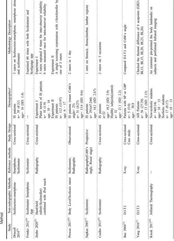
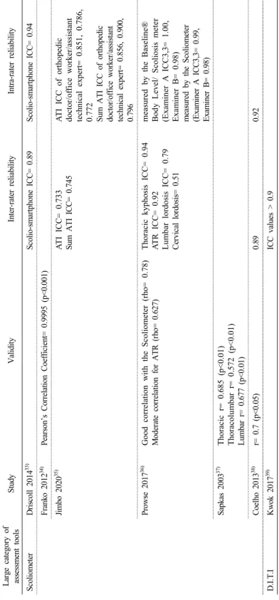
각 연구의 특성과 타당도, 신뢰도, 결론 등은 Table 2, Table 3, Table 4에서 정리하였으며, 각각의 평가 척도를 중심으로 살펴본 결과 다음과 같다. (Table 2~4)
1. Scoliometer
척추측만계 (scoliometer)는 몸통의 비대칭이나 축 방향으로의 회전을 측정하기 위해 개발된 경사계로
12), Adam’s test와 같이 몸을 앞으로 구부린 자세에 서 측정을 실시한다. 저렴하고 측정이 빠르며 측정을 위해 전문적인 교육이 거의 필요하지 않다는 등의 장점13)을 가지고 있어 측만의 진행상황을 간단하게 수치화할 수 있는 실용적 도구로14-16) 학교 등에서 척 추측만증 선별을 위한 기본 검사로 흔히 사용된다.17,
18)
Sapkas 등과 Coelho 등의 연구에서 scoliometer를 현재 golden standard로 여겨지는 Cobb’s angle과 비 교분석한 결과 모두 양호한 신뢰수준과 타당도를 보 였으며, 가장 높은 민감도 값은 체간의 회전이 5°인 경우에서 87%로 보고되었다. Prowse 등의 연구에서 사용된 Baseline® Body Level/Scoliosis meter는 scoliometer와 양호한 상관관계가 있었으며 (rho=0.78), 방사선 촬영을 통해 얻은 Cobb’s angle과는 중간 정 도의 상관관계를 보였다 (rho=0.627). 흉추, 요추, 경 추에 대한 신뢰도는 높았으나 그에 비해 골반 및 어 깨 기울기에 대한 신뢰성은 비교적 낮은 것으로 나 타났다.
이 외 scoliometer를 스마트폰 등의 모바일 기기와
연구에서는 39명의 척추측만증 환자를 대상으로 3가 지 비방사선적 방법 (Scolioscreen-smartphone, smartphone 단독, scoliometer 단독)을 비교한 결과, 스마트폰을 단독으로 측만증 측정에 사용한 경우 관 측자 간, 관측자 내 일관성이 떨어졌으며, 스마트폰 을 끼워 측정이 용이하도록 하는 고무 의료 장치인 scolioscreen-smartphone의 조합과 scoliometer의 단 독 사용은 모두 유사하게 우수한 신뢰성과 일관성을 제공하였다. Franko 등은 스마트폰에 내장된 경사계 를 활용해 scoliometer의 기능을 모방한 iPhone 앱 (Scoliguage)의 유효성을 검증하였다. Scoliguage 앱 과 표준 scoliometer의 판독치를 비교분석한 결과, 앱의 사용이 scoliometer에 비해 측정에 소요되는 시 간이 증가하지 않았으며, 피어슨 상관계수는 0.9994 – 0.9996으로 양호한 타당도를 보여주었다. Jimbo 등은 iPod touch와 같은 모바일 장치와 결합해 사용 하기 위해 개발된 i-Scolioroller를 Cobb’s angle 20°
이상 환자들의 석고 몸통 몰드에 적용하여, 79,2%의 민감도와 70.0%의 특이도를 보고하였다.
2.
적외선 체열 촬영(D.I.T.I)
컴퓨터 적외선 체열 촬영 (Digital Infrared Thermographic Imaging; D.I.T.I)은 인체의 피부 표 면에서 정상적으로 방출되는 극미량의 적외선을 센 서를 통해 미세하게 감지하고 컴퓨터를 통해 영상으 로 변환하여 신체의 이상유무를 진단하는 검사방법 이다.19)
체표의 온도는 자율신경계에 의해 신체의 양쪽에 균일하게 조절되어 혈액의 흐름이 피부를 통해 대칭 적인 온도 패턴을 생성하므로20) 체온의 비대칭은 질 병의 진단에 중요한 기준이 된다. 일반적으로 D.I.T.I 상 좌우 온도차 (ΔT)가 0.5°C 이상일 경우, 비정상 으로 판단한다.21) 흉요추부에서 적외선 체열 촬영의 정상 소견은 극돌기를 따라 고온 현상이 대칭적으로 나타나는 것이며, 비정상 소견으로는 특징적인 체열
Table 2. Summary of the included studies. Table summarizes non-radiographic methods, Reference methods, Study design, Patients demographics and Method StudyNon-radiographic MethodsReference methodsStudy DesignDemographicsaMethodology Description Driscoll 201433)Scolioscreen-smartphoneSmartphone, ScoliometerCross-sectionalIS patients n= 39 (8:31) age= 16 (SD: 1.4)
3 raters on Scolioscreen-smartphone, smartphone alone and scoliometer Franko 201234)Scoliometer Smartphone AppScoliometerCross-sectional―Examined 60 times with both Scoliometer and Scoliguage app Jimbo 202035) Hand-held roller(i-Scolioroller) combined with iPod touch
Radiography Cross-sectionalExperiment I Plater torsos of IS patients n= 10 (1:9) age: 10 – 14 Experiment II IS patients n= 112 (15:97) age: 6 – 17
Experiment I 3 raters repeated 4 times for inter-observer reliability 8 raters measured once for intra-observer reliability Experiment II A single screening examination with i-Scrolioroller by one of 3 raters Prowse 201736)Body Level/Scoliosis meterScoliometer, RadiographsObservationalIS patients (mean Cobb’s angle= 25) n= 31 (4:27) age= 13.6 (SD: 0.6) 2 raters in 1 day Sapkas 200337) ScoliometerRadiography(Cobb’s angle, Risser stage)ProspectiveIS patients n= 291(47:244) age= 14.1 (SD: 2.67)
1 rater on thoracic, thoracolumbar, lumbar regions Coelho 201338) ScoliometerRadiographyCross-sectionalIS patients n=32 age= 18.2 (SD: 3.9) Healthy subjects(control) n= 32 age= 21.1 (SD: 2.2) 2 raters on 3 occasions Bae 200423)D.I.T.IX-rayCross-sectional Case studyPatients with SP or LBP n= 5 (5:0) age= 38.8
Compared D.I.T.I and Cobb’s angle Yang 201621)D.I.T.IX-rayCross-sectionalAIS Patients n= 56 (27:29) age= 13 (SD: 2.13)
Checked the thermal difference of 6 acupoints (GB21, BL13, BL15, BL20, BL23, BL26) Kwok 201739) Infrared Thermography―Cross-sectionalNon-scoliotic students n= 14(0:14) age= 10 – 13 Scoliotic students n= 17 (0:17) age= 10 – 13 An examiner pin-pointed the body landmarks on subjects and performed Infrared imaging
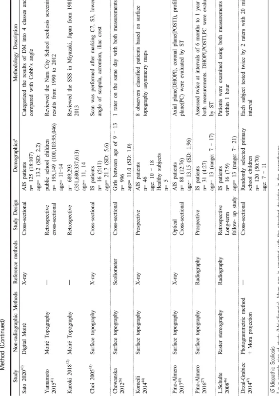
Table 2. Summary of the included studies. Table summarizes non-radiographic methods, Reference methods, Study design, Patients demographics and Method (Continued) StudyNon-radiographic MethodsReference methodsStudy DesignDemographicsaMethodology Description Sato 202040)Digital MoiréX-rayCross-sectionalAIS patients n= 125 (18:107) age= 13.2 (SD: 2.2) Categorized the results of DM into 4 classes and compared with Cobb’s angle Yamamoto 201541)Moiré Topography―Retrospective cross-sectionalpublic school children n= 195,149 (100,103:95,046) age= 11–14
Reviewed the Nara City School scoliosis screening results from 1990 to 2012 Kuroki 201842) Moiré Topography―Retrospectiven= 689,293 (351,680:337,613) age= 11, 14 Reviewed the SSS in Miyazaki, Japan from 1981 to 2013 Choi 200543) Surface topographyX-rayCross-sectionalIS patients n= 16 (5:11) age= 21.7 (SD: 5.6)
Scan was performed after marking C7, S3, lower angle of scapula, acromion, iliac crest Chowanska 201226)Surface topographyScoliometerCross-sectionalGirls between age of 9 – 13 n= 996 age= 11.0 (SD: 1.0)
1 rater on the same day with both measurements Komeili 201444)Surface topographyX-rayProspectiveAIS patients n= 46 age: 10 – 18 Healthy subjects n= 5 8 observers classified patients based on surface topography asymmetry maps Pino-Almero 201745)Surface topographyX-rayOptical Cross-sectionalAIS patients n= 88 (12:76) age= 13.15 (SD: 1.96)
Axial plane(DHOPI), coronal plane(POSTI), profile plane(PC) were evaluated by ST Pino-Almero 201627)Surface topographyRadiographyProspectiveIS patients n= 31 (4:27) age= 13 (range: 7 – 17) Assessed twice at intervals of 6 months to 1 year with both measurements. DHOPI,POSTI,PC were evaluated by ST L.Schulte 200846)Raster stereographyRadiographyRetrospective Long-term follow- up study
IS patients n= 16 (7:9) age= 13 (range: 7– 21)
Patients were examined using both measurements within 1 hour Drzal-Grabiec 201447)Photogrammetric method + Mora projection―Cross-sectionalRandomly selected primary school children n= 120 (50:70) age: 7 – 11
Each subject tested twice by 2 raters with 20 min interval IS Idiopathic Scoliosis a n = sample size of study (Male:Female), Mean age is reported with the standard deviation in the parentheses. SP Shoulder Pain, LBP Lower Back Pain, D.I.T.I Digital Infrared Themographic Imaging, AIS Adolescent Idiopathic Scoliosis, DM Digital Moiré, SSS school scoliosis screening DHOPI horizontal plane deformity index, POSTI posterior trunk symmetry index, PC columnar profile
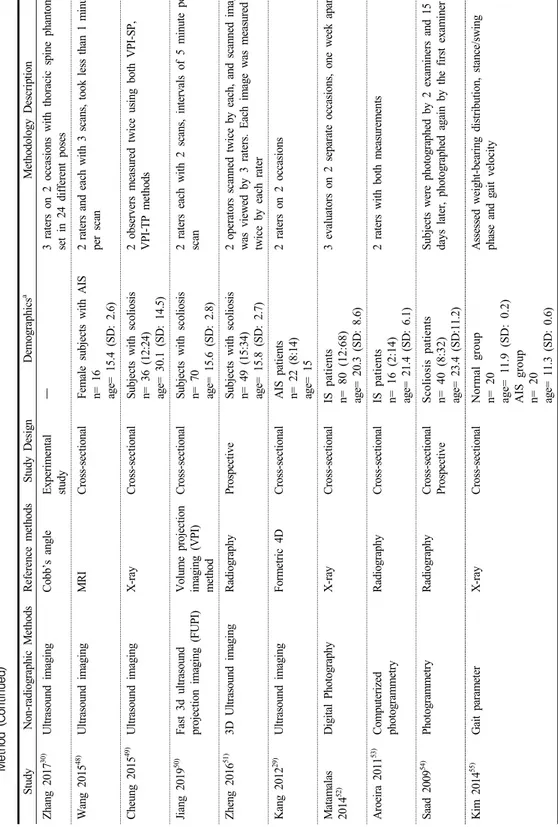
Table 2. Summary of the included studies. Table summarizes non-radiographic methods, Reference methods, Study design, Patients demographics and Method (Continued) StudyNon-radiographic MethodsReference methodsStudy DesignDemographicsa Methodology Description Zhang 201730)Ultrasound imagingCobb’s angleExperimental study―3 raters on 2 occasions with thoracic spine phantom set in 24 different poses Wang 201548)Ultrasound imagingMRICross-sectionalFemale subjects with AIS n= 16 age= 15.4 (SD: 2.6) 2 raters and each with 3 scans, took less than 1 minute per scan Cheung 201549)Ultrasound imagingX-rayCross-sectionalSubjects with scoliosis n= 36 (12:24) age= 30.1 (SD: 14.5)
2 observers measured twice using both VPI-SP, VPI-TP methods Jiang 201950)Fast 3d ultrasound projection imaging (FUPI)Volume projection imaging (VPI) method
Cross-sectionalSubjects with scoliosis n= 70 age= 15.6 (SD: 2.8) 2 raters each with 2 scans, intervals of 5 minute per scan Zheng 201651) 3D Ultrasound imagingRadiography ProspectiveSubjects with scoliosis n= 49 (15:34) age= 15.8 (SD: 2.7)
2 operators scanned twice by each, and scanned image was viewed by 3 raters. Each image was measured twice by each rater Kang 201229)Ultrasound imagingFormetric 4DCross-sectionalAIS patients n= 22 (8:14) age= 15 2 raters on 2 occasions Matamalas 201452)Digital PhotographyX-rayCross-sectionalIS patients n= 80 (12:68) age= 20.3 (SD: 8.6)
3 evaluators on 2 separate occasions, one week apart Aroeira 201153)Computerized photogrammetryRadiographyCross-sectionalIS patients n= 16 (2:14) age= 21.4 (SD: 6.1) 2 raters with both measurements Saad 200954) PhotogrammetryRadiographyCross-sectional Prospective Scoliosis patients n= 40 (8:32) age= 23.4 (SD:11.2)
Subjects were photographed by 2 examiners and 15 days later, photographed again by the first examiner Kim 201455)Gait parameterX-rayCross-sectionalNormal group n= 20 age= 11.9 (SD: 0.2) AIS group n= 20 age= 11.3 (SD: 0.6) Assessed weight-bearing distribution, stance/swing phase and gait velocity
Table 2. Summary of the included studies. Table summarizes non-radiographic methods, Reference methods, Study design, Patients demographics and Method (Continued) StudyNon-radiographic MethodsReference methodsStudy DesignDemographicsaMethodology Description Cho 201856)Machine learning based gait analysis testX-rayCross-sectionalTeenage scoliosis patients n= 24 age= 15.2 (SD: 2.5) Normal subjects(control) n= 18 age= 15.7 (SD: 2.6)
All subjects completed a 10m gait course for 10 times Ovadia 200757)Low intensity electromagnetic scan sensoring the spatial position of spinous process
X-rayProspectiveAIS patients n= 124 (35:89) age= 13 (SD: 3.17)
4 independent sites, 6 independent examiners, repeated twice for each patient Kokabu 201958) 3-D depth sensor imaging systemRadiographyProspective cohort studySubjects with suspected AIS n= 170 (21:149) age= 14.3 (range: 8 – 18) Subjects back was scanned by a 3D depth sensor VPI-SP volume projection imaging-spinous process, VPI-TP volume projection imaging-transverse process,
Table 3. Validity and reliability of the included studies Large category of assessment toolsStudyValidityInter-rater reliabilityIntra-rater reliability ScoliometerDriscoll 201433) Scolio-smartphone ICC= 0.89Scolio-smartphone ICC= 0.94 Franko 201234)Pearson’s Correlation Coefficient= 0.9995 (p<0.001) Jimbo 202035) ATI ICC= 0.733 Sum ATI ICC= 0.745ATI ICC of orthopedic doctor/office worker/assistant technical expert= 0.851, 0.786, 0.772 Sum ATI ICC of orthopedic doctor/office worker/assistant technical expert= 0.856, 0.900, 0.796 Prowse 201736)Good correlation with the Scoliometer (rho= 0.78) Moderate correlation for ATR (rho= 0.627)Thoracic kyphosis ICC= 0.94 ATR ICC= 0.92 Lumbar lordosis ICC= 0.79 Cervical lordosis= 0.51
measured by the Baseline® Body Level/ Scoliosis meter (Examiner A ICC3,3= 1.00, Examiner B= 0.98) measured by the Scoliometer (Examiner A ICC3,3= 0.99, Examiner B= 0.98) Sapkas 200337)Thoracic r= 0.685 (p<0.01) Thoracolumbar r= 0.572 (p<0.01) Lumbar r= 0.677 (p<0.01) Coelho 201338)r= 0.7 (p<0.05)0.890.92 D.I.T.IKwok 201739) ICC values > 0.9 ICC Intra-class correlation coefficient, ATI Angle of trunk inclination, ATR Axial thoracic rotation
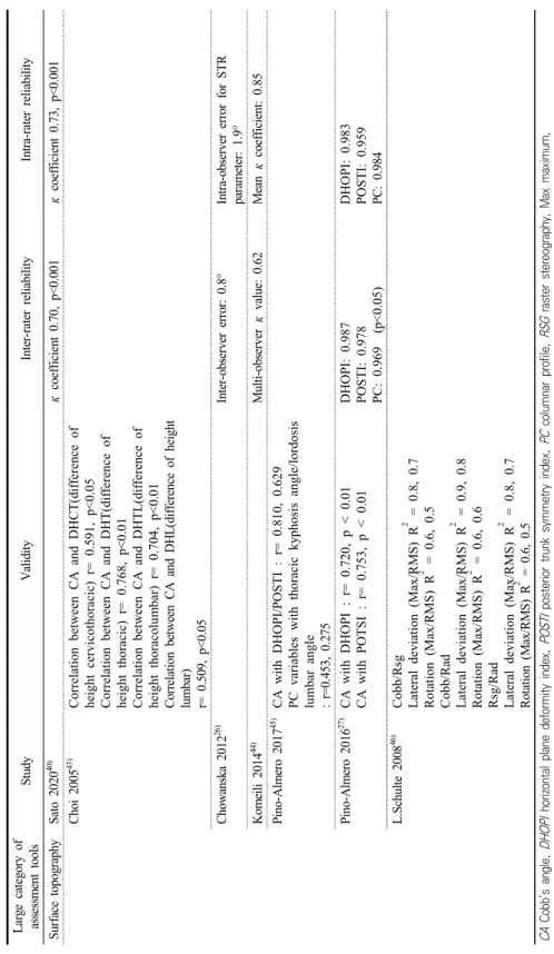
Table 3. Validity and reliability of the included studies (Continued) Large category of assessment toolsStudyValidityInter-rater reliabilityIntra-rater reliability Surface topographySato 202040) κ coefficient 0.70, p<0.001κ coefficient 0.73, p<0.001 Choi 200543) Correlation between CA and DHCT(difference of height cervicothoracic) r= 0.591, p<0.05 Correlation between CA and DHT(difference of height thoracic) r= 0.768, p<0.01 Correlation between CA and DHTL(difference of height thoracolumbar) r= 0.704, p<0.01 Correlation between CA and DHL(difference of height lumbar) r= 0.509, p<0.05 Chowanska 201226)Inter-observer error: 0.8°Intra-observer error for STR parameter: 1.9° Komeili 201444)Multi-observer κ value: 0.62Mean κ coefficient: 0.85 Pino-Almero 201745)CA with DHOPI/POSTI : r= 0.810, 0.629 PC variables with thoracic kyphosis angle/lordosis lumbar angle : r=0.453, 0.275 Pino-Almero 201627)CA with DHOPI : r= 0.720, p < 0.01 CA with POTSI : r= 0.753, p < 0.01DHOPI: 0.987 POSTI: 0.978 PC: 0.969 (p<0.05)
DHOPI: 0.983 POSTI: 0.959 PC: 0.984 L.Schulte 200846)Cobb/Rsg Lateral deviation (Max/RMS) R2 = 0.8, 0.7 Rotation (Max/RMS) R2 = 0.6, 0.5 Cobb/Rad Lateral deviation (Max/RMS) R2 = 0.9, 0.8 Rotation (Max/RMS) R2 = 0.6, 0.6 Rsg/Rad Lateral deviation (Max/RMS) R2 = 0.8, 0.7 Rotation (Max/RMS) R2 = 0.6, 0.5 CA Cobb’s angle, DHOPI horizontal plane deformity index, POSTI posterior trunk symmetry index, PC columnar profile, RSG raster stereography, Max maximum, RMS root mean square, Rad radiography
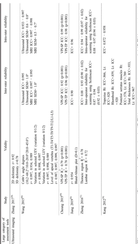
Table 3. Validity and reliability of the included studies (Continued) Large category of assessment toolsStudyValidityInter-rater reliabilityIntra-rater reliability Ultrasound imagingZhang 201730)2D deformity r= 0.92 3D deformity r=0.78 Wang 201548)Cobb’s angle degrees (10,2~68.2°/10.0~20.0°/20.0~45.0°) r= 0.997, 0.934, 0.989 Variation in selected UEV (variation 0/1/2) r= 0.998, 0.998, 0.997 Variation in selected LEV (variation 0/1/2) r= 0.997, 0.998, 0.989 Level of apical vertebra (T1-T4/T5-T8/T9-T12/L1-L5) r= 0.987, 0.997, 0.999, 0.997 Ultrasound ICC= 0.995 Ultrasound SEM= 1.0° MRI ICC= 0.997 – 0.995 MRI SEM= 1.0°
Ultrasound ICC= 0.933 – 0.997 Ultrasound SEM= 0.6 – 0.8° MRI ICC= 0.997 – 0.998 MRI SEM= 0.5 – 0.7° Cheung 201549) VPI-SP R2 = 0.79 (p<0.001) VPI-TP R2 = 0.78 (p<0.001)VPI-SP ICC: 0.92 (p<0.001) VPI-TP ICC: 0.96 (p<0.001)VPI-SP ICC: 0.99 (p<0.001) VPI-TP ICC: 0.98 (p<0.001) Jiang 201950)r= 0.954 Bland-Altman plot (D=0.1)ICC= 0.90ICC= 0.96 Zheng 201651)Thoracic region R2 = 0.78 Lumbar region R2 = 0.72ICC= 0.88 – 0.93 (0.90 ± 0.02) Inter-operator reliability for scanning using Scolioscan ICC= 0.87 – 0.94 (0.92 ± 0.03)
ICC= 0.94 – 0.99 (0.97 ± 0.02) Intra-operator reliability for scanning using Scolioscan ICC= 0.88 – 0.97 (0.94 ± 0.03) Kang 201229) Trapezius Rt: ICC=.866, Lt: ICC=.820 Rhomboid Rt: ICC=.859, Lt: ICC =.920 Posterior serratus muscles Rt: ICC=.578, Lt: ICC=.789 Total thicknesses Rt: ICC=.933, Lt: ICC=.967 ICC= 0.872 – 0.958 SEM standard error of measurement, UEV upper-end vertebra, LEV lower-end vertebra, VPI-SP Volume projection imaging spinous process method, VPI-TP Volume projection imaging transverse process method
Table 3. Validity and reliability of the included studies (Continued) Large category of assessment toolsStudyValidityInter-rater reliabilityIntra-rater reliability PhotographyMatamalas 201452)0.37 < r < 0.51Back (LRTA/SHA/AHA) ICC= 0.80, 0.80, 0.88 Front (LRTA/SHA/AHA) ICC= 0.65, 0.89, 0.85
Back (LRTA/SHA/AHA) ICC= 0.79, 0.88, 0.93 Front (LRTA/SHA/AHA) ICC= 0.78, 0.91, 0.91 Aroeira 201153) Thoracic region Kappa index= 0.92 Lumbar region Kappa imnex= 0.82 Saad 200954)Thoracic curves R= 0.619 Lumbar curves R= 0.551Thoracic: 0.942 (p<0.001) Lumbar: 0.564 (p= 0.010) Thoracolumbar: 0.879 (p<0.001)
Thoracic: 0.963 (p<0.001) Lumbar: 0.975 (p<0.001) Thoracolumbar: 0.945 (p<0.001) Gait analysisCho 201856) Cross-validation test result Accuracy of SVM to recognize scoliosis group and control group : 90.5% (if optimally selected, 95.2%) Accuracy of SVM to recognize scoliosis severity gait patterns : 81.0% (if optimally selected, 85.7%) Low intensity electromagnetic scan
Ovadia 200757)Thoracic r= 0.87 Lumbar r= 0.84 All curves r= 0.86 (p < 0.0001)
mean absolute difference between the paired coronal measurements : 6.3° (SD = 4.9°), Pearson’s correlation coefficient: 0.86 (P= 0.08) mean absolute difference between the paired sagittal measurements : 6.1° (SD = 4.9°), Pearson’s correlation coefficient: 0.87 (P= 0.11) mean absolute difference between the paired coronal measurements : 2.74° (SD = 2.4°), Pearson’s correlation coefficient: 0.86 (P= 0.11) mean absolute difference between the paired sagittal measurements : 4.83° (SD = 4.28°), Pearson’s correlation coefficient: 0.87 (P= 0.67) 3-D depth sensor imaging systemKokabu 201958)r= 0.85 (p<0.01) LRTA left/right trapezium angle ratio, SHA shoulder height angle, AHA axilla height angle, SVM support vector machine
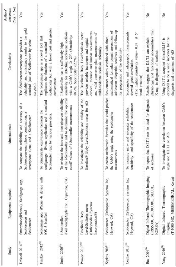
Table 4. Methodological characteristics of included studies, summarizing equipments, study aims and conclusion StudyEquipments required Aims/rationaleConclusionsAuthors' consensus (Yes / No) Driscoll 201433)Smartphone(iPhone4), Scoliguage app, Scolioscreen and Scoliometer To compare the reliability and accuracy of a Scolioscreen-smartphone combination, a smartphone alone, and a Scoliometer The Scolioscreen-smartphone provides a reliability and consistency similar to the gold standard (use of Scoliometer by spine surgeon).
Yes Franko 201234) Scoliguage app, iPhone 4s device with iOS 5 installedTo demonstrate equivalence between the scoligauage iPhone application and a standard Scoliometer read by various providers.
The Scoliguage app is a novel tool that replicates the function of a standard scoliometer but with a lower cost and greater convenience.
Yes Jimbo 202035)i-Scolioroller, iPod touch(Apple Inc., Cupertino, CA)To evaluate the intra-and inter-observer reliability of the i-Scolioroller and to determine the optimal cutoff values of i-Scolioroller measurements
I-Scolioroller has a sufficiently high sensitivity for detecting adolescent scoliosis with a Cobb’s angle ≥ 20°. (Sensitivity: 88.9%, Specificity: 62.5%)
Yes Prowse 201736)Baseline® Body Level/Scoliosis meter (Orthopaedics Systems Incorporation®)
To investigate the reliability and validity of the Baseline® Body Level/Scoliosis meter for AISThe Baseline® Body Level/Scoliosis meter provides reliable transverse and sagittal cervical, thoracic and lumbar measurements and valid transverse plan measurements of mild-moderate scoliosis deformity.
Yes Sapkas 200337)Scoliometer (Orthopaedic Systems Inc, Hayward, CA)To create mathematic formulas that could predict the Cobb’s angle using the scoliometer measurements
Scoliometer values combined with three mathematical formulas permit assessment of adolescent idiopathic scoliosis and follow-up for progression of the deformity
Yes Coelho 201338) Scoliometer (Orthopaedic Systems Inc, Hayward, CA)To measure intra- and interrater reliability, sensitivity and specificity of the scoliometerScoliometer and radiographic measurements showed good correlation. (The highest sensitivity value= 0.87 at 5° trunk rotation)
Yes Bae 200423)Digital Infrared Thermographic Imaging (IRIS5000, MEDICORE, SEOUL, KOREA)
To report that D.I.T.I can be used for diagnosis of scoliosisResults suggest that D.I.T.I can explain physiologic and functional abnormalities than X-ray, in diagnosis of scoliosis
No Yang 201621)Digital Infrared Thermographic Imaging (T-1000 HD, MESHMEDICAL, Korea)
To investigate the correlation between Cobb’s angle and D.I.T.I on AISUsing D.I.T.I, acupoint Simsu(BL15) is expected to be a valid indicator for the diagnosis and treatment of AIS
No
Table 4. Methodological characteristics of included studies, summarizing equipments, study aims and conclusion (Continued) StudyEquipments required Aims/rationaleConclusionsAuthors' consensus (Yes / No) Kwok 201739) FLIR E33 camera (FOL-18 lens; 10,800 pixels), Thermacam Researcher Professional 2.9 Software(FLIR) To explore the possibility of using IR thermography to evaluate Infra red emissions from subjects to detect abnormalities in temperature distribution in their paraspinal muscles.
The findings of this study suggest the feasibility of incorporating IR thermography as part of SSS.
Yes Sato 202040) Hump measurement system with a built-in 3D camera, personal computer (Kinect for Windows: Microsoft Corporation, Redmond, Washington)
To assess the usefulness of Digital Moiré(DM) for scoliosis screening.DM is useful as a new method for the screening of scoliosis with sufficient accuracy and reliability to replace Moiré topography. (Sensitivity= 0.98, Specificity= 0.53) Yes Yamamoto 201541)―Evaluate the accuracy of Moiré topography tool as a screening tool.Moiré topography had a high false-positive rate (66.7%), which did not improve with examiner experience.
No Kuroki 201842)―To make clear the both results and problems of SSS by Moiré topography(MT)SSS by MT seemed to be effective in detecting scoliosis although both positive predictive value and the reference rate to the second screening were low.
Yes Choi 200543)3D-surface topography (Koastron, IBS-2000, Korea)To measure correlation between 3D-surface topography and Cobb’s angle in scoliosis 3D surface topography and Cobb’s angle was highly correlated.Yes Chowanska 201226)CQ Electric System (Poland) deviceTo assess the usefulness of Surface topography(ST) for scoliosis screeningDid not reveal the advantage of ST as a scoliosis screening method in comparison with the use of scoliometer.
No D.I.T.I digital infrared thermographic Image, IR infrared, SSS school scoliosis screening, AIS adolescent idiopathic scoliosis
Table 4. Methodological characteristics of included studies, summarizing equipments, study aims and conclusion (Continued) StudyEquipments required Aims/rationaleConclusionsAuthors' consensus (Yes / No) Komeili 201444)Four VIVID 910 3D laser scanners (Konica Minolta Sensing Inc., Ramsey, NJ, USA), Polygon Editing Tool (PET version 2.21, Konica Minolta), Geomagic software (Geomagic Studio 12, Morrisville, NC, USA) Introduces a 3D markerless analysis technique for assessing torso asymmetry in AIS and a system for classifying patients based on this technique.
Distinctive patterns of asymmetry were identified with very good to excellent reliability.
Yes Pino-Almero 201745)Mobile white screen, projector, digital camera, computer with image recognition software designed in Matlab 7.9.0 (Matlab & Simulink Release 2009b. The Mathworks, Inc., Natick, MA, USA)
To study the correlation between asymmetry of back (measured by ST) and deformity of the spine (quantified by Cobb’s angle) ST cannot substitute for radiographs in the diagnosis of scoliosis but it can offer data that complement radiologic study.
No Pino-Almero 201627)Mobile white screen, projector EPSON(3LCD projector model: EMP-835), digital camera CANON, computer(MacBook Pro) with the program developed in MATLAB 7.9.0
To study if ST would be useful in the follow-up of AIS patientsST showed 90.32% agreement with radiographic method in follow-up of AIS patients.
Yes L.Schulte 200846)Formetric system (Diers International, Wiesbaden, Germany)To investigate the reliability and accuracy of raster stereography in comparison with radiography as the gold standard.
Rasterstereography accurately reflects the radiographically measured progression of scoliosis during the long-term follow-up, but these parameters are not directly comparable with the Cobb’s angle.
Yes Drzal-Grabiec 201447)Mora projection (MORA System 4th Generation)To evaluate the compatibility of external measurements of parameters characterizing scoliosis using the photogrammetric method.
The photogrammetric method gives significant results in terms of parameters characterizing the position of the shoulder blades and shoulders, as well as pelvis rotation.
Yes ST surface topography, AIS adolescent idiopathic scoliosis
Table 4. Methodological characteristics of included studies, summarizing equipments, study aims and conclusion (Continued) StudyEquipments required Aims/rationaleConclusionsAuthors' consensus (Yes / No) Zhang 201730)Ultrasound scannerDP-50 (Mindray Ltd., China) with a MHz probe, electromagnetic spatial sensing device PATRIOT (Polhemus Ltd., USA), personal computer (Intel Xeon CPU E5-1620-v3, 3.5GHz with 8G RAM) To analyze the correlation between the Cobb’s angle and spinous process angle(SPA) measured by ultrasound data
SPA and Cobb’s angle has high correlation especially for the curves with 2D deformity.Yes Wang 201548)3-D SonixTABLET, SonixGPS, C5-2/60 Convex transducer (Ultrasonix, Canada)To evaluate validity and reliability of Ultrasound imaging compare to MRIThe ultrasound imaging is a reliable and valid measurement of spinal curvature in the coronal plane using Center of Laminae (COL) method.
Yes Cheung 201549) Ultrasound scanner EUB-8500 (Hitachi Ltd., Tokyo, Japan), a computer with an Intel Core 2 Q6600 2.4-GHz processor and a video capture card NIIMAQPCI/PXI-1411 (National Instruments Corporation, Austin, TX, USA), a compact electromagnetic spatial sensing device MiniBird Model 130 (Ascension Technology Corporation, Burlington, VT, USA).
To assess the performance of newly developed freehand 3D ultrasound system.Results suggested that the ultrasound volume projection imaging method can be a promising approach for the assessment of scoliosis.
Yes Jiang 201950) A custom-designed liner 2-D ultrasound probe (width: 10 cm; frequency: 4–10 MHz), An electromagnetic spatial sensing device (driveBAY, Ascension Technology Corporation, Burlington, USA)
To develop a fast 3-D ultrasound projection imaging (FUPI) method for assessment of scoliosis.
The results indicate that the developed projection imaging method could greatly decrease the processing time while preserving the comparative image quality.
Yes Zheng 201651) The Scolioscan system (Model SCN801, Telefield Med- ical Imaging Ltd, Hong Kong)
To test the reliability of spine deformity measurement of Scolioscan and its validity compared to the Cobb’s angle from radiography in AIS patients.
Scolioscan is reliable for measuring coronal deformity for patients with AIS and appears promising in screening large numbers of patients, for progress monitoring, and evaluation of treatment outcomes.
Yes Kang 201229)Sonoace 8000 (Medison Inc, South Korea)To demonstrate the reliability of using diagnostic ultrasound imaging(USI) in the assessment of the thickness of the soft tissues of the interscapular region in AIS
USI could be a reliable method in evaluating of the thickness of the soft tissues of the interscapular region which in turn could be a useful guide to the assessment of the effects of AIS.
Yes
Table 4. Methodological characteristics of included studies, summarizing equipments, study aims and conclusion (Continued) StudyEquipments required Aims/rationaleConclusionsAuthors' consensus (Yes / No) Matmalas 201452)Digital Nikon D5100 (Nikon Corporation, Tokyo, Japan) cameraTo determine the validity of digital photography as an evaluation method for shoulder balance (ShB) in patients with idiopathic scoliosis.
Digital clinical photography appears to be a reliable method for objective clinical measurement of ShB. The correlation between clinical and radiological balance is statistically significant although moderate/weak.
No Aroeira 201153) A digital camera, Sony 7.1 megapixel (Sony, Manaus, Amazonas, Brazil), a Greika WT3750 (Greika, São Paulo, SP, Brazil) tripod, A Carci Simetograph (Carci, Americanópolis, SP, Brazil), CorelDraw13 software (CorelCorporation, Ottawa, Canada)
To develop a protocol for computerized photogrammetry for the quantification of scoliosis, and to mathematically relate this proposed method with the Cobb radiographic method.
The preliminary results presented demonstrate equivalence between the two methods. More studies are needed.
Yes Saad 200954)Photographic camera positioning (Sony P200 7.2.mp; Sony, Tokyo, Japan)The purpose of this study was to investigate the reliability and validity of photogrammetry in measuring the lateral spinal inclination angles.
Although the current study did not show the validity of photogrammetry as a measure of the lateral spinal curvature in scoliosis, high reliability coefficients were observed.
No Kim 201455)The SmartstepTM pneumatic insole, The SmartstepTM softwareTo demonstrate that relationship between scoliosis and gait factor and foot weight bearing in ambulation.
In this study Influence of scoliosis was not found on the rate of stance phase and rate of swing phase and gait velocity. Fore foot weight bearing (P = 0.019) was significantly higher in the AIS group.
No Cho 201856)IMU-based system (Human Track, Rbiotech Co., Ltd., Seoul, Korea) consisting of a gyroscope, accelerometer and magnetic sensor
This study discussed application of a machine learning approach for the automatic cognition of gait changes due to scoliosis using gait measures.
Study’s results demonstrate considerable potential in applying SVMs in gait classification for medical applications. (Accuracy of SVM to recognize scoliosis group and control group : 90.5% ) (Accuracy of SVM to recognize scoliosis severity gait patterns : 81.0% ) Yes Ovadia 200757)Ortelius800TM system (OrthoScan Technologies, Rosh Pina, Israel)To investigate the clinical value of Ortelius800TM Found the novel clinical tool to be reliable for following mild and moderate idiopathic curves in both coronal and sagittal planes.
Yes Kokabu 201958)Consumer-grade 3D depth sensor (Xtion Pro Live, ASUSTeK Computer Inc. Taipei, Republic of China), a laptop computer (Core-i5, 7200U-4 GB HP pavilion-15-au105tu, HP Inc, California, USA)
To report the potential accuracy of newly developed, asymmetry-recognition system for the surface of the human back using a 3D depth sensor This study demonstrates the outstanding ability of this new system for deciding whether additional radiography is needed to define scoliosis. This system can be an alternative to the forward bend test and scoliometer measurement in clinics. (Sensitivity: 0.97, Specificity: 0.93)
Yes SVM support vector machine
온 현상이, 만성기에는 저온 현상이 나타나며, 근육 의 경축이나 근막 통증 증후군의 경우는 병변 부위 에 지엽적인 고온 현상을 보인다.22)
Bae 등의 연구에서 5례의 척추측만증 환자의 D.I.T.I 결과, 극돌기에 보이는 고온 현상이 측만을 형성하였다. 또한 측만의 볼록한 쪽 온도보다 오목한 쪽의 온도가 낮았으며, 좌우 온도차 (ΔT)가 심한 곳 은 최소 0.6°C 이상 낮게 나타났으나, 통계적 유의성 여부는 본 연구에서 검증되지 않았다. Yang 등의 연 구에서는 척추측만증 환자의 D.I.T.I 상 6개의 혈자 리 (肩井 (GB21), 肺兪 (BL13), 心兪 (BL15), 脾兪 (BL20), 腎兪 (BL23), 關元兪 (BL26))에서 좌우 온 도차(ΔT)를 비교한 ANOVA 분석 결과, 心兪 (BL15)에서만 유의성이 확인되었다. 그러나 心兪 (BL15)에 대한 사후 검정 결과 Duncan test에서는 유의한 차이가 있었으나 Scheffe test에서는 유의성 이 확인되지 않았다. Kwok 등의 연구에서도 척추측 만증 환자의 좌우 체열 분포가 승모근, 광배근, 요방 형근 부위에서 유의한 차이를 보였다.
적외선 체열 촬영은 검사 시에 통증이나 방사선 노출의 위험이 없는 검사법으로 안전하며 반복적으 로 사용이 가능해 척추측만증의 경과 및 임상 양상 을 손쉽게 평가할 수 있다. 또한, 환자에게 증상의 개선 정도와 현상태 등을 직접 컬러화된 영상으로 제시하여 환자 본인의 이해도를 높일 수 있다는 점 에서 의미가 있다.23) 그러나 외부요인과 검사자의 숙 련도가 쉽게 결과에 영향을 미치며 비정상과 정상을 판정하는 기준이 모호하다는 등의 단점이 있어 검사 에 앞서 실행 조건을 명확히 할 필요성이 있다.24)
3. Surface Topography
체표면 영상분석 (Surface topography; ST)는 Moiré, ISIS (integrated shape investigation system)
25), Orthoscan26), Rasterstereography 등의 다양한 테 크닉을 사용하여 수행할 수 있는 외부의 신체 윤곽
27) 피사체의 등에 여러 줄의 빛을 투영한 후 특정 소 프트웨어 프로그램을 적용하여 3차원의 지형도를 얻 어 형태학적 비대칭성을 평가할 수 있다.28)
Moiré를 활용한 3편의 연구 중 Sato 등의 연구는 특발성 척추측만증 환자 126명을 대상으로 Cobb’s angle과의 비교를 통해 민감도 98%, 특이도 53%, 거짓양성율 47%, 평가자 간 신뢰도 73%, 평가자 내 신뢰도 70%로 높은 정확도와 신뢰도를 보고하였다.
Yamamoto 등이 23년간 학교 척추측만증 선별검사 로서 사용된 Moiré topography를 전향적으로 조사한 결과, 56.7-93.3%의 높은 거짓양성율을 보였으며 이 는 측정자의 경험에 의해 개선되지 않았다. 이와 유 사하게 33년간 Moiré topography의 척추측만증 선 별검사로서의 유효성을 평가한 전향적 연구에서는 척추측만증의 선별에는 유용한 것으로 나타났으나 양성예측율과 2차 선별 검사의 참고율은 낮았다. 16 명의 환자를 대상으로 한 장기간 추적연구에서 Rasterstereography는 방사선 촬영과 높은 상관관계 (R2≥0.5) 를 보였으며 이는 측만증 환자의 follow-up 에 있어 Rasterstereography가 측만의 진행상황을 안 정적으로 반영함을 나타낸다. Choi 등의 연구에서 laser scan 방식의 3D surface topography 측정값이 Cobb’s angle과 수평축 선상의 흉추비에서는 상관관 계를 보이지 않았으나 전후축 선상에서 흉부, 경흉 부, 흉요부, 요부 높이차와는 상관성이 높은 것으로 보고되었으며, 특히 흉부에서는 상관성이 가장 높게 나타났다 (r=0.768, p<0.01). 46명의 환자를 대상으 로 한 ST scan 기반 조사에서는 양호한 관찰자 내 신뢰도 (k=0.85), 중등도의 관찰자 간 신뢰도 (k=0.62) 와 높은 검사-재검사 신뢰도 (1년, Kappa value:
0.70 – 0.92)를 보고하였다. Pino-Almero 등은 31명 의 환자를 대상으로 6개월에서 1년간 ST로 척추측만 증의 진행률을 평가해 환자의 79.96% (31명 중 22 명)에서 2번째 방사선 촬영을 피하는 결과를 보고하 였다. 88명의 척추측만증 환자를 대상으로 한 동일한
도 평가에서 축 방향의 비대칭성 평가 (horizontal plane deformity index;DHOPI)와 관상면 상 비대칭 성 평가 (posterior trunk symmetry index;POSTI)의 높은 상관관계를 보였다. 척추 측만증 등급에 따른 분류에서도 DHOPI와 POSTI는 유의한 차이가 있었 으나 종단면상 비대칭성 평가 (columnar profile;
PC)는 통계적으로 유의하지 않았다. (F=2.77, p=0.068) Chowanska 등은 996명의 학생을 대상으로 ST가 학 교선별검사로서 Scoliometer를 대체할 수 있는지의 여부를 조사하였다. 관찰자 내 오차는 1.9°, 관찰자 간 오차는 0.8°며, 충분하지 않은 민감도와 특이도로 선별 절사값(cut-off value)를 설정할 수 없어 scolimeter의 대체에 적합하지 않았다.
4. Ultrasound Imaging
초음파 검사는 방사선에 대한 노출 없이 실시간으 로 간편하게 평가할 수 있는 저비용, 비침습적 검사 로 근육 및 뼈의 길이, 두께, 면적 등을 정량적으로 측정한다.29,30) 초음파 영상은 횡돌기, 추궁판, 극돌기 등과 같은 척추의 여러 부분을 2차원적 (2D)으로 포 착해 시각화 할 수 있어 척추 곡률의 평가 도구로 사 용할 수 있다.31)
척추측만증의 평가 도구로 초음파를 이용한 연구 로는 극돌기 각도 (spinous process angle; SPA) 평 가, center of laminae (COL) 방법을 통한 평가, volume projection imaging (VPL) 방법을 통한 평 가, 고속 3D 초음파 투영 영상 방법을 통한 평가, 어 깨뼈 사이 연부조직의 두께 평가 등이 이루어졌다.
척추 모형을 이용한 Zhang 등의 연구에서는 초음 파를 통해 측정한 극돌기 각도 (SPA)로 Cobb’s angle을 예측할 수 있음을 시사하였고, Wang 등, Cheung 등의 연구에서는 모두 초음파 측정과 Cobb’s angle 간의 높은 상관관계와 높은 신뢰도를 보고하였 다. (r>0.9 p<0.05, R2>0.8 p<0.001) Kang 등의 연 구에서는 척추측만증에 수반되는 어깨 연부조직의
는 ICC=0.87~0.96로 높은 신뢰 수준을 나타내었으 나 측정자간 신뢰도는 ICC=0.58~0.97로 비교적 낮 은 수준을 보였다. Zheng 등의 연구에서도 이와 유 사하게 측정자내 신뢰도 (ICC>0.94)보다 낮은 수준 의 측정자간 신뢰도 (ICC>0.88)를 보고하였는데 이 는 초음파 검사의 단점으로 꼽을 수 있는 검사자의 숙련도 차이, 결과에 대한 판독의 차이 등에 의한 것 으로 사료되며 초음파 측정의 경험 및 교육이 중요 함을 시사한다.
5. Photography
체간 사진의 촬영을 통해 척추측만증을 평가하는 방법으로 어깨의 균형을 이용한 연구, computerized photogrammetry를 활용한 연구, 측면 척추 기울기 각도 (lateral spinal inclination angle)를 이용한 연구 등이 보고되었다.
Matamalas 등의 연구에서 척추측만증에 수반되는 어깨불균형을 디지털 사진을 통해 분석한 결과 측정 자 간 신뢰도와 측정자 내 신뢰도는 모두 ICC>0.8로 높은 신뢰 수준을 보여주었으나, 방사선학적 평가방 법과의 상관관계는 0.37<r<0.51로 약한 통계학적 유 의성을 나타내었다. Aroeira 등은 척추측만증 환자 16명을 대상으로 척추 촉진을 통해 C7 – L5에 마킹 한 뒤 사진 촬영을 통해 측만각을 컴퓨터로 분석하 였다. 방사선학적 방법을 통해 얻은 Cobb’s angle과 의 평균 차이는 4.1°였으며 흉추부와 요추부에서 평 가의 일치도 Kappa 지수는 각각 0.92, 0.82였다.
Saad 등의 연구에서는 척추측만증 환자 40명의 배부 사진을 통해 측만각을 측정하고 신뢰도와 타당도를 평가하였다. ICC>0.8로 측정자내, 측정자간 신뢰도 모두 높은 신뢰수준을 보여주었으나 사진 측정법과 방사선학적 평가 사이에 선형 관계는 관찰되지 않아 타당도가 검증되지 않았다.
6.
기타행 분석, 저강도 전자파 스캔, 3D 깊이 센서 등을 이 용한 연구들이 보고되었다.
웨어러블한 관성 측정 장치 (inertial measurement units; IMU)와 컴퓨터 머신 러닝을 활용한 Cho 등의 연구에서는 정상 보행과 척추측만증 보행 패턴을 분 류하였다. 18명의 대조군과 24명의 척추측만증 환자 를 대상으로 보행 패턴을 추출해 교차 검증 테스트 를 실시하였을 때 척추측만증 그룹과 정상 그룹의 구별은 90.5%의 정확도를, 척추측만증의 중증도 인 식은 81.0%의 정확도를 보였다. Kim 등의 연구에서 는 척추측만증 환자들과 대조군이 센서가 내장된 SMARTSTEPTM을 발목 주위에 착용 후 보행하여 보행 속도, 체중 지지 분포 등 척추 기형이 보행에 미치는 영향을 분석하였다. 앞발의 체중 분포가 척추 측만증 환자들에게서 유의하게 높았으나, 보행 단계 및 보행 속도에 있어서 척추측만증의 영향은 발견되 지 않았다. Ovadia 등의 연구에서 활용한 저강도 전 자파 스캔기기 Ortelius800TM은 X-ray 촬영을 통해 얻은 Cobb’s angle과 관상면에서는 ± 5° 이하, 시상 면에서는 ± 6° 이하의 차이를 보여 두 방법 간의 유 의한 상관성을 보여주었다. Kokabu 등은 3D 깊이 센서를 통한 비대칭 인식 시스템이 Cobb’s angle 10° 이상에서 민감도 97%, 특이도 93%, 정확도 97%의 뛰어난 척추측만증 선별 능력을 보임을 보고하였다.
고 찰
척추측만증은 흔하게 접할 수 있는 척추 질환 중 하나로 건강보험심사평가원의 통계에 따르면 2019년 에 척추측만증으로 진단받은 환자는 9만 4천여명이 며, 그 중 10대 환자가 약 43%로 가장 높은 비율을 차지한 것으로 나타났다. 청소년기는 일반적으로 골 격이 미성숙하며 측만의 진행에 대한 위험이 높으므 로 측만의 조기진단과 진행상황의 평가는 임상적으로 중요하다.32) 이러한 척추측만증의 진단과 평가에는
로 가장 널리 사용되고 있으나3) 반복된 방사선 노출 을 포함한 몇가지 문제가 보고되었다. 척추 측만증 환자는 일반적으로 평균 25회의 척추 방사선 촬영을 거치며, 이 기간 동안 상대적으로 높은 양의 전리방 사선에 노출된다4).(평균 10.8cGy) Doody 등4)의 연 구에 따르면 다중 방사선 검사에 노출된 척추측만증 여성의 유방암 위험이 증가하였으며, 유방암으로 인 한 사망 위험은 일반 인구의 2배 이상이었다.
Goldberg 등5)은 척추측만증 여성의 난소 방사선 피 폭은 미래의 생식 결과에 악영향을 미친다고 보고하 였으며, Schmitz 등6)의 연구에서는 소아기의 방사선 진단이 백혈병과 전립선암에 크게 기여하는 것으로 나타났다.
이 외에도 Cobb’s angle은 측정자 내, 측정자 간 판독 편차를 보이며, 이는 각각 최대 3-5°와 6-9°인 것으로 보고되었다.2,9,10) 임상에서 측만의 진행은 Cobb’s angle에서 5° 이상의 증가가 발견될 때 진단 되므로 측정자 간의 이러한 편차는 척추 측만증의 장기 모니터링에 있어 평가의 신뢰성과 효율성을 저 하시킬 수 있다. 또한 Cobb’s angle은 2차원 평면 상 에서 도출되어지는 값으로 척추의 3차원적 변형에 대한 정보를 제공하지 못한다.8)
현재, 한의사는 직접 X-ray나 MRI 등의 의료영상 장비를 활용할 수 없고, 의원 또는 종합병원과 같은 의료기관에 의뢰하거나 의료법에 따라 한방병원에서 양방 진료과목을 추가로 개설하여 협진을 통해 의료 영상장비 등을 활용하는 상황으로,11) 한의사의 척추 측만증 치료에 있어 빠른 진단과 피드백이 어려운 실정이다. 이러한 이유들로 한의사를 포함한 임상의 들이 사용하는데 제한이 없고 비침습적이며 방사선 노출이 없는 척추측만증 평가 도구에 대한 필요성이 지속적으로 대두되어져왔다.
지금까지의 관련 연구를 살펴보면, 시상면상 척추 의 균형을 평가하기 위한 비방사선 평가척도에 대한 체계적 문헌 고찰 연구가 발표된 바 있다33). 그러나
사선적 평가 방법들의 신뢰도 및 타당도 평가가 주 를 이루고 있어 3차원적 기형이 특징인 척추측만증 을 온전하게 평가하였다고 볼 수 없으며, 현재 척추 측만증 평가척도의 특성과 타당성을 종합적으로 살 펴본 연구는 부족하다고 볼 수 있다.
이에 본 연구는 척추측만증 환자에게 적용할 수 있는 비방사선적 평가 도구의 연구 근황과 특성을 분석하기 위해 문헌 고찰 연구를 수행하였다. 문헌 검색 및 선정 결과 국외 논문 총 32편을 최종 분석 하였으며, 여기서 Surface topography, Scoliometer, Ultrasound, 적외선 체열 촬영, Photography, Gait parameter 등의 평가 도구가 도출되었다.
Surface topography는 3차원적으로 비대칭성을 평 가할 수 있는 도구이나 고가의 장비와 전문적인 조 작 및 평가 기술을 요한다는 단점이 있으며, 선별검 사로서 유효성이 검증되지 않다는 연구 결과가 보고 된 바 있다. 초음파는 실시간으로 간편하게 척추의 곡률을 평가할 수 있는 도구로 양호한 타당도와 측 정자 내 신뢰도를 보여주었으나 몇몇 연구에서 비교 적 낮은 수준의 측정자 간 신뢰도를 보여주었다. 이 는 검사자의 숙련도와 판독의 차이 등에서 기인한 것으로 사료된다. 척추측만증 환자들에서 적외선 체 열 촬영 결과, 좌우온도차가 관찰되었으나 포함된 3 개의 연구가 각각 5명, 56명, 14명을 환자를 대상으 로 하여 표본이 충분히 크지 않았으며 신뢰도와 타 당도 또한 보고되지 않아 유효성을 검증할 수 없었 다. Photography는 체부의 사진 촬영을 통해 척추측 만증을 평가하는 방법으로 어깨 균형을 이용한 연구 에서는 방사선학적 평가방법과 유의미한 상관관계가 없었으며, 측면 척추 기울기 각도를 이용한 연구에서 도 방사선학적 평가방법과 선형관계는 관찰되지 않 았다. Scoliometer는 저렴하고 짧은 시간 내에 간단 하게 측정할 수 있으면서도 신뢰성과 타당성이 여러 차례 검증된 척도로 본 고찰에서 다룬 6편의 연구에 서 모두 양호한 타당도와 신뢰도가 보고되었다. 특히
한 앱 등도 활발히 개발되고 있어 임상에서 활용도 가 높을 것으로 기대된다. 이 외에도 척추측만증에 수반되는 비정상적 보행을 분석하는 보행 척도, 저강 도 전자파 스캔, 3D 깊이 센서 등의 평가 척도에 대 한 연구가 이루어졌다.
위와 같은 비방사선학적 평가 도구들은 한의사를 포함한 임상의들이 제한 없이 접근할 수 있을 뿐 아 니라, 측만의 진행 및 예후 평가에 활용함으로써 주 기적인 방사선 촬영의 횟수를 줄이고 Cobb’s angle 을 보완하는 보완책으로서의 역할을 기대할 수 있다.
하지만 아직은 충분하지 않은 통계적 검증 및 검사 자의 경험에 대한 높은 의존성 등이 단점으로 판단 된다. 이에 임상적 활용도를 높이기 위한 지속적이고 추가적인 연구 및 시도가 필요할 것으로 판단된다.
본 연구는 비방사선적 척추측만증 평가 척도를 체 계적으로 고찰함에 있어 몇 가지의 제한점이 있다.
첫째, 본 연구는 평가 척도의 연구 동향을 파악하고 새로운 진단 도구에 대한 최신지견을 얻기 위해 2000년 이후에 발표된 논문만을 포함하였다. 둘째, 국내외의 데이터베이스를 사용하여 한국어, 영어, 중 국어로 된 문헌만을 선택적으로 선정하였다. 셋째, 본 연구는 논문의 검색 및 선정 과정에 소수의 연구 자가 참가하였으며 검색기간이 짧아 보편성을 획득 하지 못했을 가능성이 있다. 넷째, 신뢰도나 타당도 가 보고되지 않은 연구들이 있었으며 같은 평가 척 도를 사용하였다 하더라도 연구마다 신뢰도와 타당 도의 범위가 넓어 각 척도별로 명확히 통합된 결론 을 제시하는데 한계가 있었다. 다섯째, 본 연구에 포 함된 연구들의 질 평가가 이루어 지지 않았다. 따라 서 향후 연구에서는 이와 같은 점들을 보완하여 비 뚤림 위험 평가, 연구의 질 평가와 메타분석등을 포 함한 보다 체계적이고 객관적인 문헌 고찰이 필요할 것으로 사료된다.
위와 같은 제한점들에도 불구하고 본 연구는 국내 외에서 현재까지 연구된 비방사선학적 척추측만증
뢰도를 통해 각 평가 도구의 질이 양호한지를 확인 하였다. 이를 통해 한의사를 비롯한 의료인들이 향후 임상 및 연구에서 방사선학적 방법의 대안으로 척추 측만증의 진단과 평가에 활용할 수 있는 평가 도구 들을 제시하였다는 점에서 본 연구의 의의가 있다.
결 론
현재 척추측만증의 진단에 가장 널리 사용되고 있 는 Cobb’s angle은 방사선 촬영을 필요로 하기 때문 에 방사선 노출에 대한 환자의 거부감이 있을 수 있 고, 측정자에 따른 편차 등이 보고된 바 있다. 따라 서 본 연구는 비방사선 평가 척도를 사용한 연구들 을 검색하여 정리, 분석하였으며 이를 통해 Surface topography, Scoliometer, 적외선 체열 촬영, Ultrasound 등의 도구들이 방사선 촬영의 횟수를 줄 이고 Cobb’s angle을 보완할 수 있는 가능성을 살펴 보았다. 하지만 본 연구에 포함된 연구들의 신뢰도와 타당도의 범위가 넓으며 대규모 임상 연구가 아직은 매우 적어 추후 연구를 통해 척추측만증 평가 도구 들에 대한 보다 면밀하고 체계적인 후속 임상 연구 가 이루어져야 할 것이다.
Acknowledgement
본 연구는 보건복지부의 재원으로 한국보건산업진 흥원의 보건의료기술 연구개발사업 지원에 의하여 이루어진 것임(HF20C0210)
참고문헌
1. Hresko MT, Idiopathic scoliosis in adolescents.
New England Journal of Medicine, 2013;368(9):
834-41.
2. Bunnell WP, The natural history of idiopathic
1986;11(8):773-6.
3. Cobb J, Outline for the study of scoliosis.
Instr Course Lect AAOS, 1948;5:261-75.
4. Doody MM, Lonstein JE, Stovall M, Hacker DG, Luckyanov N, Land CE, Breast cancer mortality after diagnostic radiography: findings from the U.S. Scoliosis Cohort Study. Spine (Phila Pa 1976), 2000;25(16):2052-63.
5. Goldberg MS, Mayo NE, Levy AR, Scott SC, Poîtras B, Adverse reproductive outcomes among women exposed to low levels of ionizing radiation from diagnostic radiography for adolescent idiopathic scoliosis. Epidemiology, 1998:271-8.
6. Schmitz-Feuerhake I, Pflugbeil S, ‘Lifestyle’
and cancer rates in former East and West Germany: the possible contribution of diagnostic radiation exposures. Radiation protection dosimetry, 2011;147(1-2):310-3.
7. Legaye J, Follow-up of the sagittal spine by optical technique. Annals of physical and rehabilitation medicine, 2012;55(2):76-92.
8. Mínguez MF, Buendía M, Cibrián RM, Salvador R, Laguía M, Martín A, et al., Quantifier variables of the back surface deformity obtained with a noninvasive structured light method: evaluation of their usefulness in idiopathic scoliosis diagnosis.
European Spine Journal, 2007;16(1):73-82.
9. Pruijs J, Hageman M, Keessen W, Van Der Meer R, Van Wieringen J, Variation in Cobb angle measurements in scoliosis. Skeletal radiology, 1994;23(7):517-20.
10. Carman D, Browne R, Birch J, Measurement of scoliosis and kyphosis radiographs.