COMPARATIVE STUDY ON THE FRACTURE
STRENGTH OF EMPRESS 2 CERAMIC AND TARGIS- VECTRIS CROWN
Young-Joo Cha, D.D.S., Jae-Ho Yang, D.D.S., M.S.D., Ph.D., Sun-Hyung Lee, D.D.S., Ph.D., Jung-Suk Han, D.D.S., M.S., Ph.D.
Department of Prosthodontics, College of Dentistry, Seoul National University
Due to an increasing interest in esthetics and concerns about toxic and allergic reactions to certain alloys, patients and dentists have been looking for metal-free tooth-colored restorations. Recent improve- ment in technology of new all-ceramic materials and composite materials has broadened the options for esthetic single crown restorations.
The aim of this investigation was to study the fracture strength of the metal-free posterior single crowns fabricated using two recently introduced systems, Empress 2 ceramic and Targis-Vectris.
Forty premolar-shaped stainless steel dies with the 1mm-wide circumferential shoulder were pre- pared. Ten cylindrical crowns having a diameter of 8.0mm and total height of 7.5mm were fabricated for each crown system respectively(PFM, Empress staining technique, Empress 2 layering technique, and Targis-Vectris).
The crowns were filled with cement and placed on the stainless steel dies with firm finger pressure. The crowns were then stored in distilled water at room temperature for 24 hours before testing. The crowns were tested for fracture strength in an Instron universal testing machine (Instron 6022). With a crosshead speed of 1mm/min the center of the occlusal surface of the crown was loaded using a 4-mm-diameter stainless steel ball until fracture occurred. The fracture surfaces of the crowns were gold coated and ex- amined using scanning electron microscopy(Jeol JSM-840 Joel Ltd., Akishima, Tokyo, Japan).
Within the parameters of this study the following conclusions were drawn:
1. The mean fracture strength for PFM crowns was 5829(±906)N; for Empress staining technique the frac- ture strength was 1697(±604)N; for Empress 2 Layering technique the fracture strength was 1781N(±
400)N, and the fracture strength for Targis-Vectris was 3093(±475)N.
2. The fracture strength of the PFM crowns was significantly higher than that of the Empress 2 and the Targis-Vectris crowns (P<0.05).
3. The fracture strength of the Targis-Vectris crowns was significantly higher than that of the Empress 2 crowns (P<0.05).
4. No statistical difference was found when Empress staining technique was compared with Empress 2 layering technique.
5. The SEM image of fracture surface of Empress 2 crown showed a very dense microstructure of the lithi- um disilicate crystals and the SEM image of fracture surface of Targis-Vectris crown showed indentations of Vectris and some fibers torn off from Vectris.
Key Words
Empress 2, Targis-Vectris, Fracture strength
J Korean Acad Prosthodont : Volume 39, Number 6, 2001
D
ue to an increasing interest in esthetics and con- cerns about toxic and allergic reactions to certain den- tal alloys, both patients and dentists have been looking for metal-free tooth-colored restorations.Recent improvement in technology of new all- ceramic materials and composite materials has broadened the options for esthetic single crown restorations.
Ceramics are rountinely used for dental restora- tions. Despite the high fracture resistance of tradi- tional metal ceramic crowns, limitations are im- posed on the systems by esthetic concerns. Ceramic materials are brittle, have limited tensile strength, and are prone to time-dependent stress failure. These shortcomings are attributable to the presence of microdefects within the material and a degradation in aqueous environment resulting from subcriti- cal crack growth (stress corrosion). In the last few years, various systems such as Dicor (Dentsply), Optec-HSP (Jeneric Pentron), In-Ceram (Vita Zahnfabrik), Empress (Ivoclar-Vivadent), Optimal Pressable Ceramics (OPC, Jeneric Pentron), recently Empress 2 (Ivoclar-Vivadent) have been intro- duced. These systems have good esthetics and im- proved physical properties.
The Empress system uses guided crystallization leucite-reinforced glass ceramics, a lost-wax process, and ceramic material that is processed using a ther- moforming procedure and a special furnace.
The Empress 2 glass-ceramic represents a new type of material that does not bear any resemblance to the leucite glass-ceramic Empress as far as materials sci- ence is concerned. Empress 2 is a lithium disili- cate glass-ceramic, and the chemical basis for the ma- terial is the SiO2-Li2O system. In the course of glass- ceramic development, lithium disilicate glass-ceramics have also been fabricated.
The first lithium disilicate glass-ceramics was developed as early as the 1950s. This development was the work of Stookey.1Following his funda- mental discovery, lithium disilicate glass-ceram-
ics became the subject of a considerable amount of research. The nucleation mechanism and the ki- netics of crystallization of the main lithium disilicate phase received the most attention.2-4A disadvantage of these lithium disilicate glass ceramics, however, was their poor chemical resistance.
Considerable progress in the development of a chemically resistant lithium disilicate-based glass- ceramic was achieved by Beall5 and Echeverria.6 Compared with Empress, Empress 2 exhibits sub- stantially improved chemical properties and high- er translucency. Morever, microcracks do not form in the microstructure. At the same time, the material is easily processed in the dental laboratory with a pressing procedure in the EP500 press furnace (Ivoclar), where the material undergoes viscous flow at 920℃. Holand et al7 analyzed the mi- crostructures of glass-ceramics of the Empress 2 and Empress systems by scanning electron mi- croscopy and concluded Empress 2 can be used to fabricate 3-unit fixed partial dentures up to the sec- ond premolar.
The glass ceramic was produced by melting a glass, which was powdered and crystallized in a sin- tering and hot-press process using a hot-press fur- nace (EP500, Ivoclar Ltd). The glass-ceramic ingot was pressed into a mold at 920℃ with a holding time of 20 minutes. The effective pressure applied through a plunger was 20 bar. The pressing process was conducted under a partial vacuum of 20 to 50 mbar in the furnace chamber. Under these conditions, the glass-ceramic became viscous and consistently flowed into the mold. The typical duration of a pressing cycle was 5 to 20 minutes, depending on the volume and complexity of the mold. Once the molding cycle ended, the mold was left to cool to room temperature. During the hot-press proce- dure and the cooling phase, the final microstructure of the glass-ceramic was formed. Subsequently, the pressed part was divested from the mold by blast- ing the mold material with corundum powder and glass beads using 1 to 2 bar pressure.
The particulate composites are commonly used to restore defects in a single tooth or as a veneer ma- terial for a tooth or prosthesis, but they are rarely used alone to make final complete-coverage crowns and fixed partial dentures. The fiber-reinforced com- posite (FRC) framework replaces the classic metal framework of a porcelain-fused-to metal prosthesis, while a particulate composite applied over this FRC substructure corresponds to the porcelain ap- plied in a traditional restoration. The FRC framework provides strength and rigidity beneath the outer lay- er of particulate composite. This two-phase polymer prosthesis combines the best characteristics of the fiber-reinforced composite (strength and rigidity) with those of the particulate composite (wear resistance and esthetics) to provide an alternative to all-ceramic or porcelain-fused-to-metal restoration. Rosenthal et al8studied the qualities that render Targis-Vectris particularly suitable for a variety of indications, including laboratory-fabricated restorations for the stress-bearing posterior regions. Many studies showed the material properties and clinical proto- col of a new material which combines a ceramic op- timized polymer with a fiber-reinforced frame- work for durable, aesthetic anterior and posterior restorations.9,10Some of fiber-reinforced compos- ite materials include Glasspan (Glasspan), Connect (Kerr), Ribbond (Ribbond), Splint-It! (Jeneric/Pentron), FibreKor (Jeneric/Pentron), and Vectris (Ivoclar/Vivadent). The improved FRC formulation is light-and heat-polymerized and contains S2 glass fibers that are preimpregnated with a bis-GMA matrix. It exhibits the same excellent physical prop- erties of the earlier polycarbonate FRC but has bet- ter handling characteristics. Mechanical tests of this new material have shown that it has up to seven times the strength of particulate composite. It is also much more rigid than particulate composite.11-13 This formulation also has improved optical properties.
Advances in resin composite technology have en- hanced and supplemented the FRC technology.
Some of these improved materials include Sculpture
(Jeneric/Pentron), Artglass (Kulzer/Jelenko), Poly(mer)glass (Kulzer/ Jelenko), Targis (Ivoclar/Vivadent), Ceromer (Ivoclar/Vivadent), and belleGlass HP (belle de St Claire/Kerr). These products employ new polymer formulations, have improved filler particle distribution, and can be polymerized with intense light, vacuum, and heat.
All of these factors have improved their wear re- sistance and elasticity, which, in turn, has resulted in increased impact and fracture resistance. This new generation of composite materials, used as the overlay or veneer providing the anatomic shape and contour over the FRC framework, provides the potential for a metal-free and ceramic-free fixed partial dentures with long-term durability and ser- viceability.
The aim of this investigation was to study the frac- ture strength of the metal-free posterior single crowns fabricated using two recently introduced sys- tems, Empress 2 ceramic and Targis-Vectris.
MATERIAL AND METHODS
Forty premolar-shaped stainless steel dies with the 1mm-wide circumferential shoulder were prepared (Fig. 1). Ten cylindrical crowns having a diameter of 8.0mm and total height of 7.5mm were fabricated for each crown system respectively (Fig. 2).
All clinical and technical steps in the fabrication of PFM, Empress 2 and Targis-Vectris crowns strict- ly followed the procedures recommended by the man- ufacturers (Fig. 3).
Conventional PFM crowns served as a control group. Crown copings (thickness 0.5mm) were cast in a nickel-chromium alloy (Verabond, AALBA Dent Inc.) and veneered with Vita VMK 95(Vita Zahnfabrik, Germany) dental ceramics. Crown cop- ings were made using a silicone template of the waxed coping. The silicone template was seated on the dies and filled with molten wax. Wax coping sizes were controlled with digital calipers. Identical crown sizes were controlled by grinding or adding
Table Ⅰ. Crown systems tested and pretreatment and cementation procedures used
Crown system Pretreatment Cement
PFM crowns Sandblasting Panavia 21
Empress staining technique crowns Etching and silane Variolink Ⅱ Empress 2 layering technique crowns Etching and silane Variolink Ⅱ
Targis-Vectris crowns Roughening and silane Variolink Ⅱ
porcelain and were checked with digital calipers to an accuracy of 0.1mm. After self-glazing, the crowns were cemented on stainless steel dies with resin cement (Panavia 21, Kuraray Co. Ltd., Japan).
The Empress staining technique crowns were made using a silicone template of the previously
waxed complete crown shape measuring 8.0mm in diameter and 7.5mm in height. The wax pat- terns were embedded in Empress 2 special invest- ment material using Empress ring base and paper ring provided by the manufacturer. The wax was eliminated in a burnout furnace at 850℃ and crowns were pressed in the system’s pressing furnace (EP500, Ivoclar Ltd., Liechtenstein). After cooling, the crowns were devested by blasting away the in- vestment using glass beads (50�100
μ
m) at 2-bar pres- sure. Sprues were removed and crowns were fitted on the dies and colored with two layers of shading porcelain and one layer of glazing porcelain.The Empress 2 layering technique crown cop- ings (thickness 0.8mm) were made using the template technique as described above. For the Empress 2 lay- ering technique, crown copings were heat pressed as described above, and the pressed crown cop- ings were reduced with diamond instruments in a Fig. 1. Stainless steel die used as an abutment.
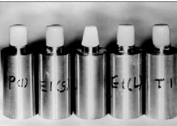
Fig. 2.Ten cylindrical crowns were fabricated for each crown system.
Fig. 3. PFM(left), Empress staining, Empress 2 layering core (center), Empress 2 layering, and Targis-Vectris crown (right) are shown.
handpiece to thickness of 0.8mm. The copings were veneered with Empress 2 layering porcelain to cre- ate the final crown shape. After glazing, the crowns were cemented on the stainless-steel dies with dual-cured resin cement (Variolink Ⅱ, Ivoclar/
Vivadent, Liechtenstein).
Targis-Vectris crowns were made using Vectris Single (Ivoclar/Vivadent, Liechtenstein) for con- structing the substructure. The Vectris Single was cured in Vectris VSI curing unit. Cured substructure was ground up to 1mm below from margin and then sandblasted, silane treated. The substructure was built up all around with layered Targis. Each layer was polymerized in the Targis Power unit. The Targis- Vectris crowns were finished with diamond burs, sil- icone wheel, Robinson brush, and polishing buffers with polishing paste.
The cement was mixed according to the manu- facturer’s instructions. The crowns were filled with cement and placed on the stainless steel dies with firm pressure. The excess cement was removed, and finger pressure was immediately applied to the crown for 10 minutes. In the case of Variolink Ⅱ, this was polymerized step by step for 40 seconds per seg-
ment by light. The crowns were then stored in distilled water at room temperature for 24 hours be- fore testing. The crowns were tested for fracture strength in an Instron universal testing machine (Instron 6022)(Fig. 4). With a crosshead speed of 1mm/min, the center of the occlusal surface of the crowns was loaded using a 4-mm-diameter stainless steel ball until fracture occurred (Fig. 5).
The fracture surfaces of the crowns were gold coat- ed and examined using scanning electron mi- croscopy (Jeol JSM-840 Joel Ltd., Akishima, Tokyo, Japan).
A statistical analysis was done using the one- way ANOVA and Duncan’s multiple range test.
RESULTS
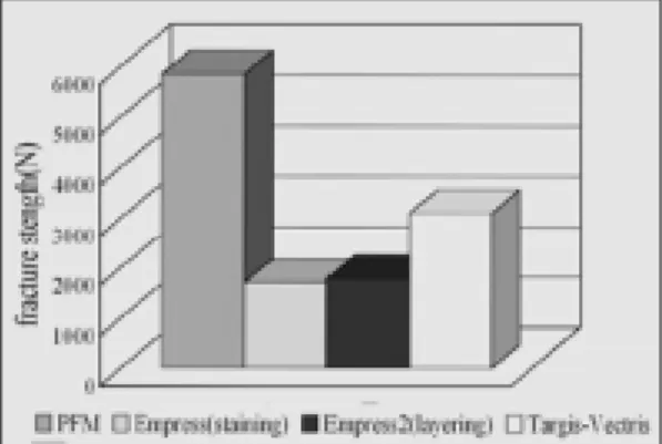
The mean load at complete fracture of the crowns is shown in Table Ⅱ and Fig. 6.
The PFM crowns fractured at a mean force of 5829(±906)N. The ceramic veneer was sheared off the metal coping, partly exposing the metal sub- structure. All Empress 2 crowns showed a com- plete fracture of the ceramic. The mean fracture strength was 1697(±604)N for the Empress staining technique and 1781N(±400)N for the Empress 2 lay- ering technique (Fig. 7). For all Targis-Vectris crowns tested, the Vectris coping remained intact.
Fig. 4.Fracture stength test on the Instron universal test- ing machine (Instron 6022).
Fig. 5. Crown was loaded by a 4-mm-diameter stainless steel ball.
Only the Targis fractured (Fig. 8). The mean fracture strength for Targis-Vectris crowns was 3093(±
475)N.
The fracture strength of the PFM crowns was significantly higher than that of the Empress 2 and Targis-Vectris crowns (P<0.05). The fracture strength of the Targis-Vectris crowns was significantly high- er than that of the Empress 2 crowns (P<0.05). No sta- tistical difference was found when Empress stain- ing technique was compared with Empress 2 layering technique (Table Ⅲ, Ⅳ).
Scanning electron microscopic (SEM) images of frac- Fig. 6.Mean fracture strength for PFM, Empress staining
technique, Empress 2 layering technique, and Targis- Vectris crown luted with adhesive resin cement.
Fig. 7.Typical fracture patterns of PFM (left), Empress staining (center), and Targis-Vectris (right) crown.
Fig. 8. Appearance of fractured Targis-Vectris crowns.
Table Ⅱ. Loads at complete fracture of crowns (N)
PFM Crown Empress staining Empress 2 layering Targis-Vectris
5935 1164 1876 2810
5225 2494 1556 4050
4685 1524 2476 2535
7420 1231 1325 2715
6090 3010 1824 3305
5355 1379 2132 2860
6625 1751 1605 3520
5755 1434 1107 3140
4565 1227 2062 2625
6635 1756 1850 3375
5829(±906) 1697(±604) 1781(±400) 3093(±475)
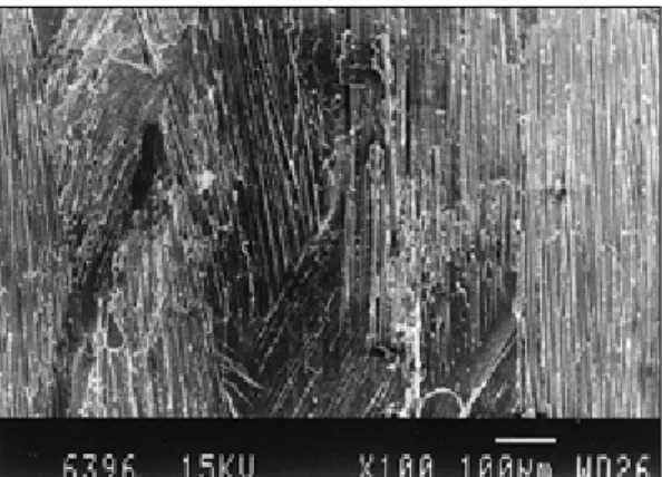
tured surface of a PFM, an Empress staining tech- nique, an Empress 2 layering technique, and a Targis-Vectris crown with two magnifications(×100,
×1,000) of the same specimen are shown in Fig. 9 thorough Fig. 16. The SEM image of fracture surface of Empress 2 crown showed a very dense mi- crostructure of the lithium disilicate crystals and Targis-Vectris crown showed indentations of Vectris and some fibers torn off from Vectris.
DISCUSSION
The testing of the fracture strength of crowns is not a standard method like a bending test of a geo- graphically well-defined bar. Many factors influence the results : crown thickness, porosity, prepara- tion design, luting agent, direction of the applied load, location of load application and radius of the load- ing stylus.14-17Static testing gives no clues about the long-term material properties under fatiguing stresses. However, compressive-strength studies
of crown systems, within their limits, give an idea for the load-bearing capacity in simulated clinical sit- uations. The results of in vitro strength studies may give helpful information for the design of clinical studies. The use of such testing methods on- ly provides criteria for further clinical evaluations, because such tests do not accurately represent the clin- ical environment and various intraoral force.
The preparation design of the abutments used in this study - a 90-degree shoulder with round inner angles- is recommended for Empress 2 ceramic and Targis-Vectris crowns in vivo. The circular shoulder width was standardized at 1.0mm. In this study, stainless steel dies were used as abutments.
The advantages are the possibility of a standardized preparation and the ensuring of identical physi- cal quality of materials. However, abutments made of stainless steel do not reproduce the actual force distribution that may occur on crowns cemented to natural teeth. Chemo-mechanical interaction be- tween the dentin and the luting composite, re- Table Ⅲ. Oneway ANOVA for fracture strength
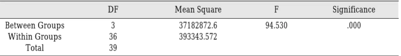
DF Mean Square F Significance
Between Groups 3 37182872.6 94.530 .000
Within Groups 36 393343.572
Total 39
Table Ⅳ. Duncan’s multiple range test
N Subset for alpha=.05
1 2 3
Empress crown
(staining) 10 1697.00
Empress 2 crown
(layering) 10 1781.30
Targis-Vectris
crown 10 3093.50
PFM crown 10 5829.00
Significance .765 1.000 1.000
Means for groups in homogenous subsets are displayed.
quired for adhesive bonding, also cannot be tested in this type of simulation. Yoshinari and Derand18 used bovine dentin abutments for the mechanical test- ing of different all-ceramic crowns, which, like steel or resin dies, cannot accurately simulate human teeth in vivo. Sobrinho et al19used brass dies for test- ing of the fatigue properties of all-ceramic crown sys- tems because the brass die did provide a repro- ducible support.
The thickness of the crown and core is important.
The small variations can affect the strength of the restoration.20But Koh and Yang21founded no statistical difference in flexural strength for Empress 2 ce- ramic according to the thickness. In this study, a sil- icone mold was used to produce a wax crown shape with the same dimension for Empress 2 crowns.
All crowns were fitted loosely to avoid tensile stress resulting from crown positioning and seating dis- crepancies during cementation. All Empress 2 and Targis-Vectris crowns were adhesively luted to the dies. The manufacturers advise the etching and silane coating of the inner restoration surface for Empress 2, the roughening and silane coating of the inner restoration surface for Targis-Vectris, and the use of an enamel and dentin bonding system with a dual-polymerizing adhesive resin. Many stud- ies showed a strong enhancement of the fracture strength of all-ceramic crowns bonded to dies or teeth versus nonbonded crowns. Ludwig and Joseph22re- ported an increase in fracture resistance of up to 200%
using adhesive luting procedures. Yoshinari and Derand18reported that there were significant dif- ferences of the fracture strength between all-ce- ramic crowns luted with zinc phosphate cement and adhesive resin cement. But a number of prob- lems associated with dentine bonding and the clin- ical behavior of resin composite cements are still un- solved and further research will use natural teeth as an abutment and resin cement as a luting agent.
In this study, the point of force application for frac- ture was the center of the occlusal surface of the crown
shapes. The biting force of posterior teeth can vary between 245 N23and 540 N.24It may be stated that Targis-Vectris as well as Empress 2 crowns have suf- ficient strength to allow clinical testing of these metal-free crowns. The static testing method gives no information about the long-term behavior of the materials with regard to fatigue stresses.
Another variable that can contribute to failure of a ceramic restoration is the environment. Some studies have shown that fracture strength of ce- ramics decreased when tested in water as com- pared to a dry environment.25-28
Probster29compared the fracture strengths of two all-ceramic crowns and metal ceramic incisor crowns tested with loading perpendicular to the long axis:
946 N for In-Ceram; 814 N for surface-colored Empress; and 1494 N for metal ceramic. Incisor crowns tested with the load inclined30yielded val- ues of 380 N for In-Ceram with 0.5mm core, 450 N for In-Ceram with 0.7mm core, 220 N for surface col- ored Empress, and 160 N for veneered Empress. For premolar crowns tested with perpendicular loading31: 1609 N for In-Ceram with 0.7mm core and 1557 N for metal ceramic. In their study, the fracture strength of premolar crowns tested with inclined load- ing after cyclic prestressing was 1060 N for In- Ceram with 0.5mm core and 891 N for Empress. Paek and Yang32compared the fracture strengths of five kinds of all-ceramic crowns and evaluated the effects of cements on the fracture strength of all-ceramic crowns. Hwang and Yang33reported that the fracture strength of Celay In-Ceram Alumina crowns had a significantly higher fracture strength than conven- tional In-Ceram Alumina crowns
Kappert34reported that the biaxial flexural strength for Empress 2, In-Ceram alumina, Empress, and Vitadur N was 433 MPa, 430 MPa, 130 MPa, and 120 MPa respectively. Sorensen et al35examined the fracture toughness and fracture strength of Empress 2 and reported that Empress 2 was able to sup- port two times the load for Empress.
Behr et al36examined in vitro whether adhesive pos-
terior inlay fixed partial dentures made with Targis- Vectris system have fracture strength and satisfac- tory marginal adaptation, which can occur under clin- ical conditions. Loose et al37examined the fracture strength and marginal adaptation of posterior fixed partial dentures made with Targis-Vectris and In- Ceram. Fiberglass reinforced systems showed sig- nificantly higher fracture strength than In-Ceram af- ter thermal cycling and mechanical loading. In this study Targis-Vectris crowns showed higher fracture strength than Empress 2 crowns with 0.8mm core thickness.
CONCLUSIONS
Empress 2 ceramic and Targis-Vectris crowns were fabricated under controlled conditions, luted on the stainless steel dies, and loaded to failure. PFM crowns served as controls for comparison. Within the parameters of this study, the following conclusions were drawn:
1. The mean fracture strength for PFM crowns was 5829 (±906)N; for Empress staining technique the fracture strength was 1697 (±604)N; for Empress 2 layering technique the fracture strength was 1781 (±400)N, and the fracture strength for Targis- Vectris was 3093 (±475)N.
2. The fracture strength of the PFM crowns was sig- nificantly higher than that of the Empress 2 and the Targis-Vectris crowns (P<0.05).
3. The fracture strength of the Targis-Vectris crowns was significantly higher than that of the Empress 2 crowns (P<0.05).
4. No statistical difference was found when Empress staining technique was compared with Empress 2 layering technique.
5. The SEM image of fracture surface of Empress 2 crown showed a very dense microstructure of the lithium disilicate crystals and the SEM image of fracture surface of Targis-Vectris crown showed indentations of Vectris and some fibers torn off from Vectris.
REFERENCES
1. Stookey SD. Chemical machining of photosensitive glass. Ind Eng Chem 1953; 45:115-118.
2. Borom MP, Turalo AM, Doremus RH. Stength and microstructure in lithium disilicate glass-ce- ramics. J Am Ceram Soc 1975;58:385-391.
3. James P. Kinetics of crystal nucleation in silicate glasses. J Non-Cyst Sol 1985; 73:517-540.
4. McMillan PW, Philips SV, Patridge G. The struc- ture and properties of a lithium zinc silicate glass- ceramic. J Mater sci 1966;1:269-279.
5. Beall GH.Glass-ceramics:Recent developments and application. In:Weinberg MC(ed). Nucleation and crystallization in liquids and Glasses. Ceram Trans 1993;30:189-203.
6. Echeverria LM. New lithium disilicate glass-ceramic.
Bol Soc Esp Ceram VID 1992;5:183-188.
7. Holand W, Schweiger M, Frank M, Rheinberger V.
A comparison of the microstructure and proper- ties of the IPS Empress 2 and IPS Empress glass ce- ramics. J Biomed Mater Res 2000;53(4):297-303.
8. Rosenthal L. A new system for posterior restora- tions:a combination of ceramic optimized polymer and fiber-reinforced composite. Pract Periodont Aesthet Dent 1997 Jun-Jul;9(5suppl):6-10.
9. Fahl N JR, Casellini RC. Ceromer/FRC technolo- gy: The future of biofunctional adhesive aesthet- ic dentistry. Signature 1997;4(2):7-13.
10. Zanghellini G. Fiber-reinforced framework and Ceromer restorations:a technical review. Signature 1997;4(1):1-5.
11. Karmaker AC, DiBenedetto AT, Goldberg AJ.
Fiber reinforced composite materials for dental ap- pliances. Presented to the Society of Plastic Engineers ANTEC, Indianapolis, IN, 1996 May:5- 9.
12. Freilich MA, Karmaker AC, Burstone CJ, Goldberg AJ. Flexure strength of fiber-reinforced composites designed for Prosthodontic application(abstract 999).J Dent Res 1997;76:138.
13. Freilich MA, Karmaker AC, Burstone CJ, Goldberg AJ. Flexure strength and handling characteristics of fiber-reinforced composites used in Prosthodontics (abstract 1361).J Dent Res 1997;76:184.
14. Friedlander LD, Munoz CA, Goodacre CJ, Doyle MG, Moore BK. The effect of tooth preparation de- sign on breaking strength of Dicor crowns. Part I.
Int J Prosthodont 1990;3:159-168.
15. Voss R, Eichner K. Orientierende Untersuchungen uber die Festigkeit metallkeramischer Kronen auss neuen Werkstoffen. Dtsch Zahnarstl Z 1978;
33:456-460.
16. Coca I, Schwicherath H: Zur Beanspruchung von Kronen im Frontzahnbereich. Dtsch Zahnarstl Z 1987;42:338-341.
17. Dickinson AJG. A comparative study of the strength of aluminous porcelain and all ceramic crowns. J Prosthet Dent 1989;61:297-304.
18. Yoshinari M, Derand T. Fracture strength of all-ce- ramic crowns. Int J Prosthodont 1994;7:329-338.
19. Sobrinho LC, Cattell MJ, Glover RH, Knowles JC.
Investigation of the dry and wet fatigue properties of three all-ceramic crown systems. Int J Prosthodont 1998;11:255-262.
20. Riley EJ. Ceramo-metal restoration. State of the sci- ence. Dent Clin North Am 1977;21:669-182.
21. Koh JW, Yang JH. Influence of thickness of Empress 2 ceramic on fracture strength. J Korean Acad Prosthod 2000;38(4):446-460.
22. Ludwig K, Joseph K. Untersuchungen zur Bruchfestigeit von Empress-Kronen in Abhangigkeit von den Zementiermodalitat-en. Quintessenz Zahntech 1994;20:247-256.
23. Kolber KH, Ludwig K. Maximale Kaukraft als Berechnungsfaktor Zahntechnischer Konstruktionen.
Dent Labor 1983;31:55-60.
24. Sonnenburg M, Fethke K, Riedel S, Voelker H.
Zur Berechnungsfaktor Zahne des menschlichen Kiefers. Zahn Mund Kieferheilkd 1978;66:125- 132.
25. Sherril CA, O’Brien WJ, Transverse strength of alu- minous and feldspathic porcelain. J Dent Res 1974;53:683-690.
26. Myers ML, Ergle JW, Fairhurst CW, Ringle RD.
Fatigue characteristics of a high-strength porcelain.
Int J Prosthodont 1994;7:253-257.
27. Southan DE, Jorgensen KD. The endurance limit of dental porcelain. Aust Dent J 1974;19:7-11.
28. Fairhurst CW, Lockwood PE, Ringle RD, Twiggs SW. Fatigue parameters of a model feldspathic porcelain(abstract 573). J Dent Res 1993;72:175.
29. Probster L. Compressive strength of two mod- ern all-ceramic crowns. Int J Prosthodont 1992;5:409- 414.
30. Schwickerath H, Was der Zahntechniker beacht- en sollte Herstellung von vollkermischem Zahnersatz. Dental Labor 1992; 40:1501-1506.
31. Grey NJA, Piddock V, Wilson MA. In vitro com- parison of conventional crowns and a new all-ce- ramic system. J Dent 1993;21:47-51.
32. Paek SJ, Yang JH. A study on the fracture strength of all-ceramic crowns. J Korean Acad Prosthodont 1995;33:611-625.
33. Hwang JW, Yang JH, Lee SH, Chung HY. A study on fracture strength of conventional and copy- milled In-Ceram crowns. J Korean Acad Prosthod 1997; 35:417-427.
34. Kappert HF. Examination report to Ivoclar AG, Schaan, January 1998.
35. Sorensen JA, Cruz M, Mito WT. Research evalu- ations of a lithium disilicate restorative system: IPS Empress 2. Signature 1999;4:4-10.
36. Behr M, Rosentritt M, Leibrock A, Schneider- Feyrer S, Handel G. In-vitro study of fracture strength and marginal adaptation of fibre-reinforced adhesive fixed partial inlay dentures. J Dent 1999
;27(2):163-168.
37. Loose M, Rosentritt M, Leibrock A, Behr M, Handel G. In vitro study of fracture strength and marginal adaptation of fibre-reinforced compos- ite versus all-ceramic fixed partial dentures. Eur J Prosthodont Restor Dent 1998 Jun;6(2):55-62.
Reprint request to:
DR. JAE-HOYANG
DEPT. OFPROSTHODONTICS, COLLEGE OF DENTISITY, SEOUL NATIONAL UNIVERSITY.
28-1 YEONGUN-DONG, CHONGNO-GU, SEOUL KOREA 110-749, Tel:+82-2-760-2661, Fax:+82-2-760-3860
FIGURES ①
Fig. 10.SEM image of fractured PFM crown (×1,000).
Fig. 11. SEM image of fractured Empress staining technique crown (×100).
Fig. 12.SEM image of fractured Empress staining technique crown with high magnification (×1,000).
Fig. 13. SEM image of fractured Empress 2 layering tech- nique crown of core/dentine interface (×100).
Fig. 14.SEM image of fractured Empress 2 layering tech- nique crown (×1,000).
Fig. 9.SEM image of fractured PFM crown (×100).
FIGURES ②
Fig. 15. SEM image of fractured Targis-Vectris crown (×100).
Fig. 16.SEM image of fractured Targis-Vectris crown with high magnification (×1,000).