ABSTRACT
Purpose: Papillary thyroid cancer (PTC) is the most common endocrine cancer worldwide.
Tumor size on observation, together with lymph node metastasis, serves as a determinant of surgery. However, not all patients with PTC experience an increase in the size of tumor. We investigated various clinicopathological factors associated with the size of tumor to discern the group that can be observed without the need for early surgery.
Methods: The records of 1,401 patients diagnosed with PTC (excluding the follicular variant) between 2015 and 2017, were reviewed. Clinicopathological features suspected to have a link to tumor growth were included, such as diabetes mellitus (DM), use of metformin, a surgical history of breast, ovary, uterine, or familial non-medullary thyroid cancer, B-type Raf kinase (BRAF) V600E mutation, and lymphovascular invasion (LVI).
Results: Size of tumor was found to be related to sex, age, hypertension, ovary-uterine surgery, LVI and psammoma bodies. However, after adjusting for the effects of other factors on tumor size, both age and LVI were found to be significantly related to tumor size, with age also being significantly related to LVI.
Conclusion: Age and LVI are significant factors in the enlargement of tumors, unlike several other features, including DM, breast cancer history, familial cancer history, Graves's disease, Hashimoto's thyroiditis, and BRAF mutation.
Keywords: Papillary thyroid cancer; Lymph nodes; Lymphatic metastasis
INTRODUCTION
Papillary thyroid cancer (PTC) is the most common endocrine malignancy in the world, and its prevalence is continuously increasing (1). It has been claimed that compared with a high prevalence of thyroid cancer in South Korea, where an increased rate of screening is carried out, the incidence of thyroid cancer detected in other countries is just the tip of the iceberg (2). However, if other countries also had increased screening, PTC could be found to be even more prevalent worldwide to the extent of being considered an epidemic (3).
The size of thyroid cancer in patients with PTC does not always increase. According to observation trials, tumor enlargement by 3 mm or more occurred in 6.4%–6.7% and 8%–15.9% of patients with thyroid cancer at 5 and 10 years, respectively (4-6).
Original Article
Received: Jul 31, 2019 Revised: Sep 24, 2019 Accepted: Oct 1, 2019 Correspondence to Jeonghun Lee
Department of Surgery, Ajou University School of Medicine, 164 World cup-ro, Yeongtong-gu, Suwon 16499, Korea.
E-mail: servent-lee@hanmail.net Copyright © 2019. Korean Association of Thyroid and Endocrine Surgeons; KATES This is an Open Access article distributed under the terms of the Creative Commons Attribution Non-Commercial License (https://
creativecommons.org/licenses/by-nc/4.0/).
ORCID iDs Sunseok Yoon
https://orcid.org/0000-0001-7626-9637 Jeonghun Lee
https://orcid.org/0000-0003-4925-9104 EuyYoung Soh
https://orcid.org/0000-0002-8483-6416 Conflict of Interest
No potential conflict of interest relevant to this article was reported.
Author Contributions
Conceptualization: Sunseok Yoon, Jeonghun Lee. Data curation: Sunseok Yoon. Formal analysis: Sunseok Yoon. Investigation:
Sunseok Yoon. Methodology: Sunseok Yoon. Software: Sunseok Yoon. Supervision:
Jeonghun Lee, EuyYoung Soh. Validation:
Jeonghun Lee. Visualization: Sunseok Yoon, EuyYoung Soh. Writing - original draft:
Sunseok Yoon. Writing - review & editing:
Jeonghun Lee, EuyYoung Soh.
Sunseok Yoon , Jeonghun Lee , EuyYoung Soh
Department of Surgery, Ajou University School of Medicine, Suwon, Korea
Which Clinicopathological Factors
Are Related to Tumor Size in Papillary
Thyroid Cancer?
In observation trials tumor size, together with lymph node (LN) metastasis, objectively determines whether a patient should undergo surgery. Tumor size has been included in many risk stratifications for thyroid cancer, such as the Age, histologic Grade of the tumor, Extent of extrathyroidal invasion or distant metastases, and Size of the primary tumor (AGES), Age, presence of distant Metastases, Extent and Size of the primary tumor (AMES), Metastasis, patient Age, Completeness of resection, local Invasion, and tumor Size (MACIS), European Organization for Research on Treatment of Cancer (EORTC), and American Thyroid Association (ATA) classifications. Furthermore, it was reported to be the most important prognostic factor for recurrence and LN metastases. Finding a predictor of enlargement of tumor will therefore help in the treatment of PTC patients.
Among other variables, young age was reported to be an independent predictor of disease progression, which is defined as tumor enlargement or new LN metastases (7). However, other factors that may be related to tumor enlargement have not yet been investigated well in thyroid cancer. For example, among many other factors, diabetes mellitus (DM) has been reported to contribute to development of cancer through multiple mechanisms, including hyperglycemia, oxidative stress, and chronic inflammation. Also, use of metformin as a first-line treatment of type 2 DM is known to affect cancer biology through inhibiting the proliferation, metabolism, and angiogenesis of cancer cells (8-10). Other clinical factors involving sex hormones have also been reported to influence tumor size (11).
We, therefore, investigated various clinicopathological factors correlated with tumor size to discern which group of patients may be observed, without the need for early surgery, so that the primary treatment will be optimally effective.
MATERIALS AND METHODS
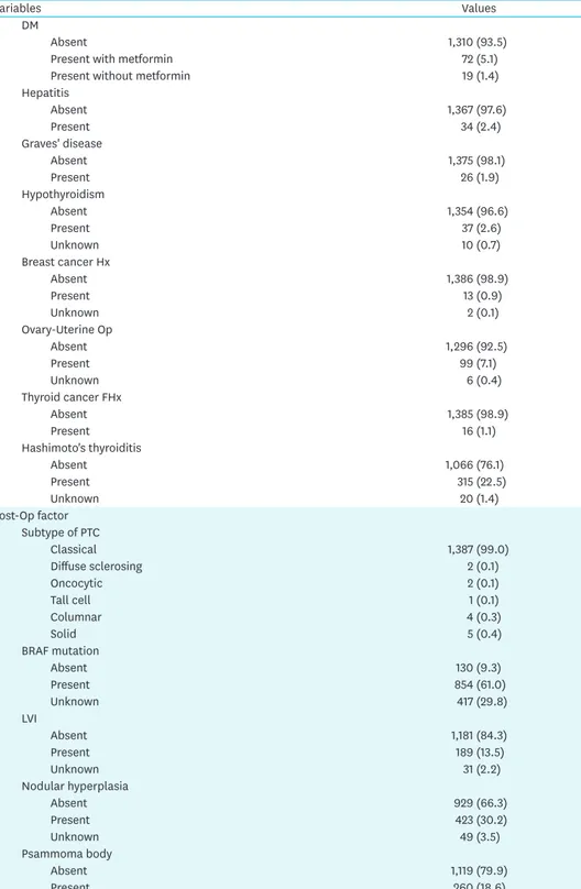
The records of 1,401 patients treated for PTC at Ajou University Hospital between 2015 and 2017 were reviewed. Clinicopathological factors were divided in two groups, pre- and post-operation factors. Those are summarized in Table 1. The follicular variant of PTC was excluded, because discerning it from noninvasive follicular neoplasm with papillary-like nuclear features is challenging (12). Of the patients, 154 (11.0%) had one or more first- degree relatives with differentiated thyroid cancer. Among them, sixteen (1.1%) patients were diagnosed as having familial non-medullary thyroid cancer (FNMTC), this being more stringently defined as patients having three or more first-degree relatives with differentiated thyroid cancer. Patients with previous ovary-uterine surgery were defined as those having undergone ovarian or uterine surgery due to myoma, ovarian cystic torsion, endometriosis, and ovarian teratoma. The V600E B-type Raf kinase (BRAF) mutation was analyzed according Table 1. Clinical and pathological features of 1,401 patients
Variables Values
Age at diagnosis 45.5±0.31
Pre-Op factor Sex
Female 1,061 (75.7)
Male 340 (24.3)
HTN
Absent 1,154 (82.4)
Present 247 (17.6)
(continued to the next page)
Variables Values DM
Absent 1,310 (93.5)
Present with metformin 72 (5.1)
Present without metformin 19 (1.4)
Hepatitis
Absent 1,367 (97.6)
Present 34 (2.4)
Graves' disease
Absent 1,375 (98.1)
Present 26 (1.9)
Hypothyroidism
Absent 1,354 (96.6)
Present 37 (2.6)
Unknown 10 (0.7)
Breast cancer Hx
Absent 1,386 (98.9)
Present 13 (0.9)
Unknown 2 (0.1)
Ovary-Uterine Op
Absent 1,296 (92.5)
Present 99 (7.1)
Unknown 6 (0.4)
Thyroid cancer FHx
Absent 1,385 (98.9)
Present 16 (1.1)
Hashimoto's thyroiditis
Absent 1,066 (76.1)
Present 315 (22.5)
Unknown 20 (1.4)
Post-Op factor Subtype of PTC
Classical 1,387 (99.0)
Diffuse sclerosing 2 (0.1)
Oncocytic 2 (0.1)
Tall cell 1 (0.1)
Columnar 4 (0.3)
Solid 5 (0.4)
BRAF mutation
Absent 130 (9.3)
Present 854 (61.0)
Unknown 417 (29.8)
LVI
Absent 1,181 (84.3)
Present 189 (13.5)
Unknown 31 (2.2)
Nodular hyperplasia
Absent 929 (66.3)
Present 423 (30.2)
Unknown 49 (3.5)
Psammoma body
Absent 1,119 (79.9)
Present 260 (18.6)
Unknown 22 (1.6)
The Clinical and pathological features of 1,401 patients treated for PTC at Ajou University Hospital are summarized.
Values are presented as the number of cases (%) or mean±standard error.
Op = operation; HTN = hypertension; DM = diabetes mellitus; Hx = history; FHx = familial history; PTC = papillary thyroid cancer; BRAF = B-type Raf kinase; LVI = lymphovascular invasion.
Table 1. (Continued) Clinical and pathological features of 1,401 patients
to the procedure of Hayashida et al. (13), and Hashimoto's thyroiditis was diagnosed on pathological examination. DM patients were divided into 2 groups, according to whether they were taking metformin. Lymphovascular invasion (LVI) was defined as the presence of tumor cells within a vascular space, meeting the following criteria: 1) tumor cells surrounded by red cells or lymphocytes, 2) tumor cells surrounded by endothelial cell lining, and 3) attachment of tumor cells to vascular walls. It is difficult to prove LVI, because both the thinness of the lymphatic vascular wall and the invasiveness of tumor mask the evidence of lymphatic invasion (14).
Comparisons of clinicopathological features were performed using Student's t-test and the χ2 test of proportions. The relationships between 2 variables were examined using correlation testing, and multivariate regression analysis was performed to control for baseline differences and confounding variables. All tests were 2-sided, with a Cronbach's α level of 0.05. All calculations were performed using SPSS 18.0 software (SPSS Inc., Chicago, IL, USA).
RESULTS
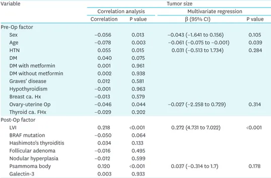
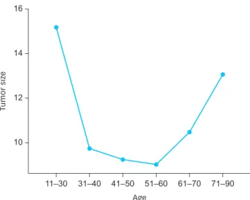
Tumor size was shown to be related to sex, age, hypertension, ovary-uterine surgery, psammoma bodies and LVI. However, after the effects of other factors were adjusted for, tumor size was found to be significantly related to both age and LVI (Table 2). Tumors increase in size at both younger age and older age, as opposed to in middle age (Table 3), as well as in cases with LVI. Age and LVI were also significantly related to each other (Tables 4 and 5). When both mean tumor size and proportion of LVI were plotted according to age, the two graphs showed a similar U shape (Figs. 1 and 2). In other words, at younger and older
Table 2. The relationship between tumor size and other clinical and pathological factors
Variable Tumor size
Correlation analysis Multivariate regression
Correlation P value β (95% CI) P value
Pre-Op factor
Sex −0.056 0.013 −0.043 (−1.641 to 0.156) 0.105
Age −0.078 0.003 −0.061 (−0.075 to −0.001) 0.039
HTN 0.055 0.015 0.031 (−0.513 to 1.734) 0.284
DM 0.040 0.075
DM with metformin 0.001 0.961
DM without metformin 0.002 0.938
Graves' disease 0.012 0.581
Hypothyroidism −0.001 0.963
Breast ca. Hx −0.013 0.579
Ovary-uterine Op −0.046 0.044 −0.027 (−2.258 to 0.729) 0.314
Thyroid ca. FHx −0.029 0.202
Post-Op factor
LVI 0.218 <0.001 0.272 (4.731 to 7.022) <0.001
BRAF mutation −0.050 0.064
Hashimoto's thyroiditis 0.034 0.133
Follicular adenoma −0.016 0.495
Nodular hyperplasia −0.012 0.599
Psammoma body 0.120 <0.001 0.037 (−0.314 to 1.7) 0.178
Galectin-3 0.003 0.933
Tumor size was shown to be related to sex, age, HTN, ovary-uterine surgery, LVI, and psammoma bodies.
However, after the effects of other factors were adjusted for, tumor size was found to be significantly related to both age and LVI.
CI = confidence interval; Op = operation; HTN = hypertension; DM = diabetes mellitus; Hx = history; FHx = familial history; LVI = lymphovascular invasion; BRAF = B-type Raf kinase.
ages tumor sizes were larger and LVI was more frequent. Other factors, including DM and metformin usage, were not found to be related to size of tumor in the present study, although they have been reported to be related to the biological behavior of cancer cells in other studies (8,9).
DISCUSSION
In PTC patients, tumor size was found to be a prognostic factor for survival, and a decisive factor for observation without early surgery. Although LN metastasis was also a decisive Table 3. The comparison of tumor sizes according to age and LVI
Variable Tumor size (mean±SE) P value
Age 11–30 114 15.1±1.06 <0.001
31–40 404 9.7±0.37 0.921
41–50 435 9.2±0.29 1.000
51–60 298 9.0±0.33 0.998
61–70 108 10.4±0.69 0.603
71–90 40 12.2±1.23 0.016
LVI Absent 1,180 9.1±0.17 <0.001
Present 189 15.3±0.81
Tumors increase in size at both younger age and older age, as opposed to in middle age, as well as in cases with LVI.
LVI = lymphovascular invasion; SE = standard error.
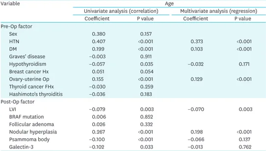
Table 4. The relationship between age and other clinical and pathological factors
Variable Age
Univariate analysis (correlation) Multivariate analysis (regression)
Coefficient P value Coefficient P value
Pre-Op factor
Sex 0.380 0.157
HTN 0.407 <0.001 0.373 <0.001
DM 0.199 <0.001 0.103 <0.001
Graves' disease −0.003 0.911
Hypothyroidism −0.057 0.035 −0.032 0.171
Breast cancer Hx 0.051 0.054
Ovary-uterine Op 0.155 <0.001 0.129 <0.001
Thyroid cancer FHx −0.030 0.259
Hashimoto's thyroiditis −0.036 0.183
Post-Op factor
LVI −0.079 0.003 −0.070 0.003
BRAF mutation 0.006 0.852
Follicular adenoma 0.026 0.332
Nodular hyperplasia 0.267 <0.001 0.198 <0.001
Psammoma body −0.100 <0.001 −0.066 0.137
Galectin-3 −0.102 0.033 −0.013 0.762
Age and LVI were also significantly related to each other.
Op = operation; HTN = hypertension; DM = diabetes mellitus; Hx = history; FHx = familial history; LVI = lymphovascular invasion; BRAF = B-type Raf kinase.
Table 5. The comparison of mean ages in significantly related factors
Variable Age (mean±SE)
Absent Present P value
HTN 43.1±0.31 56.2±0.68 <0.001
DM 44.7±0.31 55.0±1.243 <0.001
Ovary-uterine Op 45.01±0.32 51.5±1.004 <0.001
LVI 45.8±0.33 43.07±0.92 0.002
Nodular hyperplasia 43.3±0.35 50.5±0.57 <0.001
SE = standard error; HTN = hypertension; DM = diabetes mellitus; Op = operation; LVI = lymphovascular invasion.
factor for observation, tumor size is known to be the strongest predictor of LN metastasis (15). Ultrasonography can be used to detect LN metastasis, but this modality comes with the problem of inter-institutional differences ranging from 55.8% to 89% (16,17). Also, accuracy in detecting LN metastasis through ultrasonography at the central compartment of the neck is low, compared to accuracy at the lateral compartment, which follows the initial metastasis at the central compartment (18). Considering these limitations of the use of ultrasonography, tumor size can be considered the easiest and most useful indicator in observation trials.
When we investigated the relationship between suspicious clinicopathological features (Table 1) and tumor size, tumor size was not correlated with ovary-uterine surgery, DM, metformin use, familial thyroid cancer history, or BRAF mutations.
In this study, ovary-uterine surgery included hysterectomy, myomectomy, and ovarian cystectomy due to myoma, teratoma, or endometriosis. Uterine surgery for uterine myoma
14
Tumor size
12
10 16
61–70 71–90 51–60
41–50 31–40 11–30
Age
Fig. 1. The distribution of mean tumor size according to age.
30
LVI (%) 20
10
0 40
61–70 71–90 51–60
41–50 31–40 11–30
Age Fig. 2. The distribution of LVI according to age.
LVI = lymphovascular invasion.
is most common, because this disease occurs in women of reproductive-age (19). Uterine myoma significantly increases concentrations of both estrogen and progesterone receptors, and both estrogen and progesterone promote myoma (20,21). Estrogen receptors are also present in thyroid cancer, and sex hormones, especially estrogen, influence thyroid cancer cell growth (11). For this reason, this history was included in this study, together with breast cancer history. However, ovary-uterine surgery was not related to tumor size in PTC in the present study.
Epidemiological and biological data have indicated that many cancers are related to DM (22). Potential mechanisms include insulin resistance, hyperinsulinemia, insulin-like growth factor-1 (IGF-1), hyperglycemia, and dyslipidemia through mitogen-activated protein kinase, phostphoinositide-3-kinase/Akt, and Janus Kinase/signal transduction and/or activation of transcription 3 signaling pathways by IGF-1, adipokines, and cytokines (8). However, in the present study we could not confirm any association between tumor size and DM, nor did we see a decrease in tumor size in PTC with metformin usage. Even in the sub-analysis of patients older than 50 years, DM or use of metformin was not found to be related to tumor size. There may be other mechanisms that support and control tumor growth and progression in thyroid cancer as compared to other cancers.
FNMTC has been reported in about 5% of the cases, whereas its rate in the Korean population has been reported to be 9.6% in a previous study (23). However, its relatively low prevalence of 1.2% of the cases in our study can be explained by the more stringent definition of FNMTC as being present when a patient had three or more first-degree relatives with differentiated thyroid cancer. Generally, FNMTC is more aggressive than sporadic differentiated thyroid cancer, and also has more extrathyroidal extension, LN metastasis, multifocality, and recurrence than sporadic differentiated thyroid cancer (24,25).
In our study, tumor size in FNMTC was similar to that in sporadic differentiated thyroid cancer, as reported in previous studies (24,25), despite FNMTC being more aggressive than the sporadic form.
BRAF mutation is the most frequent genetic mutation in PTC, present in more than 45%
of cases (26). BRAF mutation is associated with aggressive behavior such as extrathyroidal extension, extent of tumor and extent of spread to lymph nodes, and increased number of metastatic LNs, but its poor prognostic effect is controversial (26,27). Considering that BRAF mutation was present in a high prevalence of cases (87%), but not associated with tumor size in our study, it can be assumed that while BRAF mutation may be associated with the development of thyroid cancer, other events together with this oncogene may be necessary for cancer proliferation.
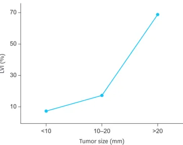
However, tumor size was found to be clearly associated with age at surgery and LVI in this study, and age was also correlated with LVI. The association between LVI and tumor size was similar to that reported in other forms of cancer (28,29). We found LVI in 13.5% of patients with PTC, and as tumor size increased, the distribution of LVI also increased from 8.7% in patients with tumors ≤1 cm to 60.7% in those with tumors >3 cm (Fig. 3). Also tumor size was found to be related to extrathyroidal extension(ETE). As tumor size increased, the proportion of ETE also increased from 15.6% in patients with tumors ≤1 cm to 40% in those with tumors >3 cm.
LVI is well known to be associated with lymphangiogenesis and LN metastasis (30). Through the mechanisms of lymphangiogenesis and tumor cell entry into the lymphatic system, LVI
is also related to tumor growth. Vascular endothelial growth factor C (VEGF-C)/VEGF-D is a significantly related mechanism in tumor growth. VEGF-C and VEGF-D are essential not only for blood vessel development, but also for lymphangiogenesis and LN metastasis (31).
The blood vessels ensure effective oxygen diffusion to cancer cells, thereby promoting tumor growth and survival. Furthermore, VEGF attracts and stimulates growth of the endothelial cells that construct new blood vessels. Therefore, interruption of VEGF and its receptor's autocrine signal can induce the arrest of tumor growth and apoptosis (32). In addition, physiological chemokine receptor ligand interaction such as CXC chemokine receptor (CXCR) 4, C-C chemokine receptor (CCR) 7, C-X-C motif chemokine ligand (CXCL) 12 (also known as stromal-cell-derived factor-1), C-C motif chemokine ligand (CCL) 21 (also known as secondary lymphoid chemokine), and lymphoepithelial cyst (LEC) is involved in LVI (33,34).
Our study has some of the limitations of any retrospective study as well as that of inter- institutional differences in definition of LVI. Diagnosing LVI is difficult, because mimickers of LVI are more common than LVI itself, which may account for its overdiagnosis (35). Other diagnostic difficulties in LVI are directly related to technical aspects of tissue preparation, such as staining techniques and number of blocks examined, and to pathologists'
experience and specialization (29,36). Therefore, a careful historical assessment is required to identify these features, and strict criteria should be followed to identify LVI (28,36).
Finally, LVI is not determined preoperatively in patients with thyroid cancer, unlike those with gastric cancer, and important decisive factor for selecting an additional surgical resection group. In future investigations, we hope to discover preoperatively available biomarkers for LVI. On the other hand, there is a statistical significance between young age and tumor size. It might be a selection bias because younger age group doesn’t undergo a health checkup routinely, which means at the time of diagnosis, younger group might be symptomatic like palpable mass.
In summary, of several suspicious preconditions which could affect tumor size, such as DM, breast cancer history, and familial cancer history, LVI was found to be significantly correlated with enlarge tumor size. Identification of a biomarker for LVI may be necessary to discern the low-risk group from high-risk group in PTC.
50
LVI (%) 30
10 70
Tumor size (mm)
>20 10–20
<10
Fig. 3. The distribution of LVI according to tumor size.
LVI = lymphovascular invasion.
CONCLUSION
Age and presence of LVI is a significant factor for enlargement of tumor size in PTC, unlike several other factors that were examined, including DM, breast cancer history, familial cancer history, Grave's disease, Hashimoto's thyroiditis, and BRAF mutation.
REFERENCES
1. Grebe SK, Hay ID. Follicular cell-derived thyroid carcinomas. Cancer Treat Res 1997;89:91-140.
PUBMED | CROSSREF
2. Ahn HS, Kim HJ, Welch HG. Korea's thyroid-cancer “epidemic”--screening and overdiagnosis. N Engl J Med 2014;371:1765-7.
PUBMED | CROSSREF
3. Pellegriti G, Frasca F, Regalbuto C, Squatrito S, Vigneri R. Worldwide increasing incidence of thyroid cancer: update on epidemiology and risk factors. J Cancer Epidemiol 2013;2013:965212.
PUBMED | CROSSREF
4. Miyauchi A. Clinical trials of active surveillance of papillary microcarcinoma of the thyroid. World J Surg 2016;40:516-22.
PUBMED | CROSSREF
5. Ito Y, Miyauchi A, Inoue H, Fukushima M, Kihara M, Higashiyama T, et al. An observational trial for papillary thyroid microcarcinoma in Japanese patients. World J Surg 2010;34:28-35.
PUBMED | CROSSREF
6. Ito Y, Miyauchi A. Appropriate treatment for asymptomatic papillary microcarcinoma of the thyroid.
Expert Opin Pharmacother 2007;8:3205-15.
PUBMED | CROSSREF
7. Ito Y, Miyauchi A, Kihara M, Higashiyama T, Kobayashi K, Miya A. Patient age is significantly related to the progression of papillary microcarcinoma of the thyroid under observation. Thyroid 2014;24:27-34.
PUBMED | CROSSREF
8. Zelenko Z, Gallagher EJ. Diabetes and cancer. Endocrinol Metab Clin North Am 2014;43:167-85.
PUBMED | CROSSREF
9. Algire C, Amrein L, Zakikhani M, Panasci L, Pollak M. Metformin blocks the stimulative effect of a high- energy diet on colon carcinoma growth in vivo and is associated with reduced expression of fatty acid synthase. Endocr Relat Cancer 2010;17:351-60.
PUBMED | CROSSREF
10. Zhou G, Myers R, Li Y, Chen Y, Shen X, Fenyk-Melody J, et al. Role of AMP-activated protein kinase in mechanism of metformin action. J Clin Invest 2001;108:1167-74.
PUBMED | CROSSREF
11. Rajoria S, Suriano R, George A, Shanmugam A, Schantz SP, Geliebter J, et al. Estrogen induced metastatic modulators MMP-2 and MMP-9 are targets of 3,3′-diindolylmethane in thyroid cancer. PLoS One 2011;6:e15879.
PUBMED | CROSSREF
12. Seethala RR, Baloch ZW, Barletta JA, Khanafshar E, Mete O, Sadow PM, et al. Noninvasive follicular thyroid neoplasm with papillary-like nuclear features: a review for pathologists. Mod Pathol 2018;31:39-55.
PUBMED | CROSSREF
13. Hayashida N, Namba H, Kumagai A, Hayashi T, Ohtsuru A, Ito M, et al. A rapid and simple detection method for the BRAF(T1796A) mutation in fine-needle aspirated thyroid carcinoma cells. Thyroid 2004;14:910-5.
PUBMED | CROSSREF
14. Mai KT, Truong LD, Ball CG, Olberg B, Lai CK, Purgina B. Lymphatic endothelial cancerization in papillary thyroid carcinoma: hidden evidence of lymphatic invasion. Pathol Int 2015;65:220-30.
PUBMED | CROSSREF
15. Ito Y, Fukushima M, Higashiyama T, Kihara M, Takamura Y, Kobayashi K, et al. Tumor size is the strongest predictor of microscopic lymph node metastasis and lymph node recurrence of N0 papillary thyroid carcinoma. Endocr J 2013;60:113-7.
PUBMED | CROSSREF
16. Rosário PW, de Faria S, Bicalho L, Alves MF, Borges MA, Purisch S, et al. Ultrasonographic differentiation between metastatic and benign lymph nodes in patients with papillary thyroid carcinoma. J Ultrasound Med 2005;24:1385-9.
PUBMED | CROSSREF
17. Shimamoto K, Satake H, Sawaki A, Ishigaki T, Funahashi H, Imai T. Preoperative staging of thyroid papillary carcinoma with ultrasonography. Eur J Radiol 1998;29:4-10.
PUBMED | CROSSREF
18. Noguchi S, Noguchi A, Murakami N. Papillary carcinoma of the thyroid. I. Developing pattern of metastasis. Cancer 1970;26:1053-60.
PUBMED | CROSSREF
19. Wise LA, Palmer JR, Stewart EA, Rosenberg L. Age-specific incidence rates for self-reported uterine leiomyomata in the Black Women's Health Study. Obstet Gynecol 2005;105:563-8.
PUBMED | CROSSREF
20. Spitz IM. Clinical utility of progesterone receptor modulators and their effect on the endometrium. Curr Opin Obstet Gynecol 2009;21:318-24.
PUBMED | CROSSREF
21. Donnez J, Donnez O, Courtoy GE, Dolmans MM. The place of selective progesterone receptor modulators in myoma therapy. Minerva Ginecol 2016;68:313-20.
PUBMED
22. Onitilo AA, Engel JM, Glurich I, Stankowski RV, Williams GM, Doi SA. Diabetes and cancer I: risk, survival, and implications for screening. Cancer Causes Control 2012;23:967-81.
PUBMED | CROSSREF
23. Park YJ, Ahn HY, Choi HS, Kim KW, Park DJ, Cho BY. The long-term outcomes of the second generation of familial nonmedullary thyroid carcinoma are more aggressive than sporadic cases. Thyroid 2012;22:356-62.
PUBMED | CROSSREF
24. Wang X, Cheng W, Li J, Su A, Wei T, Liu F, et al. Endocrine tumours: familial nonmedullary thyroid carcinoma is a more aggressive disease: a systematic review and meta-analysis. Eur J Endocrinol 2015;172:R253-62.
PUBMED | CROSSREF
25. Zhou YM, Luo H, Gou JX, Zhao WJ, Dai WY, Zhu J, et al. Second generation of familial nonmedullary thyroid carcinoma: a meta-analysis on the clinicopathologic features and prognosis. Eur J Surg Oncol 2017;43:2248-56.
PUBMED | CROSSREF
26. Xing M. BRAF mutation in papillary thyroid cancer: pathogenic role, molecular bases, and clinical implications. Endocr Rev 2007;28:742-62.
PUBMED | CROSSREF
27. Elisei R, Ugolini C, Viola D, Lupi C, Biagini A, Giannini R, et al. BRAF(V600E) mutation and outcome of patients with papillary thyroid carcinoma: a 15-year median follow-up study. J Clin Endocrinol Metab 2008;93:3943-9.
PUBMED | CROSSREF
28. Betge J, Pollheimer MJ, Lindtner RA, Kornprat P, Schlemmer A, Rehak P, et al. Intramural and extramural vascular invasion in colorectal cancer: prognostic significance and quality of pathology reporting. Cancer 2012;118:628-38.
PUBMED | CROSSREF
29. Huh JW, Kim HR, Kim YJ. Lymphovascular or perineural invasion may predict lymph node metastasis in patients with T1 and T2 colorectal cancer. J Gastrointest Surg 2010;14:1074-80.
PUBMED | CROSSREF
30. Sun W, Lan X, Zhang H, Dong W, Wang Z, He L, et al. Risk factors for central lymph node metastasis in CN0 papillary thyroid carcinoma: a systematic review and meta-analysis. PLoS One 2015;10:e0139021.
PUBMED | CROSSREF
31. Karkkainen MJ, Haiko P, Sainio K, Partanen J, Taipale J, Petrova TV, et al. Vascular endothelial growth factor C is required for sprouting of the first lymphatic vessels from embryonic veins. Nat Immunol 2004;5:74-80.
PUBMED | CROSSREF
32. Dias S, Hattori K, Zhu Z, Heissig B, Choy M, Lane W, et al. Autocrine stimulation of VEGFR-2 activates human leukemic cell growth and migration. J Clin Invest 2000;106:511-21.
PUBMED | CROSSREF
33. Kriehuber E, Breiteneder-Geleff S, Groeger M, Soleiman A, Schoppmann SF, Stingl G, et al. Isolation and characterization of dermal lymphatic and blood endothelial cells reveal stable and functionally specialized cell lineages. J Exp Med 2001;194:797-808.
PUBMED | CROSSREF
34. Streit M, Detmar M. Angiogenesis, lymphangiogenesis, and melanoma metastasis. Oncogene 2003;22:3172-9.
PUBMED | CROSSREF
35. Yamamoto S, Kawakami S, Yonese J, Fujii Y, Ohkubo Y, Suyama T, et al. Lymphovascular invasion is an independent predictor of prostate-specific antigen failure after radical prostatectomy in patients with pT3aN0 prostate cancer. Int J Urol 2008;15:895-9.
PUBMED | CROSSREF
36. Compton C, Fenoglio-Preiser CM, Pettigrew N, Fielding LP. American Joint Committee on Cancer Prognostic Factors Consensus Conference: Colorectal Working Group. Cancer 2000;88:1739-57.
PUBMED | CROSSREF