506
서 론
피하기종은 치과치료 도중 발생할 수 있는 흔치 않은 합병 증의 하나이다. 조직 하방으로 압축공기가 들어가면 부종이 발생하며 이 부위를 촉진 시 염발음(crepitus)이 나타나게 되고, 유입된 공기가 근막공간을 따라 다른 공간으로 퍼질 수 있는 위험성이 있다. 치과치료 술식과 연관되어 피하기 종이 발생한 여러 증례들이 보고되어 왔으며 구강 내 수술, 발치, 근관치료, 수복치료 등 많은 술식들이 포함되어 있다.
대부분의 증례보고에서 압축공기가 분사되는 핸드피스나 3-way 시린지의 사용이 주된 원인으로 나타나고 있다.1-3
본 증례보고는 파절선 관찰을 하던 도중에 나타난 피하기
종에 관한 것으로, 그 원인과 관리, 예방법에 대해 고찰해보 고자 한다.
증례보고
42세 여자 환자가 왼쪽 아래 어금니가 씹을 때 아프다는 주소로 연세대학교 치과대학병원 보존과에 내원하였다. 특 이 할만한 의과적 병력은 없었으며 약 10년 전 하악 좌측 제1대구치의 근관치료, 포스트, 및 금관 수복을 한 병력이 있었다. 임상검사상 이 치아는 타진에 반응을 보였고 근심 치근 주위로 방사선 투과상 병소가 관찰되었으며 탐침 결과 근심협측 부위의 치주낭 깊이가 매우 깊은 것으로 나타났
파절선 관찰 도중 발생한 피하기종: 증례보고
김민영∙박성호∙신유석∙김의성*
연세대학교 치과대학 치과보존학교실, 현미경센터
Subcutaneous emphysema during fracture line inspection: case report
Min-Young Kim, Sung-Ho Park, Yoo-Seok Shin, Euiseong Kim*
Department of Conservative Dentistry, Microscope Center, Yonsei University College of Dentistry, Seoul, Korea
The development of subcutaneous emphysema is a well-known complication that has been reported after den- tal extraction, endodontic treatment, or restorative preparation. Gaseous invasion, leading to swelling, crepi- tus on palpation, is commonly restricted to the connective tisssues immediately adjacent to the entry site.
However, the use of compressed air- and water-cooled turbines may allow large amounts of air and water to be driven through the fascial planes into the mediastinum, pleural space, or even the retroperitoneum.
This case report is about the patient who presented with subcutaneous emphysema that occurred after fracture line inspection. Possible cause, treatment, and prevention of emphysema will be discussed. [J Kor Acad Cons Dent 2011;36(6):506-509.]
Key words:
Dental treatment; Subcutaneous emphysema-Received 30 June 2011; revised 23 August 2011; accepted 29 August 2011- ABSTRACT
Kim MY, DDS, Graduate student; Park SH, DDS, MSD, PhD, Professor; Shin YS, DDS, MSD, Clinical assistant professor; Kim E, DDS, MSD, PhD, Professor, Department of Conservative Dentistry, Microscope Center, Yonsei University College of Dentistry, Seoul, Korea
*Correspondence to Euiseong Kim, DDS, MSD, PhD.
Professor, Department of Conservative Dentistry, Microscope Center, Yonsei University College of Dentistry, 50 Yonsei-ro, Seodaemun-gu, Seoul, Korea 120-752
TEL, +82-2-2228-8700; FAX, +82-2-313-7575; E-mail, [email protected]
Case Report
Kim MY et al. JKACD Volume 36, Number 6, November, 2011
pISSN 1225-0864 / eISSN 2093-8179 http://dx.doi.org/10.5395/JKACD.2011.36.6.506
Copyright � 2011 Korean Academy of Conservative Dentistry
507
Case Report
JKACD Volume 36, Number 6, November, 2011 Subcutaneous emphysema during fracture line inspection
다. 근심 치근의 수직 치근 파절 가능성이 높을 것으로 판단 되었으며 환자에게 예후 불량함에 대해 설명했다. 정확한 진단을 위해 진단 수술을 고려해 보았으나 하악 좌측 제1대 구치는 협측으로 치은퇴축이 심하여 근심 치근이 밖으로 많 이 노출되어 있었기 때문에 현미경 하에서 노출된 치근면의 파절선을 관찰해보기로 하였다(Figure 1).
메틸렌 블루의 효과적인 염색을 위해 노출된 치근 표면을 3-way 시린지로 건조시켰고 메틸렌블루 용액을 적용하였 다(Figure 2). 하지만 주변 조직으로부터 나오는 치은열구 삼출액과 출혈이 시야를 흐리게 만들었고 다시 건조 및 염 색을 시행하였다. 이러한 과정이 수차례 반복되었는데, 갑 자기 환자가 얼굴이 붓는 것 같다고 호소하였다. 안모 관찰 결과, 좌측 협부와 안와 하부에 갑작스러운 부종이 나타난 상태였으며 구강 내 관찰 시 하악 좌측 제1대구치의 협측 전정부에도 부종이 나타나 있었다(Figure 3). 구강 외 부종
이 나타난 부위를 촉진 시 염발음을 들을 수 있었으며 통증, 발열, 발적, 림프절증 및 기타 전신 증상은 없었다. 검사소 견들을 토대로 피하기종으로 진단 하였으며 일주일간 항생 제 및 진통제 복용을 지시하였다. 일주일 후 검진 시, 부종 은 모두 가라앉은 상태였고 환자도 2 - 3일 지나면서 부었 던 것이 다 가라앉았다고 하였다.
갑작스러운 피하기종의 발생으로 인하여 중단되었던 치료 가 이어서 진행되었다. 현미경하에서 관찰 시 치근면의 파절 선은 발견할 수 없었기 때문에 치주-근관 복합병소의 가능 성을 설명한 후 근심 근관의 재근관치료와 치주치료를 함께 시행하였으며 주기적인 경과관찰 이후에 보철치료를 고려하 기로 하였다. 6개월의 경과관찰 결과 치근단 병변의 감소가 확인되었고 치주낭 깊이도 정상범주를 유지하였으며 환자의 주관적 증상 역시 없었기 때문에 전장관으로 수복하였다.
고 찰
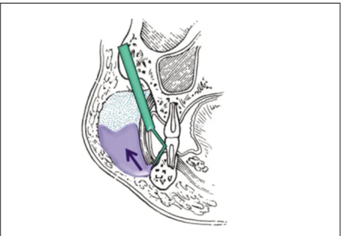
피하기종은 치과치료 후에 나타날 수 있는 비교적 빈도가 적은 합병증으로 대부분 압축공기가 분사되는 3-way 시린 지나 핸드피스의 사용 후에 나타난다.4,5 분사된 압축공기는 구강내의 붕괴된 장벽(barrier)을 통해 유입되며 주변 결합 조직을 분리시키며 진행하게 되고 근막공간을 통해 다른 인 접부위로 퍼질 수 있다.6본 증례에서도 근심치근의 협측 치 은퇴축이 심하여 부착치은이 매우 얇게 남아있음을 확인할 수 있다. 현미경 하에서 관찰하기 이전에 치주탐침을 시행하 였을 때 full depth였는데 부착치은이 매우 얇게 남아있었던 점을 고려한다면 프로브(probe)의 끝이 골막 위로 미끄러졌 을 수 있으며 이는 장벽(barrier)을 붕괴시켜 공기가 유입될 수 있는 통로를 만들어 준 것일 수도 있다(Figure 4).
연세대학교 치과대학병원 보존과에서 수년 전에 발생한 유사한 증례에서도 치은 퇴축에 의해 부착 치은이 매우 얇 게 남은 것을 관찰할 수 있다. 이 증례에서는 5급 와동 수복
Figure 2. Extraoral and intraoral photographs after onset of subcutaneous emphysema
: Sudden onset of swelling on left infraorbital, buccal, vestibular area.
Figure 1.Preoperative clinical photograph and radiograph : Buccal surface of mesial root is exposed due to severe gingival recession. Periapical radiograph revealed periapical radiolucency around mesial root of #36.
Figure 3.Methylene blue staining procedure.
508
Case Report
Kim MY et al. JKACD Volume 36, Number 6, November, 2011
을 위해 치은열구사(retraction cord)를 삽입 후 핸드피스 로 우식 제거를 하던 도중 피하기종이 발생했다. 뚜렷하지 는 않지만 우측 안면부의 부종이 나타났으며 염발음을 들을 수 있었다.
피하기종의 가장 뚜렷한 증상은 부종과 염발음이다. 부종 은 여러 문헌에서 술식 직후에 나타난다고 하나 일정 시간 이후에 나타난 증례도 보고되고 있다.7그리고 염발음은 피 하기종의 특징적인 증상으로서 갑작스런 부종을 나타낼 수 있는 혈관부종(angioedema)이나 아나필락시스 반응으로 부터 감별 진단하는데 도움을 준다. 만약 유입된 공기가 근 막공간을 통해 다른 부위로 퍼지게 된다면 부가적인 증상들 이 발생할 수 있다. 기관주위공간, 중격동으로 유입 시에는 호흡곤란이 나타나며 폐중격동으로 유입시에는 기흉에서 나타나는 햄먼징후(Hammam’s sign)가 나타나고 심장주 위공간으로 유입시에는 electrocardiogram의 변화가 나타 날 수 있다.5,8
대부분의 피하기종은 이번 증례에서와 같이 3내지 5일 이 내에 저절로 소실이 되며 일주일 안에는 완전한 회복이 일 어나 주의 깊은 관찰과 보존적인 처치만으로도 충분하며 항 생제와 진통제, 또는 스테로이드 제재를 처방한다.9,10 항생 제를 복용하는 이유는 유입된 공기 내에 구강 내 세균이 포 함되어 있을 가능성 때문이며 이차적인 감염을 예방하기 위 함이다. 하지만 항생제나 진통제, 스테로이드 제재의 복용 이 반드시 필요한 가에 대하여는 아직 논란의 여지가 있으 며 피하기종의 치료에 있어 명확한 장점을 지니는가에 대하 여는 확실히 밝혀낼 수 없다.5
유입된 공기가 다른 부위로 퍼져 호흡곤란과 같은 전신적 인 증상을 나타낸다면 즉각적인 외과적 처치가 필요할 수 있다. 먼저 방사선 사진을 통해 유입된 공기의 위치와 범위 를 확인하여야 하며 진단수술, 기관절개술 또는 흉관의 삽 입이 필요할 수 있다.11
결 론
피하기종은 치과치료 중 일어날 수 있는 비교적 흔치 않은 합병증이다. 하지만 이는 생명을 위협하는 합병증으로 발전 할 수도 있기 때문에 치과의사는 피하기종의 적절한 진단과 처치에 대해 숙지하고 있어야 할 것이다. 구강 내에 장벽이 붕괴된 부위가 있다면 압축공기가 분사되는 기구의 사용 시 주의해야 할 것이며 피하기종이 발생했다면 이에 필요한 조 치를 빠르고 적절하게 취할 수 있도록 해야 할 것이다.
Conflict of Interest: No potential conflict of interest relevant to this article was reported.
REFERENCES
1. Gamboa Vidal CA, Vega Pizarro CA, Almeida Arriagada A. Subcutaneous emphysema secondary to dental treatment: case report. Med Oral Patol Oral Cir Bucal 2007;12:76-78.
2. Smatt Y, Browaeys H, Genay A, Raoul G, Ferri J.
Iatrogenic pneumomediastinum and facial emphysema after endodontic treatment. Br J Oral Maxillofac Surg 2004;42:160-162.
3. Zemann W, Feichtinger M, Ka¨rcher H. Cervicofacial and mediastinal emphysema after crown preparation: a rare complication. Int J Prosthodont 2007;20:143-144.
4. Heyman SN, Babayof I. Emphysematous complications in dentistry, 1960-1993: an illustrative case and review of the literature. Quintessence Int 1995;26:
535-543.
5. McKenzie WS, Rosenberg M. Iatrogenic subcutaneous emphysema of dental and surgical origin: a literature review. J Oral Maxillofac Surg 2009;67:1265-1268.
6. Szubin L, La Bruna A, Levine J, Komisar A.
Subcutaneous and retropharyngeal emphysema after dental procedures. Otolaryngol Head Neck Surg 1997;
117:122-123.
7. Arai I, Aoki T, Yamazaki H, Ota Y, Kaneko A.
Pneumomediastinum and subcutaneous emphysema after dental extraction detected incidentally by regular medical checkup: a case report. Oral Surg Oral Med Oral Pathol Oral Radiol Endod 2009;107:33-38.
8. Reiche-Fischel O, Helfrick JF. Intraoperative life- threatening emphysema associated with endotracheal intubation and air insufflation devices: report of two cases. J Oral Maxillofac Surg 1995;53:1103-1107.
9. Horowitz I, Hirshberg A, Freedman A. Pneumome- diastinum and subcutaneous emphysema following surgi- cal extraction of mandibular third molars: three case reports. Oral Surg Oral Med Oral Pathol 1987;63:25-28.
10. Aragon SB, Dolwick MF, Buckley S. Pneumome- diastinum and subcutaneous cervical emphysema dur- ing third molar extraction under general anesthesia. J Oral Maxillofac Surg 1986;44:141-144.
11. Gulati A, Baldwin A, Intosh IM, Krishnan A.
Pneumomediastinum, bilateral pneumothorax, pleural effusion, and surgical emphysema after routine apicec- tomy caused by vomiting. Br J Oral Maxillofac Surg 2008;46:136-137.
Figure 4. Schematic of the pathogenesis of subcutaneous emphysema.
509 509 국문초록
파절선 관찰 도중 발생한 피하기종: 증례보고
김민영∙박성호∙신유석∙김의성*
연세대학교 치과대학 치과보존학교실, 현미경센터
피하기종은 치과치료 시 나타날 수 있는 합병증의 하나로서, 발치, 근관치료, 수복치료 등과 연관되어 발생할 수 있다. 피하 로 유입된 공기는 부종과 염발음을 일으키며 일반적으로는 주변 결합조직에 의해 그 범위가 한정되나 많은 양의 압축공기가 유 입될 경우 근막공간을 통해 중격동, 흉막공간, 심지어 후복막에까지 영향을 미칠 수 있어 생명을 위협하는 합병증으로 발생할 수도 있다.
이번 증례는 파절선을 관찰하던 도중에 피하기종이 발생한 환자에 대한 증례로서 그 원인과 관리, 예방법에 관해 고찰해보고 자 한다.
주요단어: 치과치료; 피하기종
Case Report
JKACD Volume 36, Number 6, November, 2011 Subcutaneous emphysema during fracture line inspection