The Functional Analysis of MLKs-JNK Pathways in Osteoclast Formation ◀ SY Ji, et al
파골세포 형성에 있어서 MLKs-JNK 경로의 역할 규명
숙명여자대학교 약학대학
지선영¶·정 정¶·노아롱새미·문미란·임미정*
= Abstract =
The Functional Analysis of MLKs-JNK Pathways in Osteoclast Formation
Sun Young Ji
¶, Zheng Ting
¶, A Long Sae mi Noh, Miran Moon, Mijung Yim
* College of Pharmacy, Sookmyung Women's University, Seoul, KoreaObjectives: Osteoclasts (OCs) are bone-resorbing multinucleated cells derived from hematopoietic progenitors of the monocyte-macrophage lineage. OC precursors, such as bone marrow-derived macrophages (BMMs), are formed in the presence of macrophage colony-stimulating factor (M-CSF) and differentiate into OCs in response to M-CSF and receptor activator of nuclear factor κB ligand (RANKL). In this study, we investigated the role of mixed lineage kinases (MLKs)-c-Jun amino-terminal kinase (JNK) pathways in OC formation.
Methods: We performed an OC formation assay and reverse transcription polymerase chain reaction (RT-PCR) analysis.
Results: We first explored the role of JNK on osteoclst formation using mouse bone marrow (BM) culture system.
We found that OC formation was impaired when the JNK inhibitor was added either in early or late stage, suggesting the requirement for JNK activation during OC formation. MLKs are serine/threonine kinases that regulate signaling by the JNK. Since the JNK activity is specifically required for osteoclastogenesis, we examined the messenger ribonucleic acid (mRNA) levels of MLKs in BMs, BMMs and OCs by RT-PCR. Among MLKs, the level of MLK3 mRNA expression is highest in BMs, BMMs and OCs. Moreover, we found that the mRNA expression of MLK2 and MLK3 is increased with the differentiation of BMs to BMMs, and is sustained in OCs. Finally we investigated the role of MLK3 in OC differentiation using gene knock-down techniques. The silencing of MLK3 in BMMs partly attenuated RANKL-induced OC differentiation.
Conclusions: These data suggest that JNK and MLK3 may positively regulate OC formation.
[Korean Journal of Bone Metabolism, 19(1): 21-28, 2012]
Key Words: JNK, MLKs, Osteoclast
Vol. 19, No. 1, 2012
OR O R IG I GI IN NA AL L A AR RT TI IC CL LE E
Received: March 26, 2012, Revised: April 21, 2012, Accepted: April 23, 2012
*Address for Correspondence: Mijung Yim, College of Pharmacy, Sookmyung Women's University, Cheongpa-ro 47-gil 100, Yongsan-gu, Seoul, 140-742, Korea.
Tel: +82-2-710-9572, Fax: +82-2-710-9871, e-mail: [email protected]
¶지선영과 정정은 본 논문에 대해 동일하게 기여하였습니다.
○CCThis is an Open Access article distributed under the terms of the Creative Commons Attribution Non-Commercial License (http://creativecommons.org/license/by-nc/3.0/).
서 론
현재 우리나라는 빠른 속도로 초고령화 사회에 진입하고 있으며, 노인성 질환도 크게 증가하고 있다. 그 중 골다공증 은 골량이 감소하고 미세구조의 이상으로 인해 결과적으로 뼈의 강도가 약해져서 부러지기 쉬운 상태가 되는 질환이다.
골다공증은 노인의 삶의 질을 급격히 저하시키는 문제점을 지님에도 불구하고 발병율이 매우 높아 그에 따른 대책이 시급한 실정이다.
뼈의 항상성은 새로운 골 기질의 형성과 오래된 골의 기질 을 용해시켜 골흡수 분해를 담당하는 파골세포 (osteoclast) 의 적절한 균형에 의해 유지된다.1,2 조골 및 파골세포는 밀 접한 관계를 맺고 있어, 파골세포의 분화는 조골세포에 의 해 엄격하게 조절된다.1,2 즉, 조골세포는 파골세포 분화인자 인 macrophage colony-stimulating factor (M-CSF, or CSF-1) 및 receptor activator of nuclear factor κB ligand (RANKL, osteoprotegerin ligand [OPGL], osteoclast differentiation factor [ODF], tumor necrosis factor-related activation induced cytokine [TRANCE])를 통해 파골세포의 분화를 조절함으로써 체내 골형성과 골흡수의 동적인 평형을 유지한다.2-5 여러 요인으 로 인해 이 균형이 깨지면 골의 생성보다 흡수가 증가하여 결국 골 손실 (bone loss)이 일어나게 된다.
파골세포 분화에 관여하는 다양한 신호전달 분자가 알려져 있다. 그 중 c-Jun amino-terminal kinase (JNK)는 파골세포의 형성과 유지에 반드시 필요한 것으로 보고되었다.6-8 세포에 대한 다양한 자극은 14종의 mitogen-activated protein kinase kinase kinase (MAP3K)를 활성화시키고, 이는 mitogen- activated protein kinase kinase (MKK) 4 또는 MKK7를 인산 화시켜 최종적으로 JNK를 활성시킨다. 파골전구세포에서 JNK의 활성화를 억제하면 RANKL 자극에 의한 파골세포 의 분화가 저해되었으며, 이는 파골세포 분화의 중요 전사인 자인 activator protein (AP)1과 nuclear factor of activated T-cells, cytoplasmic (NFATc)1을 매개한 것임이 밝혀졌다.6,7 또한 JNK는 파골세포에서 AP1의 구성성분인 c-jun의 활 성화를 통해 세포사를 억제하여 수명연장에 관여하는 것으 로 나타났다.8,9 JNK를 조절하는 상위 신호와 파골세포와의 연관성도 일부 보고되었다. 파골세포에 MAP3K의 일종인 mitogen-activated protein kinase kinase kinase (MEKK) 1을 과 잉발현시키면 JNK의 활성화가 관찰되는 동시에 세포사가 억제되었다.9 또한 파골전구세포에서 MKK7의 억제는 JNK 의 활성을 감소시켰으며 이에 따라 파골세포 형성도 억제되
었다.10 이상의 보고로 미루어 JNK는 파골세포의 형성과 유 지에 매우 중요한 역할을 담당하고 있는 것으로 생각되나, JNK를 조절하는 상위 신호인 MAP3K와 파골세포 형성과 의 연관성에 대해서는 아직 많은 부분이 알려져 있지 않다.
따라서 본 연구자는 JNK를 활성화시키는 14종의 MAP3K 신호 중 mixed lineage kinases (MLKs)에 주목하여 파골세포 형성에서 MLK의 역할을 규명하고자 하였다.
MLKs (MLK1, MLK2 및 MLK3)는 serine/threonine kinase 로 MKK7을 통해 JNK를 활성화시킨다. MLKs는 초기에 신경세포의 세포사를 조절하는 것이 알려졌으며, 따라서 신 경 질환에 대한 잠재적인 약물 타겟으로 여겨지고 있다.11 실 제로 반합성 MLK 저해제 (CEP1347)가 파킨슨 질환 치료제 로 임상 시험이 진행중이다. 한편 최근의 연구는 MLKs가 면 역세포에서도 중요한 역할을 담당할 가능성이 있음을 시사하 고 있다.12 Lipopolysaccharide (LPS)를 처리한 단구세포에서 MLK의 억제는 tumor necrosis factor-alpha (TNF-alpha)의 분비를 감소시켰다.13 또한 MLK 저해제는 LPS와 poly (I:C) 자극에 의한 수지상세포의 분화와 항원 제시 기능을 저해 하였으며, TNF-alpha, interleukin (IL)-10, IL-6와 같은 사이토 카인의 분비를 감소시켰다.14
본 연구자는 파골세포 형성에 있어서 MLKs의 역할을 마 우스 bone marrow-derived macrophages (BMMs) 세포를 사 용하여 검토하였기에 보고하는 바이다.
연구대상 및 방법
1. 마우스 골수세포의 배양
Imprinting control region (ICR) 마우스 (6-9주, 수컷)를 경추 탈골한 후 70% 에탄올로 소독하였다. 경골 부분의 피 부를 절개하여 부착 근육을 떼어냈다. 경골 원심부를 절단 하고 슬개골을 탈골시켜 경골을 적출하였다. 뼈 양끝을 조 금 잘라 한 쪽 끝에 25G의 주사바늘을 꽂고 alpha minimum essential medium (α-MEM)을 흘려보내 골수세포를 시험관 에 모았다. 원심 분리한 후 α-MEM에 현탁하고 2배의 Gey's solution을 가해 적혈구를 제거했다. 원심 분리한 후 10%
fetal bovine serum (FBS)가 함유된 α-MEM으로 재현탁했다.
2. 파골세포의 분화유도
초기 배양한 골수세포를 M-CSF 10 ng/mL로 하룻밤 배 양한 후 부유세포를 M-CSF 30 ng/mL로 3일간 추가 배양 하여 BMM 세포를 얻었다. BMM 세포를 회수 후 1 × 10⁴
The Functional Analysis of MLKs-JNK Pathways in Osteoclast Formation ◀ SY Ji, et al
cells/well에 RANKL 50 ng/mL과 M-CSF 30 ng/mL 존재하 에서 4-6일간 배양했다. 배양이 끝난 세포는 10% formalin 으로 10분간 고정한 후 ethanol-aceton (1:1)로 1분간 재고정 하여 tartrate-resistant acid phosphatase (TRAP) 염색하였다.
현미경하에서 관찰하여 3개 이상의 핵을 가진 TRAP+ 세포 를 다핵 파골세포로 판정하였다.
3. Reverse transcription polymerase chain reaction (RT-PCR) 분석
Total ribonucleic acid (RNA) 추출은 easy-Blue (iNtRON Biotechnology, Sungnam, Korea)를 이용하였다. Comple- mentary deoxyribonucleic acid (cDNA)는 1 mg의 total RNA 를 oligo(dT) primer, 10 mM deoxyribonucleotide triphosphate (dNTP), 1 unit RNase inhibitor 그리고 4 unit Script reverse transcriptase (Fermentas, York, UK)로 42℃에서 60분 처리하 여 합성한 후, 70℃에서 10분 가열함으로써 반응을 중지 시 켰다. PCR은 Table 1에 명시된 프라이머를 사용하여 94℃
3분간 처리한 후, 94℃ 30초, 58℃ 45초, 72℃ 1분의 과정 을 32회 또는 28회 (β-actin) 반복하였다. PCR 후, 증폭된 cDNA는 1% agarose gel에서 분리하였고 ethidium bromide 로 염색하여 ultraviolet (UV) 상에서 관찰하였다.
4. Small interfering RNA (siRNA)를 이용한 MLK3 유전자 발현 저하
마우스 MLK3 siRNAs (Cat. no. SI01299893)는 Qiagen GmbH (Hilden, Germany)에서 구입하였다. 대조군 siRNA 로는 Allstars Negative Control siRNA (Cat. no. 1027280) (Qiagen GmbH, Hilden, Germany)를 사용하였다. BMM 세 포에 siRNA (20 nM)를 LipofectamineTM 2000 CD (Cat. no.
12566-014; Invitrogen, Carlsbad, USA)로 도입시켰으며, 하루 동안 배양한 후 RT-PCR 분석과 파골세포 형성 실험에 사용 하였다.
5. 통계분석
실험결과는 평균 ± 표준편차로 표기하였고, Student's t-test 로 분석하여 P 값이 0.05 미만일 때 통계적으로 유의하다고 판단하였다.
결 과
1. JNK 저해제에 의한 파골세포 형성 억제
성숙 파골세포는 골수세포로부터 유래하며 파골전구세포 를 거쳐 형성된다 (Fig. 1A). 즉, 마우스 골수세포를 M-CSF 로 처리하여 파골전구세포인 BMMs가 형성되는 첫 번째 단 계와 (①), 파골전구세포를 M-CSF와 RANKL로 처리하여 성숙 파골세포로 분화시키는 두 번째 단계 (②)를 거쳐 최종 적으로 파골세포가 형성된다. 기존의 많은 연구를 통해 JNK Table 1. Primer sequences for RT-PCR
Primer name Sequence
MLK1
sense 5'-CCTGGTAGATGGATACAA-3' MLK1
antisense 5'-CGGGTAGAGTTGCATTCA-3' MLK2
sense 5'-GAGCCTTCTCCTTGTGTT-3' MLK2
antisense 5'-GCCCTCCATGTCCATGT-3' MLK3
sense 5'-CGGTCCCGCATGGATGAA-3' MLK3
antisense 5'-CCTGGGCACCCATGTCTT-3' CD11b
sense 5'-CTTAAAGCTCTTCTGGTCACAGCC-3' CD11b
antisense 5'-GTATTCTCCTTGCAGTTTTGGTGC-3' F4/80
sense 5'-GAATCTTGGCCAAGAAGAGAC-3'
F4/80
antisense 5'-GAATTCTCCTTGTATATCATCAGC-3' CTR sense 5'-TTTCAAGAACCTTAGCTGCCAGAG-3'
CTR antisense 5'-CAAGGCACGGACAATGTTGAGAAG-3'
CathepsinK
sense 5'-CTTCCAATACGTGCAGCAGA-3' CathepsinK
antisense 5'-ACGCACCAATATCTTGCACC-3' β-actin
sense 5'-TGTGATGGTGGGAATGGGTCAG-3' β-actin
antisense 5'-TTTGATGTCACGCACGATTTCC-3' MLK, mixed lineage kinase; CTR, calcitonin receptor; RT-PCR, reverse transcription polymerase chain reaction
를 매개하는 신호전달 경로가 두 번째 단계인 RANKL 처리 에 의한 성숙 파골세포로의 분화에 필수적이라는 것이 밝혀 졌다.6-8 그러나 파골세포 분화의 첫 번째 단계인 파골전구세 포의 형성에 있어서 JNK의 역할은 보고된 바 없다. 따라서 이를 알아보기 위해 JNK 저해제인 SP600125 (SP)를 파골 세포 분화 단계별로 처리하여 파골세포 형성 여부를 조사하 였다 (Fig. 1B). 구체적으로는 마우스 골수세포에 JNK 저해 제를 처리하여 파골전구세포 (BMM)를 형성한 후 RANKL 처리한 군과 (①+SP), 마우스 골수세포를 파골전구세포 (BMM)로 분화시킨 후 RANKL과 JNK 저해제를 처리한 군 (②+SP)으로 나누어 성숙 파골세포 형성 정도를 비교하 였다. 기존에 보고된 바와 같이 JNK 저해제는 파골전구세포 (BMM)로부터 성숙 파골세포로의 분화를 완벽하게 억제하
였다 (②+SP). 또한 골수세포에 JNK 저해제를 처리하여 파 골전구세포를 형성한 경우에도 RANKL에 의한 성숙 파골 세포 형성이 크게 감소하였다 (①+SP). 이는 JNK 저해제가 파골전구세포로의 분화를 억제한 때문으로 생각되며, 이상의 결과로부터 JNK가 성숙 파골세포 분화뿐 아니라 파골전구 세포의 분화에도 중요한 역할을 담당하고 있음을 짐작할 수 있다.
2. Bone marrow (BM), BMM, osteoclast (OC)에서 MLKs 의 발현
세포 내에서 JNK를 활성화시키는 다양한 상위 신호전달 경로가 존재하며, 그 중 MLKs는 MKK7을 통해 JNK를 활 성화시키는 것으로 알려져 있다.11 파골세포 분화에 있어서 MLKs의 역할을 분석하기 위해 골수, 파골전구 및 성숙 파 골세포에서의 MLKs의 발현을 RT-PCR 방법으로 분석하였 다 (Fig. 2). 먼저 MLK1, MLK2와 MLK3 mRNA 발현을 각 각 골수, 파골전구 및 성숙 파골세포를 사용하여 조사하였다.
세 종류의 MLK 중 MLK3가 모든 세포에서 가장 높이 발 현되고 있음을 알 수 있다. 반면 MLK1과 MLK2는 모든 세 포에서 상대적으로 낮은 발현량을 보였다.
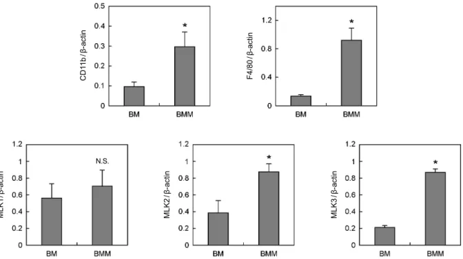
다음은 파골세포 분화 단계에 따른 MLKs 발현 변화를 RT-PCR 방법으로 분석하였다 (Fig. 3). 먼저 골수세포와 파골전구세포에서의 MLKs 발현 비교에 앞서, 파골전구세 포의 형성을 마커 유전자인 CD11b와 F4/80의 발현으로 확 인하였다. 골수세포에서와 달리 파골전구세포에서 CD11b와 F4/80이 높게 발현되어, 파골전구세포가 올바르게 형성되었 음을 알 수 있다. 골수세포가 파골전구세포로 분화함에 따 라 MLK1의 발현은 크게 변화하지 않으나, MLK2와 MLK3 의 발현은 증가하였다. 골수세포에 비해 파골전구세포에서 MLK2은 약 2배, MLK3는 약 4배 발현이 증가하였다. 이는 MLK2와 MLK3가 파골전구세포로의 분화에 관여할 가능성 을 시사하는 결과이다. 다음은 파골전구세포와 성숙 파골세 포에서 MLKs 발현을 비교하였다 (Fig. 4). 성숙 파골세포의 형성은 마커 유전자인 calcitonin receptor (CTR)와 Cathepsin K의 발현 증가로 확인하였다. 성숙 파골세포에서 MLK1, MLK2와 MLK3의 발현은 크게 변화하지 않는 것으로 나타 났다. 즉, MLK1은 파골전구세포에서와 동일하게 성숙 파골 세포에서 발현이 유도되지 않았으며, 파골전구세포에서 발현 이 증가한 MLK2와 MLK3는 성숙 파골세포에서도 그 발현 이 계속 유지되었다. 이상의 결과를 통해 MLKs 중 MLK2 와 MLK3가 파골전구세포 및 성숙 파골세포의 분화에 관여 Fig. 1. Effects of JNK inhibitor on osteoclatstogenesis. (A)
Schematic design for treatment of JNK inhibitor. (B) Bone marrow cells were cultured with 10 ng/mL M-CSF overnight. Non-adherent cells were harvested and cultured with 30 ng/mL M-CSF in the absence or presence of SP for 3 days. Floating cells were removed and adherent cells were used as BMMs (①). BMMs were cultured with 30 ng/mL M-CSF and 200 ng/mL RANKL in the absence or presence of SP for 4 days (②). Cultured cells were fixed and stained for TRAP. TRAP+ multinucleated cells containing more than three nuclei were counted as osteoclasts. Data represent means
± SD of triplicate samples in a representative experiment. *P <
0.05 versus vehicle. (JNK, c-Jun amino-terminal kinase; M-CSF, macrophage colony-stimulating factor; SP, SP600125 [a JNK inhibitor] BMM, bone marrow-derived macrophage; RANKL, receptor activator of nuclear factor κB ligand; TRAP, tartrate- resistant acid phosphatase; MNC, multinucleated cells)
A
B
The Functional Analysis of MLKs-JNK Pathways in Osteoclast Formation ◀ SY Ji, et al
Fig. 2. The mRNA expression levels of MLKs in BMs, BMMs and OCs. Whole cell lysates were harvested from cultured cells. Total RNA were then isolated from the cells and cDNA templates prepared. The mRNA expression level was determined by RT-PCR using specific primers designed for MLK1, MLK2 and MLK3. Data represent means ± SD of three independent experiments. (mRNA, messenger ribonucleic acid; MLK, mixed lineage kinase; BM, bone marrow; BMM, bone marrow-derived macrophage; OC, Osteoclast;
cDNA, complementary deoxyribonucleic acid; RT-PCR, reverse transcription polymerase chain reaction)
CD11b/β-actin F4/80/β-actin
MLK1/β-actin MLK2/β-actin MLK3/β-actin
N.S.
Fig. 3. Expression pattern of MLKs in BM and BMM. (A, B) Whole cell lysates were harvested from cultured cells and analyzed by RT-PCR. Data represent means ± SD of three independent experiments. *P < 0.05 versus BM. (N.S., non-significant; MLK, mixed lineage kinase; BM, bone marrow; BMM, bone marrow-derived macrophage; RT-PCR, reverse transcription polymerase chain reaction)
B A
β-actin
MLK3/β-actin
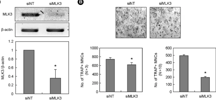
Fig. 5. Effects of MLK3 knock-down on osteoclatstogenesis. (A, B) BMMs were transfected with non-targeting siRNA or siRNA specific for MLK3. (A) Transfected BMMs were harvested from cultured cells and analyzed by RT-PCR. (B) Transfected BMMs were cultured for 4 days with 30 ng/mL M-CSF and 200 ng/mL RANKL. Cultured cells were fixed and stained for TRAP. TRAP-positive (+) multinucleated cells (MNCs) having more than 3 (n > 3) or 10 (n > 10) nuclei were counted. Data represent means ± SD of triplicate samples in a representative experiment. *P < 0.05 versus siNT. scale bar = 200 μM. (siNT, small interfering non-targeting; MLK, mixed lineage kinase;
TRAP, tartrate-resistant acid phosphatase; MNC, multinucleated cells; siRNA, small interfering ribonucleic acid; BMM, bone marrow- derived macrophage; RT-PCR, reverse transcription polymerase chain reaction; M-CSF, macrophage colony-stimulating factor; RANKL, receptor activator of nuclear factor κB ligand; TRAP, tartrate-resistant acid phosphatase)
MLK1/β-actin MLK2/β-actin MLK3/β-actin
CTR/β-actin
Cathepsin K/β-actin
N.S.
N.S.
Fig. 4. Expression pattern of MLKs in BMM and OC. (A, B) Whole cell lysates were harvested from cultured cells and analyzed by RT-PCR. Data represent means ± SD of three independent experiments. *P < 0.05 versus BMM. (N.S., non-significant; CRT, calcitonin receptor, MLK, mixed lineage kinase; BMM, bone marrow-derived macrophage; OC, Osteoclast; RT-PCR, reverse transcription polymerase chain reaction)
The Functional Analysis of MLKs-JNK Pathways in Osteoclast Formation ◀ SY Ji, et al
할 가능성이 있다고 판단하였다.
3. MLK3 유전자 Knock-down에 의한 파골세포 형성 억제
다음은 MLKs가 파골세포 형성에 관여하는지의 여부를 조사하였다. MLKs 중 MLK3가 골수세포에서 파골전구세포 로 분화함에 따라 발현량이 증가하고, 파골전구 및 성숙 파 골세포에서 가장 발현량이 높았으므로 MLK3를 대상으로 실험을 진행하였다. 파골세포 형성에 있어서 MLK3의 기 능을 직접적으로 규명하기 위해 siRNA를 사용해 MLK3를 knock-down 시킨 후 파골세포 형성의 변화를 관찰하였다 (Fig. 5). 먼저 파골전구세포에 siRNA를 도입한 후 MLK3의 발현 정도를 RT-PCR로 조사하였다. siRNA 도입 후 MLK3 mRNA 발현이 대조군에 비해 약 60% 감소한 것을 확인하 였다 (Fig. 5A). 다음은 MLK3 siRNA를 도입한 파골전구세 포를 RANKL 처리하여 성숙 파골세포로 분화시켰다 (Fig.
5B). MLK3 발현이 감소한 경우 대조군에 비해 성숙 파골세 포 형성이 유의하게 억제되었으며, 특히 10개 이상의 핵을 가지는 다핵 파골세포로의 분화가 저해되는 것으로 나타났다.
이상의 결과로 JNK의 상위 신호전달 분자인 MLK3가 성숙 파골세포 형성에 일정 부분 관여하고 있을 가능성이 제시되 었다.
고 찰
골다공증은 고관절 등의 부위에 골절을 유발하여 노인의 삶의 질을 급격히 저하시키는 문제점을 지님에도 불구하고 발병율이 매우 높은 질환 중의 하나이다. 따라서 골다공증 치료제에 대한 수요도 증가하여 골다공증에 대한 7개 주요 제약시장에서의 의약품 시장은 2016년 111억 달러 규모를 형성할 것으로 전망한다. 지금까지 개발된 골다공증 치료제 중 특히 파골세포에 작용하는 골흡수 억제제는 비용이 적게 들며 골절의 위험을 감소시키는 데 효과적인 것으로 나타나, 가장 많이 처방되고 있다. 따라서 새로운 골다공증 치료제의 개발에 있어서 파골세포의 분화 기전을 이해하는 것은 매우 중요하다고 할 수 있다.
파골세포의 형성에 있어서 JNK를 매개한 신호전달 경로 는 필수적으로, JNK1-/- 마우스 유래의 골수세포는 파골세 포로의 분화가 급격히 감소하는 것으로 보고된 바 있다.6 이 는 JNK가 파골세포 분화인자인 RANKL의 신호를 매개하 여 성숙 파골세포의 형성을 조절하기 때문으로 알려져 왔다.
본 연구자는 JNK 저해제를 사용하여 JNK가 성숙 파골세포 의 형성뿐 아니라 파골전구세포의 형성에도 중요함을 밝혔 다. 따라서 JNK는 파골전구세포 및 성숙 파골세포의 형성에 중요하다고 할 수 있다.
세포 내 JNK의 활성화를 조절하는 상위 신호전달 분자 로 MLKs가 알려져 있다. 본 연구자는 RT-PCR 분석법을 통해 골수세포에서 파골전구세포로 분화 시 MLKs 중 MLK3 발현이 가장 많이 증가하며, 성숙 파골세포에서도 그 발현이 지속되는 것을 확인했다. 또한 siMLK3를 사용한 knock-down 실험을 통해 MLK3가 파골세포 형성에 관여 할 가능성을 제시하였다. 최근 MLK3 유전자를 결손시킨 MLK3 녹아웃 마우스는 건강하고 표면적으로 특이사항이 없는 것으로 보고된 바 있다.15 이는 in vitro 실험을 통해 MLK3의 중요성을 제시한 다수의 실험결과와는 일치하지 않는 것이다. 이러한 모순은 in vivo 상에서 MLK3의 기능 을 타 MLKs가 부분적으로 보완하여 발생한 것으로 생각되 며, 추가 연구를 통해 규명이 필요하다.
본 연구를 통해 파골세포 형성을 조절하는 새로운 기전이 제시되었으며, 향후 이를 이용한 골대사 장애 치료제 등의 개발도 가능할 것으로 생각된다.
결 론
JNK를 매개한 신호전달 경로는 파골전구세포 및 성숙 파골세포의 형성에 중요하다. 또한 JNK의 상위 조절 분자 중 하나인 MLK3는 성숙 파골세포 형성에 일부 관여한다.
1. Takahashi N, Akatsu T, Udagawa N, et al. Osteoblastic cells are involved in osteoclast formation. Endocrinology 1988;
123:2600-2.
2.Suda T, Takahashi N, Udagawa N, Jimi E, Gillespie MT, Martin TJ. Modulation of osteoclast differentiation and function by the new members of the tumor necrosis factor receptor and ligand families. Endocr Rev 1999;20:345-57.
3. Wong BR, Rho J, Arron J, et al. TRANCE is a novel ligand of the tumor necrosis factor receptor family that activates c-Jun N-terminal kinase in T cells. J Biol Chem 1997;272:25190-4.
4.Yasuda H, Shima N, Nakagawa N, et al. Osteoclast differen- tiation factor is a ligand for osteoprotegerin/osteoclastogenesis-
참 고 문 헌
inhibitory factor and is identical to TRANCE/RANKL. Proc Natl Acad Sci U S A 1998;95:3597-602.
5. Lacey DL, Timms E, Tan HL, et al. Osteoprotegerin ligand is a cytokine that regulates osteoclast differentiation and activation.
Cell 1998;93:165-76.
6. David JP, Sabapathy K, Hoffmann O, Idarraga MH, Wagner EF. JNK1 modulates osteoclastogenesis through both c-Jun phosphorylation-dependent and -independent mechanisms. J Cell Sci 2002;115:4317-25.
7. Chang EJ, Ha J, Huang H, et al. The JNK-dependent CaMK pathway restrains the reversion of committed cells during osteoclast differentiation. J Cell Sci 2008;121:2555-64.
8. Otero JE, Dai S, Foglia D, et al. Defective osteoclastogenesis by IKKbeta-null precursors is a result of receptor activator of NF-kappaB ligand (RANKL)-induced JNK-dependent apoptosis and impaired differentiation. J Biol Chem 2008;
283:24546-53.
9.Ikeda F, Matsubara T, Tsurukai T, Hata K, Nishimura R, Yoneda T. JNK/c-Jun signaling mediates an anti-apoptotic effect of RANKL in osteoclasts. J Bone Miner Res 2008;23:
907-14.
10. Yamamoto A, Miyazaki T, Kadono Y, et al. Possible involve- ment of IkappaB kinase 2 and MKK7 in osteoclastogenesis induced by receptor activator of nuclear factor kappaB ligand.
J Bone Miner Res 2002;17:612-21.
11. Gallo KA, Johnson GL. Mixed-lineage kinase control of JNK and p38 MAPK pathways. Nat Rev Mol Cell Biol 2002;3:
663-72.
12.Handley ME, Rasaiyaah J, Chain BM, Katz DR. Mixed lineage kinases (MLKs): a role in dendritic cells, inflammation and immunity? Int J Exp Pathol 2007;88:111-26.
13. Ciallella JR, Saporito M, Lund S, et al. CEP-11004, an inhibitor of the SAPK/JNK pathway, reduces TNF-alpha release from lipopolysaccharide-treated cells and mice. Eur J Pharmacol 2005;515:179-87.
14.Handley ME, Rasaiyaah J, Barnett J, et al. Expression and function of mixed lineage kinases in dendritic cells. Int Immunol 2007;19:923-33.
15.Brancho D, Ventura JJ, Jaeschke A, Doran B, Flavell RA, Davis RJ. Role of MLK3 in the regulation of mitogen- activated protein kinase signaling cascades. Mol Cell Biol 2005;25:3670-81.
= 국문초록 =
연구목적: 파골전구세포는 M-CSF 존재하에서 조혈모세포로부터 형성되며, M-CSF와 RANKL 존재하에서 성숙 파골세포 로 분화된다. 본 실험에서는 성숙 파골세포 형성에 있어서 MLKs-JNK 경로의 역할을 알아보고자 하였다.
연구방법: 파골세포 형성 측정법과 RT-PCR 분석법을 사용하였다.
결 과: 먼저 파골세포 형성에 있어서 JNK의 역할을 조사한 결과, JNK 저해제는 파골전구세포와 성숙 파골세포의 형성 을 모두 억제하는 것으로 나타났다. JNK의 활성은 MLK에 의해 조절된다는 보고가 있으므로 MLKs mRNA의 발현을 RT-PCR 방법으로 조사하였다. MLKs 중, MLK3의 발현이 골수, 파골전구, 성숙 파골세포에서 가장 높은 것으로 나타났 다. 또한 MLK2와 MLK3의 발현은 골수에서 파골전구세포로 분화시 증가하였으며, 성숙 파골세포에서도 발현이 높게 유 지되었다. siRNA를 사용한 MLK3 유전자의 knock-down은 파골세포 분화를 일부 억제하였다.
결 론: JNK는 파골전구세포와 성숙 파골세포로의 분화에 필수적이며, MLK3는 파골세포 분화에 일부 관여한다.
중심단어: JNK, MLKs, 파골세포