Background and Purpose Carotid endarterectomy (CEA) is performed to prevent cere- bral infarction, but a common side effect is cerebral microinfarcts. This study aimed to iden- tify the variables related to the production of microinfarcts during CEA as well as determine their association with delayed postoperative infarction.
Methods This was a retrospective review of data collected prospectively from 548 patients who underwent CEA. The clinical characteristics of the patients and the incidence rates and causes of microinfarcts were analyzed. Microinfarcts were diagnosed by diffusion-weighted magnetic resonance imaging. The presence of delayed postoperative infarction was compared between microinfarct-positive and microinfarct-negative groups.
Results In total, 76 (13.86%) patients were diagnosed with microinfarcts. Preoperative neu- rological symptoms were significantly related to the incidence of microinfarcts [odds ratio (OR)=2.93, 95% confidence interval (CI)=1.72–5.00, p<0.001]. Shunt insertion during CEA was the only significant procedure-related risk factor (OR=1.42, 95% CI=1.00–2.19, p=0.05).
The presence of microinfarcts did not significantly increase the incidence of delayed postoper- ative infarction (p=0.204).
Conclusions In the present study, risk factors for microinfarcts after CEA included preopera- tive symptoms and intraoperative shunt insertion. Microinfarcts were not associated with de- layed postoperative infarction.
Key Words carotid artery endarterectomy, microinfarcts, risk factor.
Analysis of Risk Factors for Cerebral Microinfarcts after Carotid Endarterectomy and the Relevance of Delayed Cerebral Infarction
INTRODUCTION
The effectiveness of carotid endarterectomy (CEA) in preventing strokes has been previ- ously confirmed in both symptomatic and asymptomatic patients with carotid artery ste- nosis.1,2 However, one of the severe complications of CEA is stroke, which includes cerebral microinfarcts.3,4 Microinfarct lesions are tiny infarctions without any neurological symp- toms that can be confirmed radiologically [generally by diffusion-weighted magnetic res- onance imaging (DW-MRI)], and are mainly due to microembolisms. These lesions were found to be risk factors for cognitive impairment in the Cardiovascular Health Study Cog- nition Study5 and to be associated with a relatively poor prognosis for geriatric depression in a long-term follow-up study.6 The techniques used in carotid artery stenting (CAS) to treat carotid artery stenosis have evolved rapidly, and embolic protection filtering devices for reducing the incidence of microembolism, such as flow reversal systems and proximal arrest systems (Mo.Ma device, Medtronic Invatec, Frauenfeld, Switzerland), are currently in use.4 Similar to CAS-related procedures for reducing cerebral microembolisms during Jun Gyo Gwona
Tae-Won Kwona Yong-Pil Choa Dong-Wha Kangb Youngjin Hana Minsu Noha
a Departments of Surgery,
b Neurology, University of Ulsan College of Medicine,
Asan Medical Center, Seoul, Korea
pISSN 1738-6586 / eISSN 2005-5013 / J Clin Neurol 2017;13(1):32-37 / https://doi.org/10.3988/jcn.2017.13.1.32
Received March 17, 2016 Revised June 1, 2016 Accepted June 2, 2016 Correspondence Tae-Won Kwon, MD, PhD Department of Surgery,
University of Ulsan College of Medicine, Asan Medical Center,
88 Olympic-ro 43-gil, Songpa-gu, Seoul 05505, Korea
Tel +82-2-3010-3492 Fax +82-2-3010-6701 E-mail twkwon2@amc.seoul.kr
cc This is an Open Access article distributed under the terms of the Creative Commons Attribution Non-Com- mercial License (http://creativecommons.org/licenses/by-nc/3.0) which permits unrestricted non-commercial use, distribution, and reproduction in any medium, provided the original work is properly cited.
JCN
Open Access ORIGINAL ARTICLEGwon JG et al.
JCN
JCN
Open AccessCEA, other preoperative methods such as gentler dissection techniques are available.7 However, the reported incidence of microembolism remains high, at 25–49%, and few stud- ies have analyzed the risk factors for microembolism during CEA.8,9
Microinfarcts after CEA can result in poor outcomes, and microembolic signals are associated with an increased peri- operative stroke risk10,11 since this is associated with cogni- tive impairment and geriatric depression. The present study therefore aimed to elucidate the risk factors for microinfarcts after CEA in order to facilitate attempts to reduce its inci- dence. Additionally, we compared the infarction-free rate be- tween microinfarct and no-microinfarct groups to assess its potential association with delayed cerebral infarction.
METHODS
Patients
This study was conducted as a retrospective review of med- ical data collected prospectively from 556 patients who un- derwent CEA at our institution between January 2009 and December 2014. The study was approved by the Institutional Review Board of the Asan Medical Center (approval no. 2015- 0736).
Postoperative MRI, including DW-MRI, was performed on every patient with or without a suspected stroke. Among the 556 patients, 8 (1.44%) were excluded because of a diagnosed brain infarction with neurological symptoms within 24 h af- ter the operation, and so 548 patients were finally analyzed.
CEA was conducted in asymptomatic patients with >70%
stenosis of the carotid artery confirmed by carotid duplex so- nography or with ≥80% stenosis confirmed by computed to- mography (CT) angiography or magnetic resonance (MR) angiography if the ultrasound investigations indicated 50–
69% stenosis. In symptomatic patients, CEA was conducted in those with >50% stenosis of the ipsilateral carotid artery confirmed by carotid duplex sonography or with ≥70% ste- nosis confirmed by CT angiography or MR angiography if the ultrasound-detected stenosis was 50–69%. Symptoms re- lated to carotid artery lesions included transient or persis- tent monocular visual loss, hemispheric transient ischemic at- tacks, nondisabling stroke, and ischemic stroke during the previous 6 months in the relevant carotid artery region.12 Re- gardless of the medical treatment applied, the indication for CEA was determined solely by the patient’s symptoms and the region of carotid artery stenosis.
Carotid endarterectomy
CEA was performed by three expert vascular surgeons who had each performed this procedure more than 30 times per
year for more than 10 years. General or regional anesthesia was administered to all patients. All endarterectomies were open, with careful dissection performed of the bifurcation into internal carotid artery (ICA) and the external carotid ar- tery. Some patients underwent shunt insertion from the com- mon carotid artery to the ICA with a Javid shunt or Pruitt- Inahara shunt depending on the preference of the surgeon.
In cases of general anesthesia, the shunt was inserted to re- duce the cerebral oxygen saturation as measured by cerebral oximetry during the clamping time. In cases of regional an- esthesia, the shunt was inserted if neurological symptoms such as mental change or dysarthria appeared during the clamping time. All patients were routinely administered an intravenous bolus of unfractionated heparin (80 units per kilogram body weight) before performing ICA cross-clamp- ing. Bovine pericardial patches were applied in 513 (93.61%) of the 548 patients treated for ICA repair at our hospital. Ad- ditionally, closures were made using primary repair in 8 pa- tients, an autogenous vein patch in 13 patients, and a polytet- rafluoroethylene patch in 1 patient, while 13 patients underwent the eversion technique only. A Jackson-Pratt drain was placed on the platysma muscle layer when closing the wound in all patients. An antiplatelet agent had been applied in 332 (60.58%) patients preoperatively: 69 patients received clopidogrel (75 mg) only, 75 received acetylsalicylic acid (100 mg) only, and 188 patients received both antiplatelet agents. A statin was given to patients with a total fasting cholesterol level of higher than 200 mg/dL. Acetylsalicylic acid, clopidogrel, and a statin were administered immediately after CEA in all patients.
Patients were generally discharged on postoperative day 3 or 4, and a neurological examination was performed at the 1-month follow-up. If no complications were found, anoth- er follow-up was performed 6 months later and then annual- ly thereafter.
Diagnosis of microinfarcts and delayed postoperative infarction
Microinfarcts were diagnosed based on DW-MRI and clini- cal symptoms. MRI studies were performed on postopera- tive day 2. Average diffusion coefficient maps were calculat- ed from the DW-MRI images. Based on information from another study,8 microinfarcts were defined as tiny focal acute lesions (volume less than 0.5 cm3) observed on DW-MRI scans; the corresponding average diffusion coefficient maps were constructed by an experienced radiologist (Fig. 1). No neurological symptoms were expected in any of the patients in whom brain DW-MRI was performed. In the present study, a delayed postoperative infarction was defined as a lesion larger than 0.5 cm3 in volume with accompanying neu- rological symptoms. Such lesions were located on the ipsi-
Microinfarcts after Carotid Endarterectomy
JCN
lateral side of the CEA hemisphere and occurred later than postoperative day 2 (after the initial DW-MRI). Patients with stroke caused by cardioembolism or other determined etiology according to Trial of ORG 10172 in Acute Stroke Treatment (TOAST) classification13 were excluded based on the delayed postoperative infarction criteria.
Statistical analysis
Variables were compared between patients with and without microinfarcts. Continuous variables are presented as mean±
standard-deviation values. Continuous and categorical vari- ables were compared using Student’s t-test and the chi-square test, respectively. Multiple logistic regression was used to an- alyze factors associated with microinfarcts by comparing the microinfarct-positive and microinfarct-negative groups. Uni- variate and multivariate analyses were used to identify risk
factors associated with microinfarcts after CEA. Multivari- ate analysis was performed with factors that reached p<0.1 on univariate analysis.
The microinfarct-related, delayed postoperative infarc- tion-free rates were estimated using the Kaplan-Meier meth- od. Statistical analyses were performed using SPSS (Statisti- cal Package for the Social Sciences, version 22, IBM, Chicago, IL, USA), and p<0.05 was used as the threshold for statisti- cally significant differences.
RESULTS
Cerebral microinfarcts were found on DW-MRI in 76 pa- tients (13.86%) after CEA. The patient characteristics and clinical data of the two groups are listed in Table 1. Univari- ate analysis indicated that the risk factors for microinfarcts
Fig. 1. DW-MRI images of an 80-year-old female patient who underwent imaging 2 days after CEA. Multiple tiny foci that exhibit restricted diffu- sion (white arrows) represent microinfarct lesions. CEA: carotid endarterectomy, DW-MRI: diffusion-weighted magnetic resouance imaging.
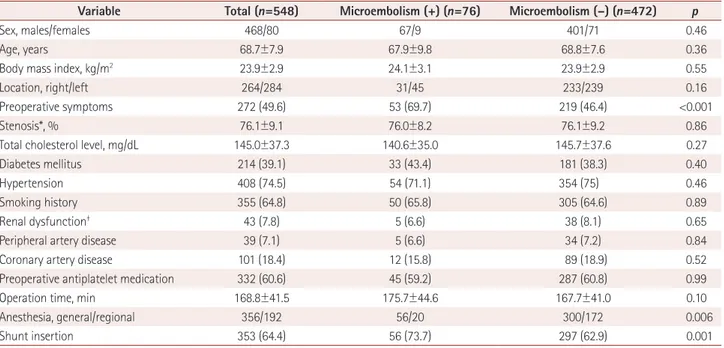
Table 1. Baseline characteristics of the study patients
Variable Total (n=548) Microembolism (+) (n=76) Microembolism (–) (n=472) p
Sex, males/females 468/80 67/9 401/71 0.46
Age, years 68.7±7.9 67.9±9.8 68.8±7.6 0.36
Body mass index, kg/m2 23.9±2.9 24.1±3.1 23.9±2.9 0.55
Location, right/left 264/284 31/45 233/239 0.16
Preoperative symptoms 272 (49.6) 53 (69.7) 219 (46.4) <0.001
Stenosis*, % 76.1±9.1 76.0±8.2 76.1±9.2 0.86
Total cholesterol level, mg/dL 145.0±37.3 140.6±35.0 145.7±37.6 0.27
Diabetes mellitus 214 (39.1) 33 (43.4) 181 (38.3) 0.40
Hypertension 408 (74.5) 54 (71.1) 354 (75) 0.46
Smoking history 355 (64.8) 50 (65.8) 305 (64.6) 0.89
Renal dysfunction† 43 (7.8) 5 (6.6) 38 (8.1) 0.65
Peripheral artery disease 39 (7.1) 5 (6.6) 34 (7.2) 0.84
Coronary artery disease 101 (18.4) 12 (15.8) 89 (18.9) 0.52
Preoperative antiplatelet medication 332 (60.6) 45 (59.2) 287 (60.8) 0.99
Operation time, min 168.8±41.5 175.7±44.6 167.7±41.0 0.10
Anesthesia, general/regional 356/192 56/20 300/172 0.006
Shunt insertion 353 (64.4) 56 (73.7) 297 (62.9) 0.001
Data are mean±standard deviation, n/n, or n (%) values.
*The North American Symptomatic Carotid Endarterectomy Trial method, †Renal dysfunction; serum creatinine level >1.5 mg/dL.
Gwon JG et al.
JCN
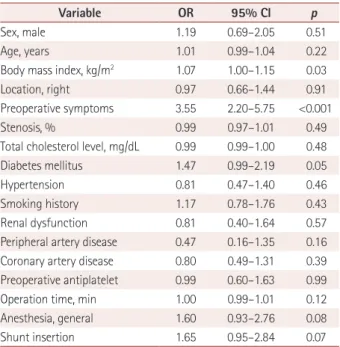
(since their probability values reached p<0.1) were body mass index [odds ratio (OR)=1.07, 95% confidence interval (CI)=1.00–1.15, p=0.03], preoperative symptoms (OR=3.55, 95% CI=2.20–5.75, p<0.001), diabetes mellitus (OR=1.47, 95% CI=0.99–2.19, p=0.05), general anesthesia (OR=1.60, 95% CI=0.93–2.76, p=0.08), and shunt insertion (OR=1.65, 95% CI=0.95–2.84, p=0.07). Multivariate analysis per- formed using these factors (Table 2) showed that preopera- tive neurological symptoms were significantly related to the incidence of microinfarcts (OR=2.93, 95% CI=1.72–5.00, p<0.001). Among the operation-related variables, shunt in- sertion during CEA was the only significant risk factor for mi- croinfarcts (OR=1.42, 95% CI=1.00–2.19, p<0.05) (Table 3).
Seven patients were diagnosed with delayed postoperative cerebral infarction among the cohort of 548 patients. An as- sessment of the association between postoperative microin- farcts and delayed postoperative infarction revealed that 2 of 76 (2.63%) microinfarct-positive patients and 5 of 472 (1.05%) microinfarct-negative patients were diagnosed
with delayed infarction after CEA. The infarction vessel ter- ritory of patients with delayed postoperative infarction who were diagnosed with microinfarcts did not coincide exactly with the microinfarct vessel territory (Fig. 2) The infarction characteristics of all those with delayed postoperative in- farction are presented in Table 4. The interval from CEA to postoperative infarction was 17.28–11.91 months (range=
5–41 months). The presence of a postoperative microinfarct did not significantly increase the incidence of delayed post- operative infarction compared with those who underwent CEA without microinfarcts in the long term follow-up (p=
0.211) (Fig. 3).
DISCUSSION
Based on our current findings and a review of the literature, we hypothesized that there are several possible causes of mi- croinfarct. One is small particles originating from unstable plaques. A surgeon cannot avoid touching the plaque when dissecting the carotid artery, and this may result in plaque fragmentation that could lead to a microembolism. Small plaque-related particles such as lipids or blood clots will arise from the plaque, and they may cause microinfarcts. Further- more, the rate of unstable carotid plaques is higher in symp- tomatic patients than in asymptomatic patients prior to sur- gery.14 The present study suggests that preoperative symptoms are relevant to microinfarcts due to the generation of such particles from unstable plaques.
The second possible cause of an microinfarct could be re- lated to excessive manipulation of the carotid artery. Stork et al.9 reported risk factors for microembolism after CEA.
That study revealed that being female and having left-side CEA are risk factors for microembolism. Those authors con- cluded that gentle manipulation was important. Female pa- tients experience a higher rate of stroke after CEA because the vessel lumen is smaller than in the male ICA, which in- Table 2. Results of univariate analysis of risk factors influencing mi-
croembolization after CEA
Variable OR 95% CI p
Sex, male 1.19 0.69–2.05 0.51
Age, years 1.01 0.99–1.04 0.22
Body mass index, kg/m2 1.07 1.00–1.15 0.03
Location, right 0.97 0.66–1.44 0.91
Preoperative symptoms 3.55 2.20–5.75 <0.001
Stenosis, % 0.99 0.97–1.01 0.49
Total cholesterol level, mg/dL 0.99 0.99–1.00 0.48
Diabetes mellitus 1.47 0.99–2.19 0.05
Hypertension 0.81 0.47–1.40 0.46
Smoking history 1.17 0.78–1.76 0.43
Renal dysfunction 0.81 0.40–1.64 0.57
Peripheral artery disease 0.47 0.16–1.35 0.16 Coronary artery disease 0.80 0.49–1.31 0.39 Preoperative antiplatelet 0.99 0.60–1.63 0.99
Operation time, min 1.00 0.99–1.01 0.12
Anesthesia, general 1.60 0.93–2.76 0.08
Shunt insertion 1.65 0.95–2.84 0.07
CEA: carotid endarterectomy, CI: confidence interval, OR: odds ratio.
Table 3. Results of multivariate analysis of risk factors influencing microembolization after CEA
Variable OR 95% CI p
Body mass index, kg/m2 1.02 0.94–1.11 0.60 Preoperative symptoms 2.93 1.72–5.00 <0.001
Diabetes mellitus 1.39 0.84–2.32 0.19
Anesthesia, general 0.75 0.27–2.04 0.57
Shunt insertion 1.42 1.00–2.19 0.05
CEA: carotid endarterectomy, CI: confidence interval, OR: odds ratio.
Fig. 2. DW-MRI images obtained on postoperative day 2 showing mi- croinfarcts in the middle cerebral artery and posterior cerebral artery border zone (white arrows), and DW-MRI images obtained 14 months postoperatively showing infarction in the middle cerebral artery terri- tory (yellow arrow). DW-MRI: diffusion-weighted magnetic resonance imaging.
Microinfarcts after Carotid Endarterectomy
JCN
creases the technical difficulty of gentle manipulation dur- ing CEA. Stork et al.9 reported that the surgeons who partic- ipated in the study were all right-handed and hence found it difficult to perform the gentle manipulation for left-side CEA. Our present study is also consistent with the hypothe- sis that shunt insertion requires more handling than cases without shunt insertion. This means that selective shunt in- sertion during CEA would be preferable over obligatory shunt insertion. Several previous studies have aimed to identify clin- ical indications for shunting when performing carotid artery cross-clamping. Shin et al.15 suggested that a reduction in the patent segments in the contralateral hemisphere or an absence of both anterior and posterior communicating arteries were independent factors for shunting being required. However, to our knowledge, there is currently no definitive evidence to support the use of selective shunting.
The potential association between postoperative cerebral infarction and microinfarcts remains unclear. Some studies found that microinfarct lesions observed on DW-MRI after CAS did not resolve, instead progressing to cerebral infarc- tions on fluid-attenuated inversion recovery MRI images at 6 months after the procedure. These silent infarcts observed on MRI may contribute to an increased risk of dementia or a threefold increase in the risk of a subsequent stroke.11,16-18 However, Wolf et al.19 found no correlation between micro- embolism and cerebral infarction and no increase in the in- farction rate in the microembolism group during a long-term follow-up. Levi et al.20 reported that microembolisms are as- sociated with an increased risk of stroke and new ischemic lesions on MRI after CEA, and they suggested that a micro- embolism results in failure of the microcirculation due to fre- quent microembolic occlusion. However, that study was lim- ited to the perioperative outcomes only, and so to assess the relationship between microembolism and stroke they assumed that this obstacle in microcirculation occurs only in the pres- ence of frequent microembolic occlusion (in excess of 50 mi- croembolic signals per hour, as measured using transcranial Doppler). In the present study we also found no association between microinfarcts and delayed postoperative infarction.
Moreover, patients with delayed postoperative infarction who had been diagnosed with microinfarcts previously had no corresponding infarction lesion or microinfarct lesion.
This study was subject to some noteworthy limitations.
First, we included only a small cohort of patients with de- layed postoperative infarction (n=7). The inclusion of only seven cases (1.27% of the total cohort) restricted the ability to identify any statistically significant relationship between microinfarcts and postoperative infarctions. Second, the origin of postoperative microinfarcts could not be distin- guished from the cardiac or intracranial vessel origin. Third, there was a lack of baseline information for pre-existing microinfarcts for all patients. Fourth, acute hemodynamic Table 4. Infarction characteristics of the patients with delayed postoperative cerebral infarction
Patient no. Microembolism Symptom Etiology Lesion location Interval between attack and CEA (months)
1 + Dysarthria Artery-to-artery embolism MCA territory 14
2 + Dysarthria Artery-to-artery embolism MCA territory
ACA–MCA border zone 24
3 – Weakness Artery-to-artery embolism ACA–MCA border zone
ACA–MCA border zone 13
4 – Sensory change Artery-to-artery embolism MCA territory 41
5 – Dysarthria Artery-to-artery embolism MCA–PCA border zone 10
6 – Dysarthria Artery-to-artery embolism MCA territory 14
7 – Dysarthria Artery-to-artery embolism MCA territory 5
ACA: anterior cerebral artery, CEA: carotid endarterectomy, MCA: middle cerebral artery, PCA: posterior cerebral artery.
1.0
0.8
0.6
0.4
0.2
0.0
0 20 40 60 80 100 120 Follow up period (month)
Cumulative delayed infarction free rate
Fig. 3. Delayed postoperative infarction-free rate according to the follow-up period.
Microinfarcts negative group Microinfarcts positive group
Gwon JG et al.
JCN
changes may contribute to the occurrence of microembo- lisms during CEA, but unfortunately we were unable to col- lect the relevant information on blood pressure when re- viewing the data.
There are many risk factors for microinfarcts after CEA, including preoperative symptoms and the intraoperative insertion of a shunt. Therefore, when preparing symptom- atic patients for CEA, selective shunt insertion should be considered and plaque stability should be evaluated. We found no association between microinfarcts after CEA and delayed postoperative infarction.
Conflicts of Interest
The authors have no financial conflicts of interest.
REFERENCES
1. North American Symptomatic Carotid Endarterectomy Trial Col- laborators. Beneficial effect of carotid endarterectomy in symptom- atic patients with high-grade carotid stenosis. N Engl J Med 1991;325:
445-453.
2. Halliday A, Mansfield A, Marro J, Peto C, Peto R, Potter J, et al. Pre- vention of disabling and fatal strokes by successful carotid endarter- ectomy in patients without recent neurological symptoms: randomised controlled trial. Lancet 2004;363:1491-1502.
3. Rothwell PM, Gutnikov SA, Warlow CP; European Carotid Surgery Trialist’s Collaboration. Reanalysis of the final results of the Europe- an Carotid Surgery Trial. Stroke 2003;34:514-523.
4. Gupta N, Corriere MA, Dodson TF, Chaikof EL, Beaulieu RJ, Reeves JG, et al. The incidence of microemboli to the brain is less with end- arterectomy than with percutaneous revascularization with distal fil- ters or flow reversal. J Vasc Surg 2011;53:316-322.
5. Lopez OL, Jagust WJ, Dulberg C, Becker JT, DeKosky ST, Fitzpatrick A, et al. Risk factors for mild cognitive impairment in the Cardiovas- cular Health Study Cognition Study: part 2. Arch Neurol 2003;60:1394- 1399.
6. Yamashita H, Fujikawa T, Takami H, Yanai I, Okamoto Y, Morinobu S, et al. Long-term prognosis of patients with major depression and si- lent cerebral infarction. Neuropsychobiology 2010;62:177-181.
7. Wilson TJ, Pandey AS, Stetler WR Jr, Davis MC, Giles DA, Chaudhary N, et al. Dual antiplatelet therapy plus postoperative heparin and dex- tran is safe and effective for reducing risk of embolic stroke during aneurysm coiling. Acta Neurochir (Wien) 2014;156:855-859.
8. Kuliha M, Roubec M, Procházka V, Jonszta T, Hrbácˇ T, Havelka J, et al.
Randomized clinical trial comparing neurological outcomes after ca- rotid endarterectomy or stenting. Br J Surg 2015;102:194-201.
9. Stork JL, Levi CR, Chambers BR, Abbott AL, Donnan GA. Possible determinants of early microembolism after carotid endarterectomy.
Stroke 2002;33:2082-2085.
10. Levi CR, Roberts AK, Fell G, Hoare MC, Royle JP, Chan A, et al. Tran- scranial Doppler microembolus detection in the identification of pa- tients at high risk of perioperative stroke. Eur J Vasc Endovasc Surg 1997;14:170-176.
11. King A, Markus HS. Doppler embolic signals in cerebrovascular dis- ease and prediction of stroke risk: a systematic review and meta- analysis. Stroke 2009;40:3711-3717.
12. Mantese VA, Timaran CH, Chiu D, Begg RJ, Brott TG; CREST Inves- tigators. The Carotid Revascularization Endarterectomy versus Stent- ing Trial (CREST): stenting versus carotid endarterectomy for carot- id disease. Stroke 2010;41(10 Suppl):S31-S34.
13. Adams HP Jr, Bendixen BH, Kappelle LJ, Biller J, Love BB, Gordon DL, et al. Classification of subtype of acute ischemic stroke. Defini- tions for use in a multicenter clinical trial. TOAST. Trial of Org 10172 in Acute Stroke Treatment. Stroke 1993;24:35-41.
14. Gaunt ME, Brown L, Hartshorne T, Bell PR, Naylor AR. Unstable carotid plaques: preoperative identification and association with in- traoperative embolisation detected by transcranial Doppler. Eur J Vasc Endovasc Surg 1996;11:78-82.
15. Shin S, Kwon TW, Cho YP, Shin JH, Yi A, Kim H, et al. Preoperative magnetic resonance angiography as a predictive test for cerebral isch- emia during carotid endarterectomy. World J Surg 2013;37:663-670.
16. Palombo G, Faraglia V, Stella N, Giugni E, Bozzao A, Taurino M. Late evaluation of silent cerebral ischemia detected by diffusion-weighted MR imaging after filter-protected carotid artery stenting. AJNR Am J Neuroradiol 2008;29:1340-1343.
17. Vermeer SE, Prins ND, den Heijer T, Hofman A, Koudstaal PJ, Bre- teler MM. Silent brain infarcts and the risk of dementia and cogni- tive decline. N Engl J Med 2003;348:1215-1222.
18. Vermeer SE, Hollander M, van Dijk EJ, Hofman A, Koudstaal PJ, Breteler MM; Rotterdam Scan Study. Silent brain infarcts and white matter lesions increase stroke risk in the general population: the Rot- terdam Scan Study. Stroke 2003;34:1126-1129.
19. Wolf O, Heider P, Heinz M, Poppert H, Sander D, Greil O, et al. Mi- croembolic signals detected by transcranial Doppler sonography during carotid endarterectomy and correlation with serial diffusion- weighted imaging. Stroke 2004;35:e373-e375.
20. Levi CR, O’Malley HM, Fell G, Roberts AK, Hoare MC, Royle JP, et al. Transcranial Doppler detected cerebral microembolism following carotid endarterectomy. High microembolic signal loads predict postoperative cerebral ischaemia. Brain 1997;120(Pt 4):621-629.