Copyright © 2011 Korean Neurological Association 43
Print ISSN 1738-6586 / On-line ISSN 2005-5013 10.3988/jcn.2011.7.1.43 CASE REPORT
J Clin Neurol 2011;7:43-46
Introduction
Lambert-Eaton myasthenic syndrome (LEMS) is well known to be a classical paraneoplastic syndrome of small cell lung car- cinoma (SCLC). Association of myasthenia gravis (MG) and SCLC is rarely known. We report a case of acetylcholine recep- tor antibody (AChR-ab) positive MG and SCLC.
Case Report
In June 1987, a 65-year-old African-American man noticed a weakness in leg muscles a week before admission to a local hospital. Weakness got worse and spread to upper arms. Short- ness of breath developed, requiring endotracheal intubation and ventilatory support. Initially, Guillain-Barré syndrome was sus- pected. However, spinal fluid evaluation was normal with pro- tein at 30 mg/dL. CK and muscle biopsy were also normal.
The patient was transferred to the University of Alabama Uni- versity Hospital. Neurologic examination showed bilateral pto- sis, marked weakness (1-2 Medical Research Council grade) in proximal muscles, moderate (3 Medical Research Council grade) weakness in distal muscles, normal reflexes except diminished ankle reflex and normal sensory function. Edrophonium test was
equivocal and another spinal fluid evaluation was also normal with protein at 21 mg/dL. Because of marked decremental re- sponse in the repetitive nerve stimulation (RNS) test, the diag- nosis of MG crisis was made. Plasmaphresis was initiated to- gether with prednisone (60 mg a day) and pyridostigmine (540 mg a day). Third spinal fluid evaluation on the third admission date was also normal with protein at 36 mg/dL. Another edro- phonium test on the 12th admission date showed a definite posi- tive response. With continued treatment for MG crisis, MG Foun- dation of America status was improved from V to IIIa in three weeks period of time.
Chest X-ray showed a nodule (2×3.5 cm) in the right upper lobe and hilar adenopathy. CT scan showed a lobulated paren- chymal mass with a soft tissue density in the right hilar area, and many scattered small nodules in the right posterior pleural space. Thymoma was not found. Transthoracic needle biopsy confirmed the diagnosis of SCLC. The patient was treated with three days of chemotherapy (CIS-Platinum and VP-16) for SCLC. One month after the chemotherapy, the patient developed septicemia and respiratory failure leading to adult respiratory distress syndrome and died two months after onset of weakness.
Nerve conduction study (NCS) on the first admission day:
Motor NCS showed mild prolongation of terminal latency and
Seropositive Myasthenia Gravis Associated with Small-Cell Lung Carcinoma
Masayuki Ohira,a Dushin Jeong,b Shin J Ohc
aDepartment of Neurology, Keiyo University Medical School, Tokyo, Japan
bDepartment of Neurology, Soonchunhang University Medical School, Cheonan, Korea
CDepartment of Neurology, University of Alabama at Birmingham, Veterans Medical Center, Birmingham, Alabama, USA
Received June 8, 2010 Revised September 1, 2010 Accepted September 1, 2010 Correspondence Shin J Oh, MD
Department of Neurology,
University of Alabama at Birmingham, Veterans Medical Center,
Birmingham, Alabama, USA Tel +1-205-934-2121 Fax +1-205-975-6758 E-mail shinjoh@uab.edu
BackgroundzzLambert-Eaton myasthenic syndrome is well known to be a classical paraneo- plastic syndrome of small cell lung carcinoma (SCLC). Three cases of seronegative myasthenia gravis (MG) and SCLC were previously reported.
Case ReportzzA 65-year-old man developed a severe progressive respiratory failure with clini- cal features of MG. Tests showed a decremental response in the repetitive nerve stimulation test, abnormal single-fiber electromyography, and positive acetylcholine receptor antibody. SCLC was confirmed by the lung biopsy.
ConclusionszzThis case represents the first case of seropositive MG and SCLC.
J Clin Neurol 2011;7:43-46 Key Wordszz acetylcholine receptor antibody, myasthenia gravis, small cell lung cancer.
Seropositive Myasthenia Gravis Associated with Small-Cell Lung Carcinoma
44 J Clin Neurol 2011;7:43-46
low compound muscle action potential (CMAP) amplitude in the median (4.8 ms/4 mV), ulnar (3.6 ms/3.0 mV), peroneal (7.0 ms/1 mV), and posterior tibial (9.1 ms/0.4 mV) nerves and mildly slow motor NCV (38.1 and 37.5 m/s) in two posterior tibial nerves. F-wave latency was mildly prolonged in all mo- tor nerve. Sensory NCS showed low CNAP amplitude and mildly slow NCV in the median (40 m/s; 8 μV), ulnar (34.3 m/s;
7 μV), and sural (33.3 m/s; 2 μV) nerves. These findings were interpreted as mild axonal motor-sensory neuropathy. NCS was not repeated.
The RNS test on the first admission day showed a low CMAP
amplitude and a marked decrement at low rate stimulation (LRS; 2-5 Hz) and high rate stimulation (HRS; 50 Hz) in the abductor digiti quinti muscle, and a low CMAP amplitude and a marked decrement at LRS in the orbicularis oculi muscle (Ta- ble 1).1 There was an improvement in decrement with edropho- nium though clinical improvement was equivocal. These were interpreted to be indicative of severe MG.1 Further deteriora- tion was noted in the RNS test on the 11th day. On the 12th day, there was minimal improvement in the RNS response. There was a definite electrophysiological and clinical improvement with edrophonium injection. On the 24rd day, the RNS test showed
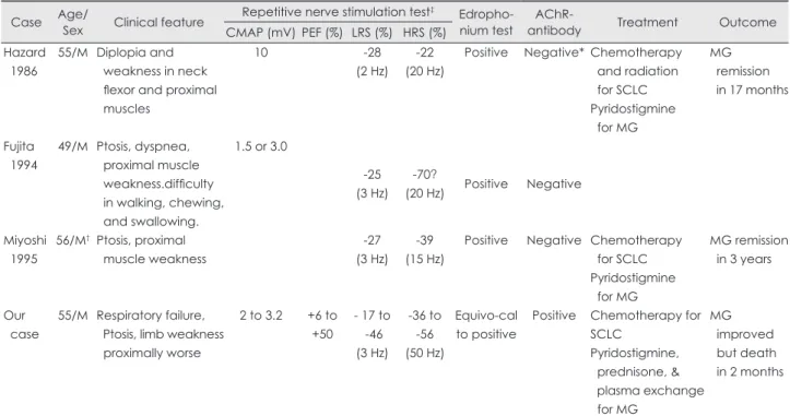
Table 2. Clinical, electrophysiological and immunological features in four cases of MG with SCLC Case Age/Sex Clinical feature Repetitive nerve stimulation test‡ Edropho-
nium test AChR-
antibody Treatment Outcome
CMAP (mV) PEF (%) LRS (%) HRS (%) Hazard
1986
55/M Diplopia and weakness in neck flexor and proximal muscles
10 -28
(2 Hz) -22 (20 Hz)
Positive Negative* Chemotherapy and radiation for SCLC Pyridostigmine for MG
MG remission in 17 months
Fujita 1994
49/M Ptosis, dyspnea, proximal muscle weakness.difficulty in walking, chewing, and swallowing.
1.5 or 3.0
-25 (3 Hz)
-70?
(20 Hz) Positive Negative
Miyoshi 1995
56/M† Ptosis, proximal muscle weakness
-27 (3 Hz)
-39 (15 Hz)
Positive Negative Chemotherapy for SCLC Pyridostigmine for MG
MG remission in 3 years
Our case
55/M Respiratory failure, Ptosis, limb weakness proximally worse
2 to 3.2 +6 to +50
- 17 to -46 (3 Hz)
-36 to -56 (50 Hz)
Equivo-cal to positive
Positive Chemotherapy for SCLC
Pyridostigmine, prednisone, &
plasma exchange for MG
MG improved but death in 2 months
*Initially not tested but negative at MG remission, †SCLC found 20 months after MG diagnosis, An artifact? ‡RNS test on ADQ muscle except APB muscle in Miyoshi’s case.
-: decrement, +: increment, AChR: acetylcholine receptor, ADQ: abductor digiti quinti muscle, CMAP: compound muscle action poten- tial, HRS: high rate stimulation, LRS: low rate stimulation, MG: myasthenia gravis, PEF: post-exercise facilitation, RNS: repetitive nerve stimu- lation, SCLC: small cell lung carcinoma.
Table 1. Repetitive nerve stimulation data
Day 1st day 11th day 12th day 24th day‡ Normal
Muscle ADQ Orbicularis oculi ADQ ADQ ADQ FCU ADQ FCU Orbicularis oculi
CMAP (mV) 3.0 0.3 2.0 3.2 3.0† 6.0 4.0 4.8 3.0 1.1
PEF (%) +50 +6 +17 0 +37
LRS (%) 2 Hz -29 -15* -50 -35 -25 -3 0 0 -7 -8 -8
3 Hz -17 -46 -38 -6 -6 -10 -7 -9 -8
5 Hz -24 -26 -24 -11 +3 0 -5 -11 -8
HRS (%) 50 Hz -36 -54 -56 -38 -18 -19
PTE (%) 5 Hz -29 -17 +5 -8 -11
MGFA V V V IIIa
*2 minutes after injection, †3 minutes after injection, ‡2 hour after pyridostigmine by mouth.
-: decrement, +: increment, ADQ: abductor digiti quinti muscle, CMAP: compound muscle action potential, FCU: flexor carpi ulnaris muscle, HRS: high rate stimulation, LRS: low rate stimulation, MGFA: MG Foundation of America, PEF: post-exercise facilitation.
Ohira M et al.
www.thejcn.com 45 normal findings except a decrement at the LRS in the flexor
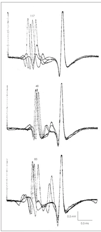
carpi ulnaris muscle. At that time, the patient was in MG Foun- dation of America status IIIa. Single-fiber electromyography in the extensor digitorum communis muscle on the 24th day showed markedly increased mean value of mean-consecutive- difference (174 μs) and frequent blocking in 7 SFPPs (Fig. 1).
AChR-ab tests were positive by two laboratories: 3.4 (nor- mal<0.7 nmol/L) and 30.55 (normal <0.03 nmol/L). Voltage gated calcium channel antibody was not tested in this patient because this test was not available in 1987.
Discussion
Our patient had seropositive MG, SCLC and mild axonal motor-sensory neuropathy. Guillain-Barré syndrome was initial- ly suspected but less likely in view of the repeated normal spi- nal fluid findings and mild axonal neuropathy in the NCS. Ax- onal neuropathy is most likely a paraneoplastic syndrome of SCLC.
The RNS tests during MG crisis in our case showed a rather typical RNS test of myasthenic crisis: low CMAP amplitude and decrement at LRS and HRS. In myasthenic crisis, this pat- tern was observed in 25% of cases.2 Lower CMAP, though with- in normal limit, and decrement at LRS and HRS are a rather characteristic pattern in severe MG.1 With clinical improve- ment, the CMAP amplitude became normal and decrement at LRS was the only abnormality in this case. This is the most co- mmon RNS pattern in MG.1 Single-fiber electromyography showed findings typical of neuromuscular transmission defect.
LEMS was ruled out by a lack of an incremental response af- ter brief exercise [post-exercise facilitation (PEF)] or at HRS.3 50% increment in PEF in the abductor digiti quinti muscle at the 11th day is slightly higher than normal 37% limit but defi- nitely lower than 60% increment, the required criteria for the diagnosis of LEMS.3 Transient increment in PEF or HRS was previously reported in a few severe MG cases.3
Fifty percentage of LEMS patients were known to have car- cinoma, mostly SCLC.4 Only three cases showing combined MG and SCLC features have been reported (Table 2).5-7 They were male and aged 49 to 56. All three cases were seronega- tive. In two cases, SCLC was found at the time when the diag- nosis of MG was made. In Myoshi’s case, SCLC was found 18 months after the diagnosis of MG. All three cases had classical oculo-proximal muscle weakness of MG.7 One case had bul- bar weakness. Ours is the only one with MG crisis. Diagnosis of MG was confirmed by edrophonium test and decrement at LRS (25-28%) and HRS (22-70%). The diagnosis of MG in Fujita’s case is questionable in view of “decrement” in 20 Hz stimulation due to artifacts and discrepancy in the first CMAP amplitude at 3 Hz (1.5 mV) and 20 Hz stimulation (3.0 mV).5
This may represent an 100% PEF typical of LEMS. With pyr- idostigmine therapy for MG and chemotherapy for SCLC, MG was in remission in two cases in 17-36 months. Our pa- tient died 2 months after the diagnosis of MG. No follow-up information was available in one case. One may argue that our case represents a case of LEMS with positive AChR-ab.
117
48
83
0.5 mV 0.5 ms
Fig. 1. Three single fiber potential pairs showing a marked jitter.
Number above single fiber potentials represent MCD value in μs.
Neuromuscular blocking is noted in the second slave potential in the bottom single fiber potential pairs. MCD: mean-consecutive-differ- ence.
Seropositive Myasthenia Gravis Associated with Small-Cell Lung Carcinoma
46 J Clin Neurol 2011;7:43-46
Lennon8 found a positive AChR-ab in 13% of LEMS patients.
However, electrophysiological findings do not support the di- agnosis of LEMS. Thus, we believe that our patient is the first one with seropositive MG and SCLC.
Conflicts of Interest
The authors have no financial conflicts of interest.
REFERENCES
1. Oh SJ, Eslami N, Nishihira T, Sarala PK, Kuba T, Elmore RS, et al.
Electrophysiological and clinical correlation in myasthenia gravis.
Ann Neurol 1982;12:348-354.
2. Goli SK, Claussen GC, Oh SJ. Repetitive nerve stimulation test in myasthenic crisis. Neurology 2009;72:A128 (Suppl 3).
3. Oh SJ, Kurokawa K, Claussen GC, Ryan HF Jr. Electrophysiological diagnostic criteria of Lambert-Eaton myasthenic syndrome. Muscle Nerve 2005;32:515-520.
4. Titulaer MJ, Verschuuren JJ. Lambert-Eaton myasthenic syndrome:
tumor versus nontumor forms. Ann N Y Acad Sci 2008;1132:129-134.
5. Fujita J, Yamadori I, Yamaji Y, Yamagishi Y, Takigawa K, Takahara J.
Myasthenia gravis associated with small-cell carcinoma of the lung.
Chest 1994;105:624-625.
6. Hazard PB, Bertorini TE, Griffin JP. Myasthenia gravis associated with small cell carcinoma of the lung. J Tenn Med Assoc 1986;79:273- 276.
7. Miyoshi R, Yamaji Y, Shima S, Fujita J, Okada H, Takahara J. [A case of small cell lung cancer that developed during therapy for myasthe- nia gravis]. Nihon Kyobu Shikkan Gakkai Zasshi 1995;33:456-462.
8. Lennon VA. Serologic profile of myasthenia gravis and distinction from the Lambert-Eaton syndrome. Neurology 1997;48:S23-S27.